CANINE iPS CELLS AND METHOD OF PRODUCING SAME

a technology of canine ips and ips cells, which is applied in the field of producing canine ips cells, can solve the problems of mice and rats, graft rejection, and inability to permit long-term follow-up examination after cell transplantation, and no report on actual establishmen

Inactive Publication Date: 2011-02-10
KYOTO UNIV
View PDF1 Cites 3 Cited by
  • Summary
  • Abstract
  • Description
  • Claims
  • Application Information

AI Technical Summary

Benefits of technology

According to the present invention, canine iPS cells can be stably produced from canine somatic cells and can be provided by using the method of production thereof, whereby long-term follow-up examination after cell or tissue transplantation in animal experiments is possible before clinically applying human iPS cells, and the results obtained by the experiments are effectively utilizable.
Additionally, production of canine iPS cells from each individual by the present invention would radically revolutionize currently available therapies in veterinary medicine, making tailor-made treatment of diseased dogs possible. Furthermore, utilizing canine iPS cells according to the present invention will make it possible to develop veterinary drugs, and to test chemical substances for their toxicity, without using individual dogs.

Problems solved by technology

However, ES cell transplantation can cause graft rejection because it is a form of allotransplantation, and has been viewed as posing ethical problems, including destructive use of human embryos and employment of abortive fetuses.
However, small animals, such as mice and rats, do not permit long-term follow-up examination after undergoing cell transplantation because of their short longevity.
To this end, it is necessary to generate a canine iPS cell; however, no reports are available on actual establishment thereof.

Method used


Image

  • CANINE iPS CELLS AND METHOD OF PRODUCING SAME
  • CANINE iPS CELLS AND METHOD OF PRODUCING SAME
  • CANINE iPS CELLS AND METHOD OF PRODUCING SAME

Examples


example 1

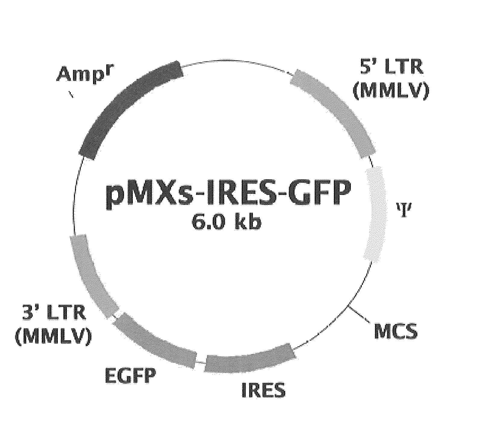
Construction of Retroviral Vectors

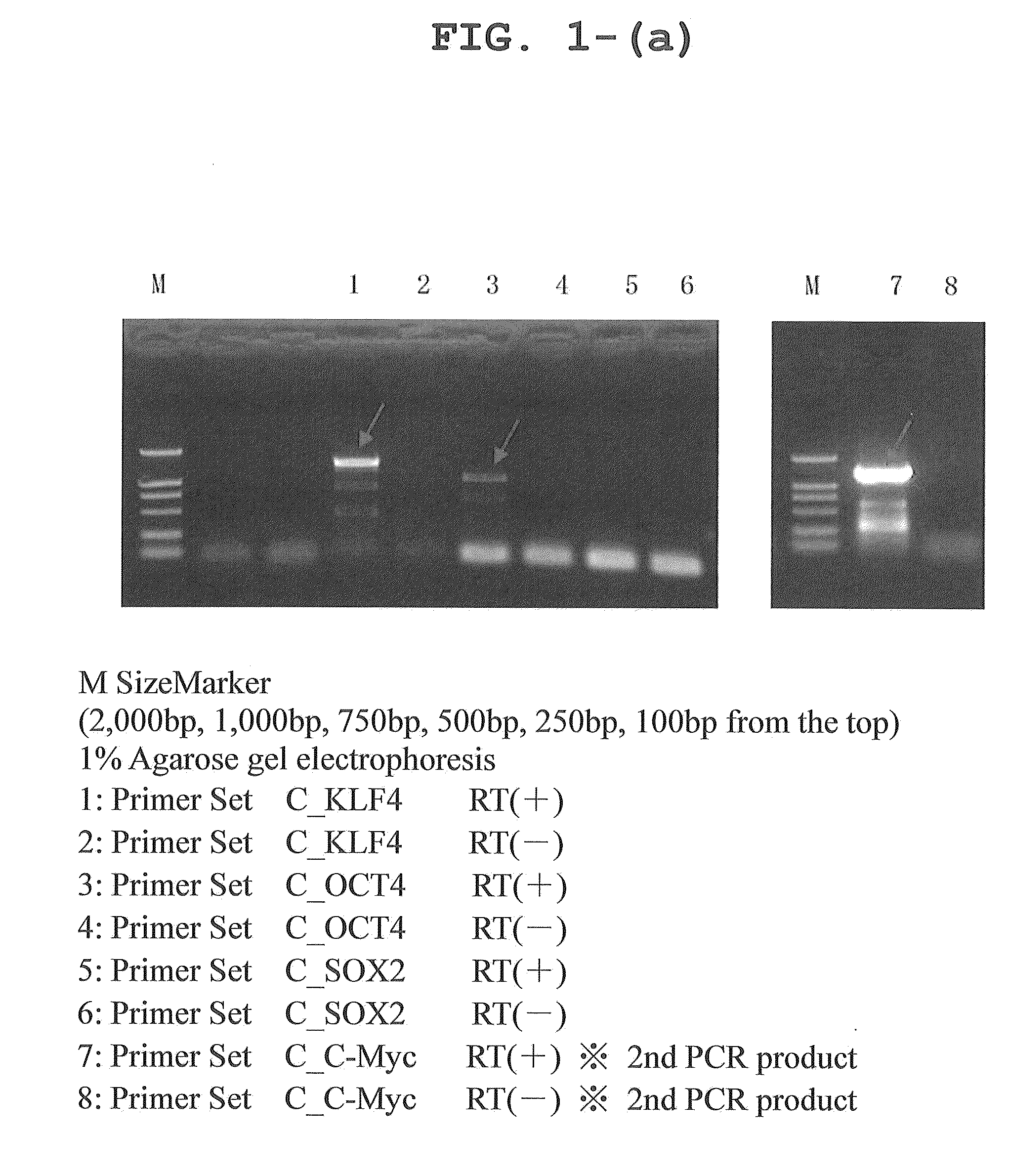
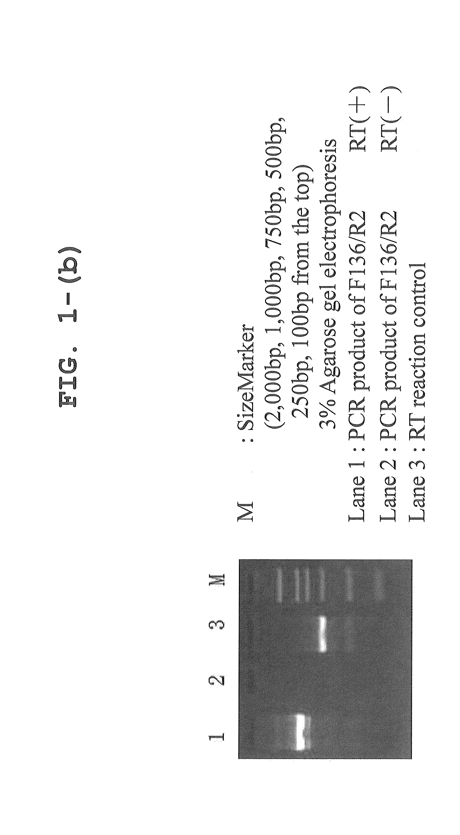
Primers were designed on the basis of cDNA sequence information registered with a public database, and each of the canine genes Oct3 / 4, Klf4, Sox2, and c-Myc was amplified by RT-PCR from RNA extracted from cerebrum, retina, gastric mucosa, skin, skeletal muscle, lung, and testis. Since Sox2 was not amplified well, a comparison was made between the predicted canine Sox2 sequence and human Sox2 sequence on the database; it was found that an extra sequence of 165 amino acids was present at the N-terminus of canine Sox2. Then, this extra portion was removed to re-design a primer, using which RT-PCR was performed with successful results of amplification of the canine Sox2 gene. The sequences of the primers used for the PCR amplification are shown in Table 1.

TABLE 1PrimerReferencePrimer SetPrimerlength(bp)Base sequence (5′→ 3′)sequenceC—KLF4KLF4-F40TTAATTAAGGATCCACCATGGCTGTCAGCGACGCTCTGCTSEQ ID NO: 9KLF4-R42GGCCTGCAGGAATTCTTAAAAGTGCCTCTTCATGTGTAAGGCSEQ ID...

example 2

Preparation of Virus

100 μL of the Fugene 6 transfection reagent (Roche) was placed in a 10 cm Petri dish containing 6×10⁴ previously seeded Plat-GP cells, and the dish was allowed to stand at room temperature for 5 minutes. Subsequently, each retroviral vector and 3 μg of pCMV-VSVG were added, and the dish was further allowed to stand at room temperature for 15 minutes, after which the dish was added to a culture broth for Plat-GP cells. The cells were cultured at 37° C. in the presence of 5% CO2 using the culture broth of a DME medium (Invitrogen) supplemented with 0.5% antibiotic and 10% FBS (final concentrations). The medium was replaced with a fresh supply 24 hours after transfection. The culture supernatant was recovered 48, 60, and 72 hours after transfection, and the recovered supernatants were combined to obtain a virus-containing liquid. The virus-containing liquid was filtered through a 0.45 μm Millipore filter, and polybrene was added at 4 μg / mL to yield a viral liquid.

example 3

Efficiency of Transfection Using Retroviral Vector

Canine fibroblasts were obtained by extirpating a fetus from a beagle dog at day 30 of gestation (purchased from ORIENTAL BIO Co., Ltd.), and shredding and enzymatically treating the fetal tissue. The canine fibroblasts were transfected with a reprogramming gene by retroviral infection, and the GFP expression level after the infection was analyzed by flow cytometry. On day 5 after the infection, about 60% of the fibroblasts became positive for GFP, demonstrating highly efficient transfer of the gene (FIGS. 2 and 3).


PUM

PropertyMeasurementUnit
lengthaaaaaaaaaa

Abstract

Provided are a method of producing canine iPS cells, comprising (a) the step of bringing into contact with each other a canine somatic cell and a nuclear reprogramming factor, and (b) the step of culturing the cell in a medium containing at least one substance selected from the group consisting of a mitogen-activated protein kinase kinase inhibitor, an activin receptor-like kinase inhibitor, a glycogen synthase kinase inhibitor, a L-type calcium channel agonist and a DNA methylation inhibitor, and a leukemia inhibitory factor, and canine iPS cells that can be obtained by the method.

Description

CROSS-REFERENCE TO RELATED APPLICATIONThis application claims the benefit of Japanese Patent Application No. 2009-185268 filed Aug. 7, 2009, the content of which is incorporated herein by reference.FIELD OF THE INVENTIONThe present invention relates to a method of producing a canine iPS cell, specifically to a method of producing a canine iPS cell by transferring a nuclear reprogramming factor to a canine somatic cell, and culturing the cell in a medium containing a defined reprogramming efficiency improver. The present invention also relates to canine iPS cells transfected with the nuclear reprogramming factor.BACKGROUND OF THE INVENTIONAn induced pluripotent stem (iPS) cell is a cell generated by transferring a defined nuclear reprogramming factor to a somatic cell to confer pluripotency to the somatic cell. The term pluripotency refers to the potential for differentiating into a wide variety of tissues; it is believed that tissue degenerative diseases such as Parkinson's disease ...

Claims


Application Information

Patent Timeline
Patent Type & Authority Applications(United States)
IPC IPC(8): C12N15/00C12N5/07
CPCC12N5/0696C12N2501/065C12N2510/00C12N2501/115C12N2501/235C12N2501/727C12N2501/999C12N2501/603C12N2501/604C12N2501/606C12N5/10C12N2501/998C12N2501/602
Inventor NAKAMURA, TATSUOSHIMADA, HIDENORI
Owner KYOTO UNIV
Why Eureka
Try Eureka