Canine iPS cells and method of producing same
a technology of canine ips and ips cells, which is applied in the field of producing canine ips cells, can solve the problems of graft rejection, mice and rats, and do not permit long-term follow-up examination after cell transplantation, and achieve the effect of stably producing and effectively utilizabl
- Summary
- Abstract
- Description
- Claims
- Application Information
AI Technical Summary
Benefits of technology
Problems solved by technology
Method used
Image
Examples
example 1
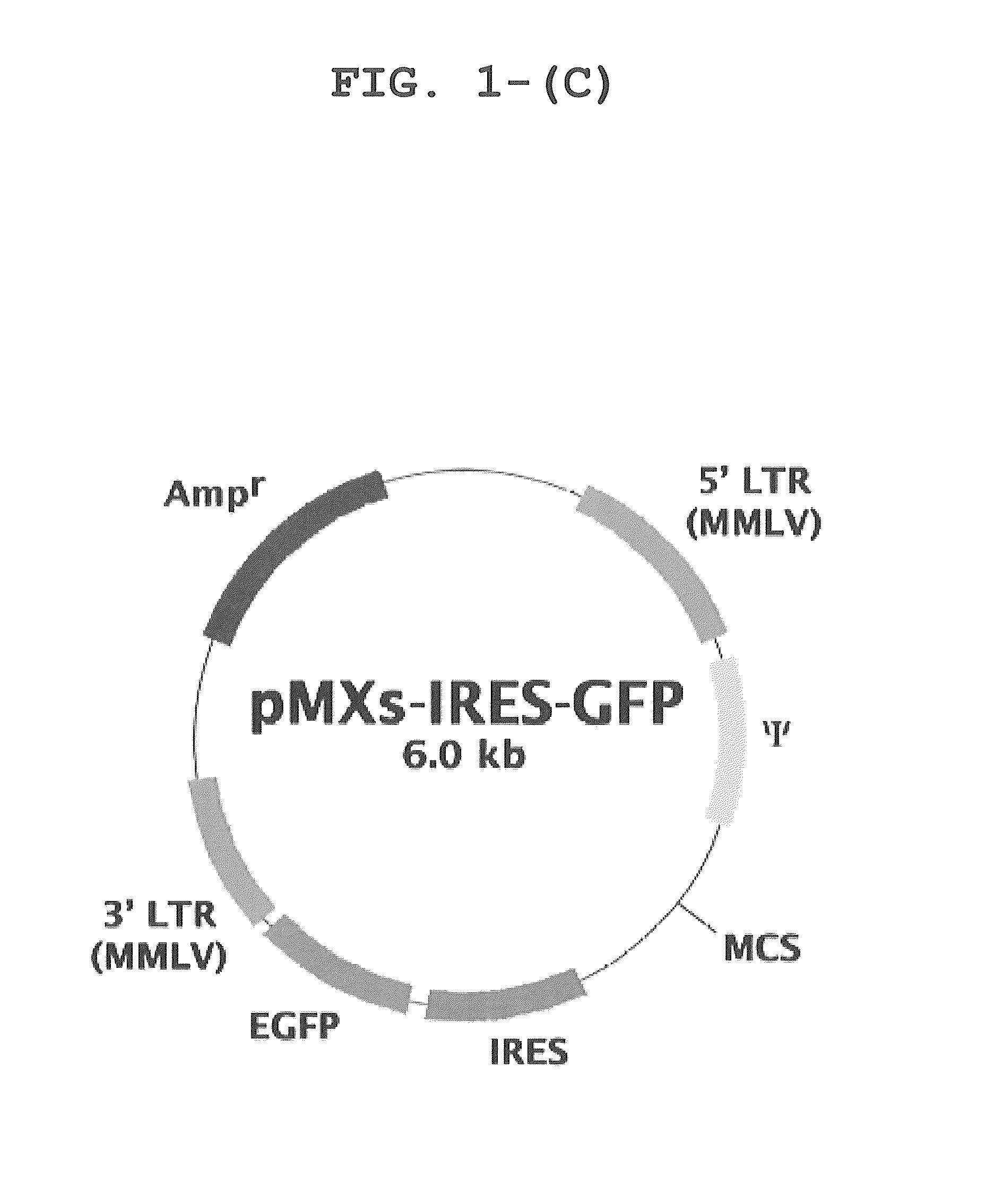
Construction of Retroviral Vectors
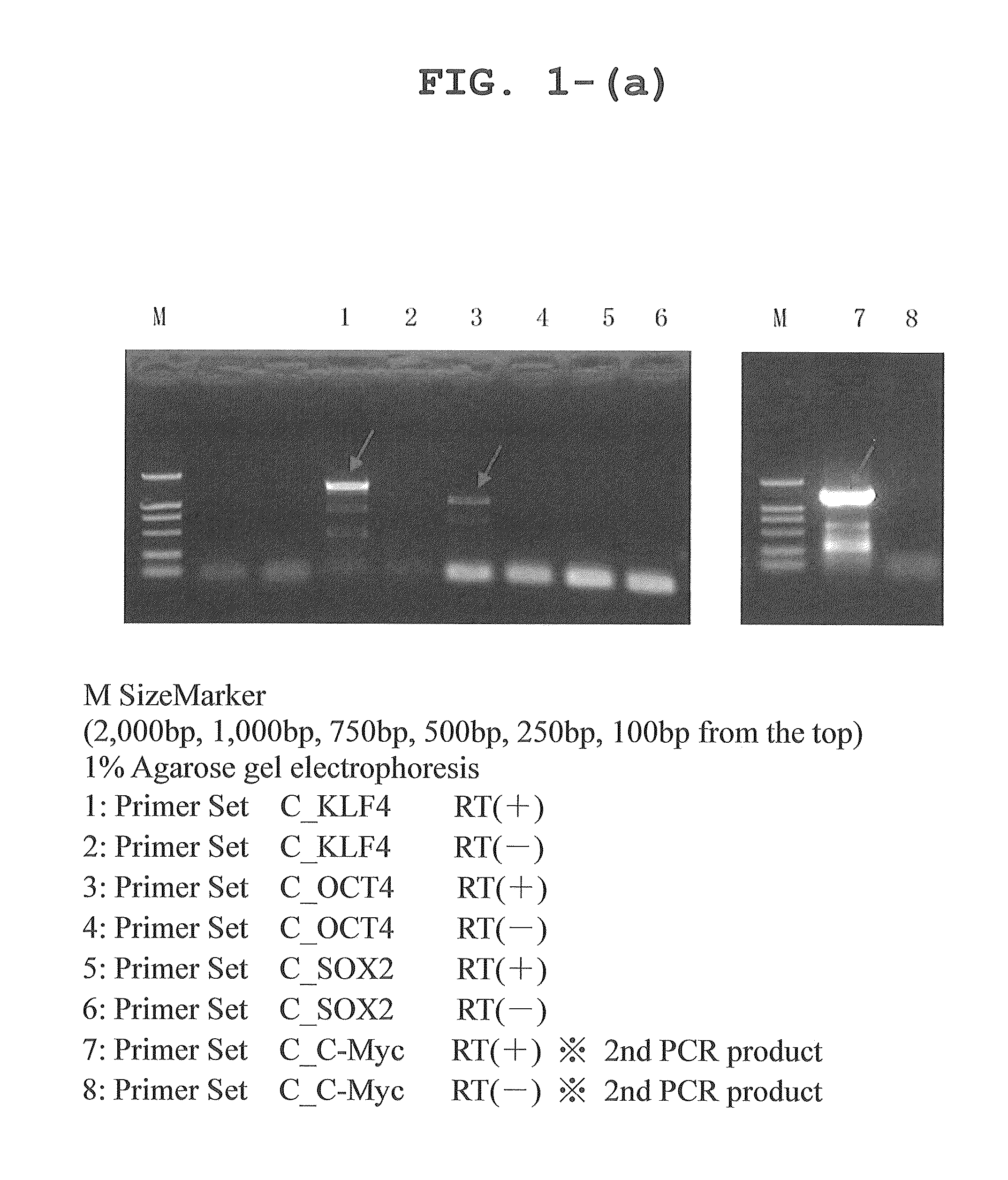
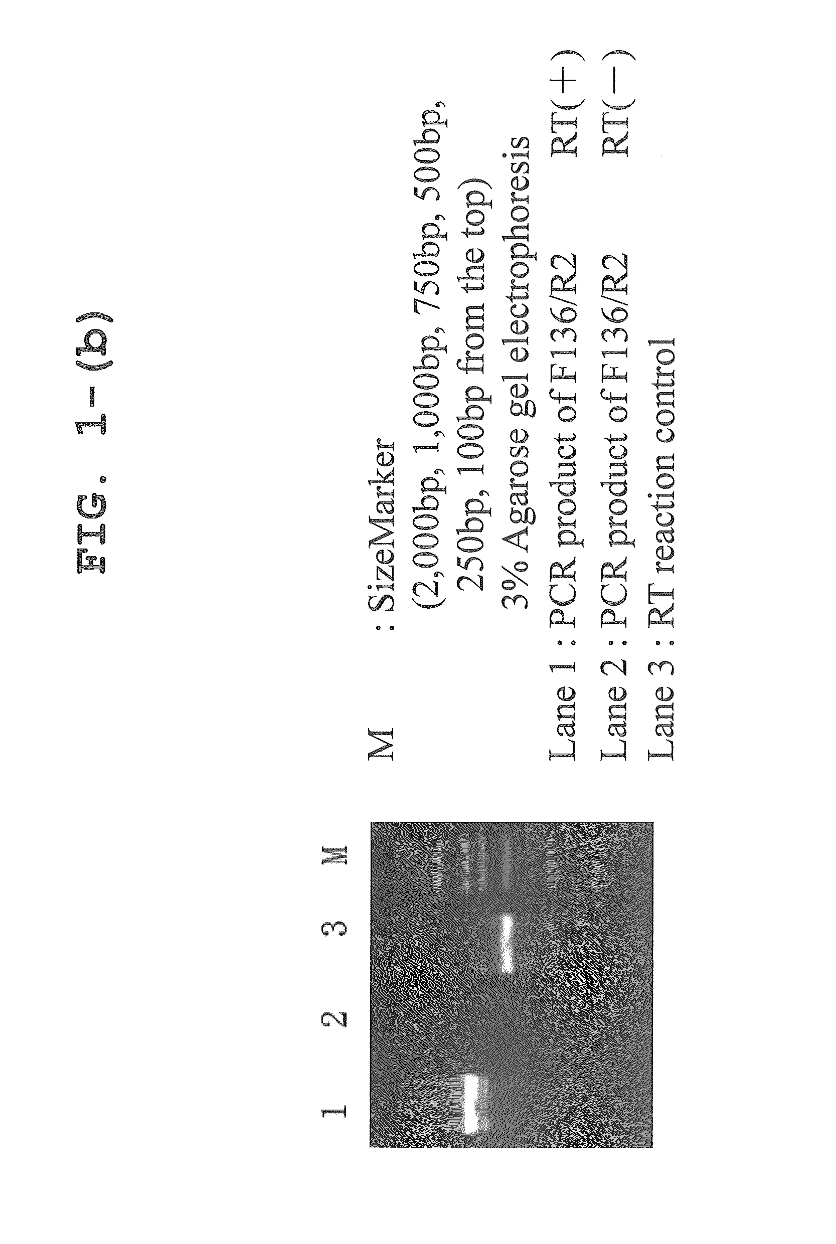
[0082]Primers were designed on the basis of cDNA sequence information registered with a public database, and each of the canine genes Oct3 / 4, Klf4, Sox2, and c-Myc was amplified by RT-PCR from RNA extracted from cerebrum, retina, gastric mucosa, skin, skeletal muscle, lung, and testis. Since Sox2 was not amplified well, a comparison was made between the predicted canine Sox2 sequence and human Sox2 sequence on the database; it was found that an extra sequence of 165 amino acids was present at the N-terminus of canine Sox2. Then, this extra portion was removed to re-design a primer, using which RT-PCR was performed with successful results of amplification of the canine Sox2 gene. The sequences of the primers used for the PCR amplification are shown in Table 1.
[0083]
TABLE 1PrimerReferencePrimer SetPrimerlength(bp)Base sequence (5′→ 3′)sequenceC—KLF4KLF4-F40TTAATTAAGGATCCACCATGGCTGTCAGCGACGCTCTGCTSEQ ID NO: 9KLF4-R42GGCCTGCAGGAATTCTTAAAAGTGCCTCTTCATGTG...
example 2
Preparation of Virus
[0086]100 μL of the Fugene 6 transfection reagent (Roche) was placed in a 10 cm Petri dish containing 6×10⁴ previously seeded Plat-GP cells, and the dish was allowed to stand at room temperature for 5 minutes. Subsequently, each retroviral vector and 3 μg of pCMV-VSVG were added, and the dish was further allowed to stand at room temperature for 15 minutes, after which the dish was added to a culture broth for Plat-GP cells. The cells were cultured at 37° C. in the presence of 5% CO2 using the culture broth of a DME medium (Invitrogen) supplemented with 0.5% antibiotic and 10% FBS (final concentrations). The medium was replaced with a fresh supply 24 hours after transfection. The culture supernatant was recovered 48, 60, and 72 hours after transfection, and the recovered supernatants were combined to obtain a virus-containing liquid. The virus-containing liquid was filtered through a 0.45 μm Millipore filter, and polybrene was added at 4 μg / mL to yield a viral liq...
example 3
Efficiency of Transfection Using Retroviral Vector
[0087]Canine fibroblasts were obtained by extirpating a fetus from a beagle dog at day 30 of gestation (purchased from ORIENTAL BIO Co., Ltd.), and shredding and enzymatically treating the fetal tissue. The canine fibroblasts were transfected with a reprogramming gene by retroviral infection, and the GFP expression level after the infection was analyzed by flow cytometry. On day 5 after the infection, about 60% of the fibroblasts became positive for GFP, demonstrating highly efficient transfer of the gene (FIGS. 2 and 3).
PUM
| Property | Measurement | Unit |
|---|---|---|
| pH | aaaaa | aaaaa |
| time | aaaaa | aaaaa |
| time | aaaaa | aaaaa |
Abstract
Description
Claims
Application Information
 - R&D
- Intellectual Property
- Life Sciences
- Materials
- Tech Scout
- Unparalleled Data Quality
- Higher Quality Content
- 60% Fewer Hallucinations
Browse by: Latest US Patents, China's latest patents, Technical Efficacy Thesaurus, Application Domain, Technology Topic, Popular Technical Reports.
© 2025 PatSnap. All rights reserved.Legal|Privacy policy|Modern Slavery Act Transparency Statement|Sitemap|About US| Contact US: help@patsnap.com


