Vascular ultrasound intima-media thickness (IMT) measurement system

a technology of imt measurement and vascular ultrasound, which is applied in the field of data processing, data storage, imaging systems, etc., can solve the problems of difficult automated recognition process, time-consuming and non-practical clinical trials, and complicated imt measurement using conventional systems

Inactive Publication Date: 2012-11-20
ATHEROPOINT
View PDF60 Cites 13 Cited by
  • Summary
  • Abstract
  • Description
  • Claims
  • Application Information

AI Technical Summary

Problems solved by technology

These challenges have complicated IMT measurement using conventional systems.
Because conventional systems may use a manual method in a ROI, it is time consuming and non-practical for clinical trials and where the need is to process hundred's of patients a day or where there is very little time for examination or where the image quality may not be very good in terms of signal-to-noise ratio (SNR) or CNR or where the proximal wall may have a lot of calcium.
The automated recognition process is difficult, given the Jugular vein in the neighborhood.
Because the ultrasound vascular scans do not always have the vessel orientation horizontal with respect bottom edge of the image, manual methods can pose a further challenge towards the region of interest estimation.(4) Guiding Method for the Calibration System: Because the calibration phase is followed by the recognition phase, the calibration system becomes very robust because the calibration processing is done in the region of interest determined by the automated recognition system.
Though, speckle reduction is common in ultrasound imaging, the way speckle reduction is used in the various embodiments described herein is very conservative.
Though conventional methods are generally suitable, conventional methods have certain problems related to accuracy, speed, and reliability.
IMT estimation having a value close to 1 mm is a very challenging task in ultrasound images due to large numbers of variabilities such as: poor contrast, orientation of the vessels, varying thickness, sudden f...

Method used


Image

  • Vascular ultrasound intima-media thickness (IMT) measurement system
  • Vascular ultrasound intima-media thickness (IMT) measurement system
  • Vascular ultrasound intima-media thickness (IMT) measurement system

Examples


Embodiment Construction

[0038]In the following description, for purposes of explanation, numerous specific details are set forth in order to provide a thorough understanding of the various embodiments. It will be evident, however, to one of ordinary skill in the art that the various embodiments may be practiced without these specific details.

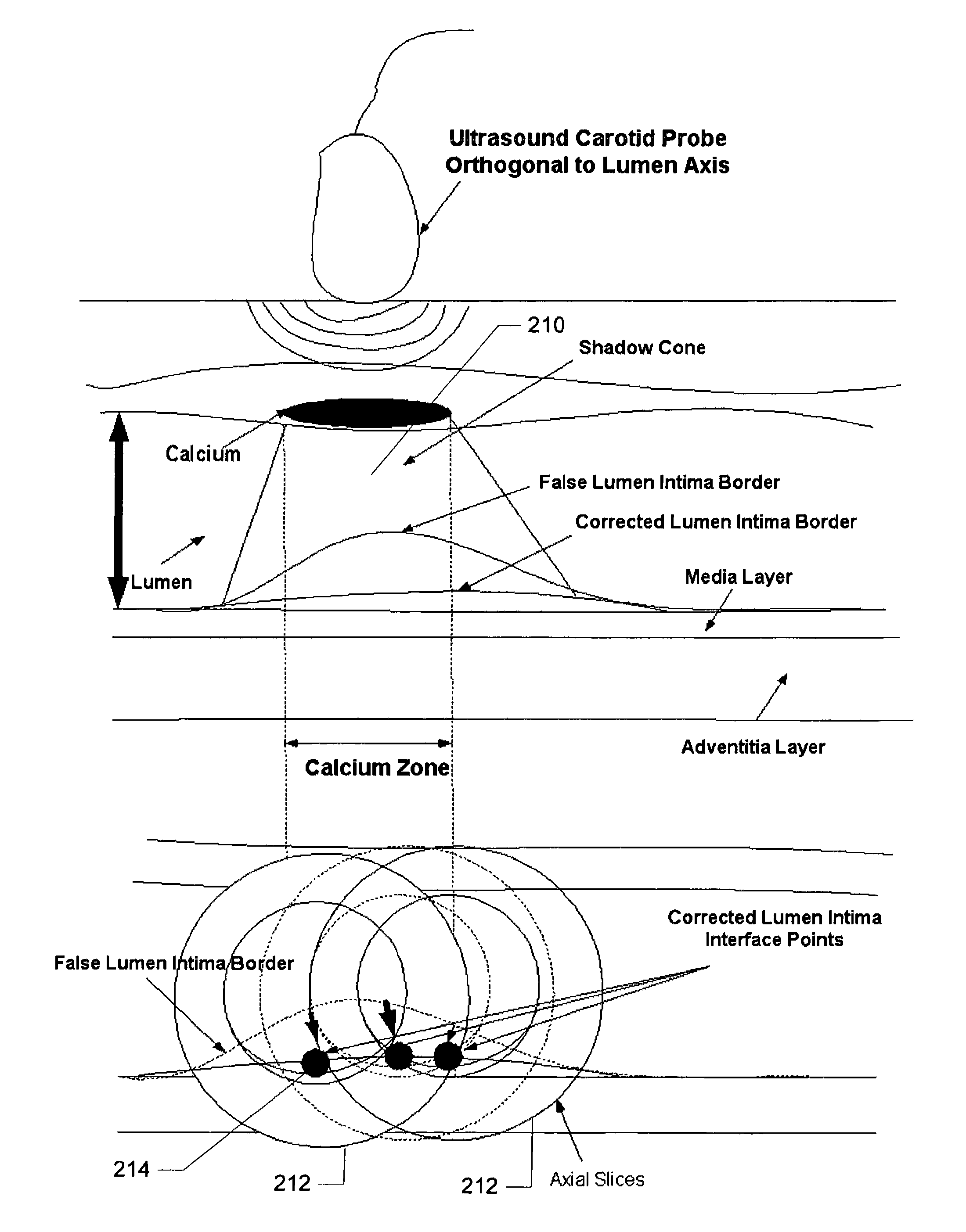
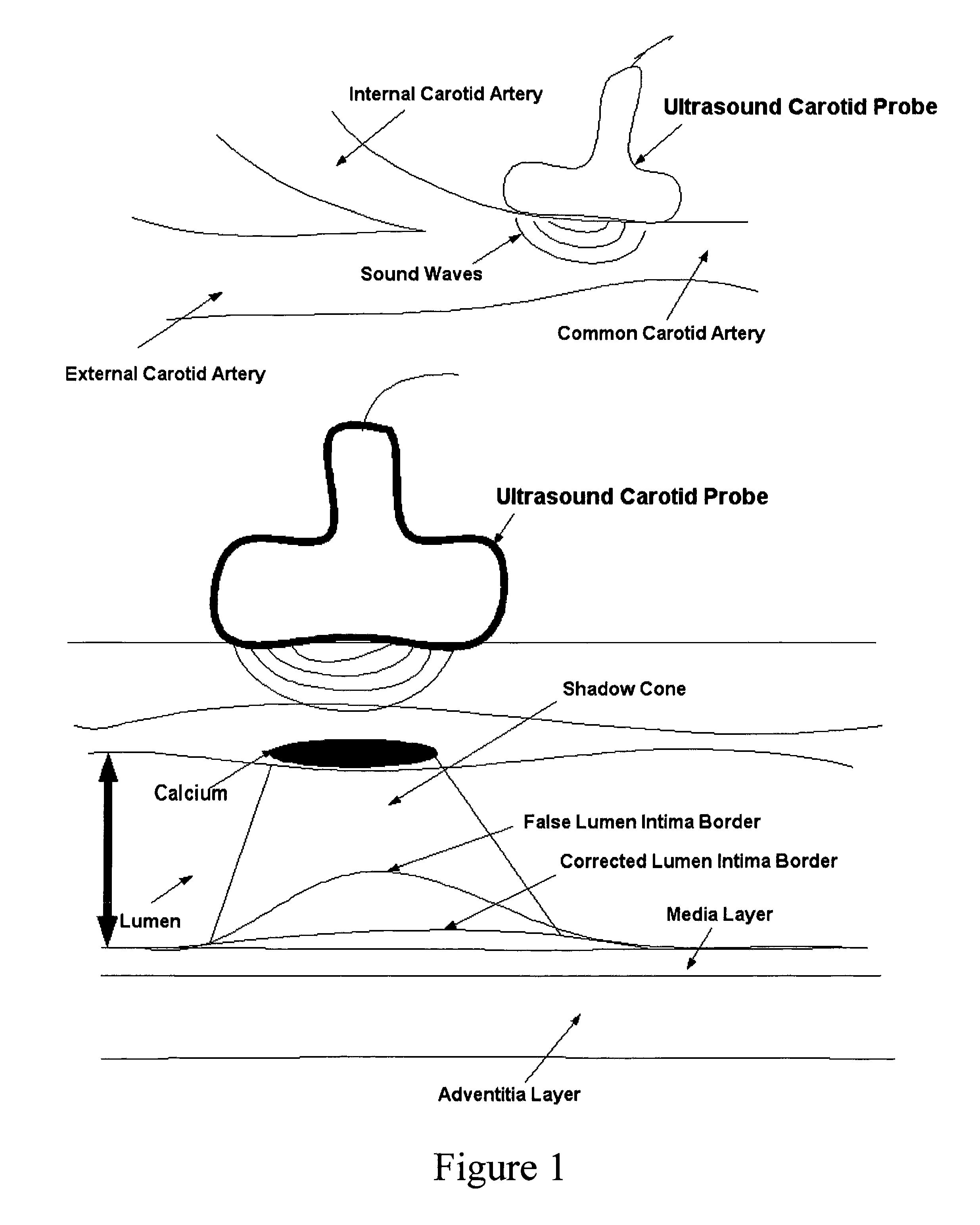
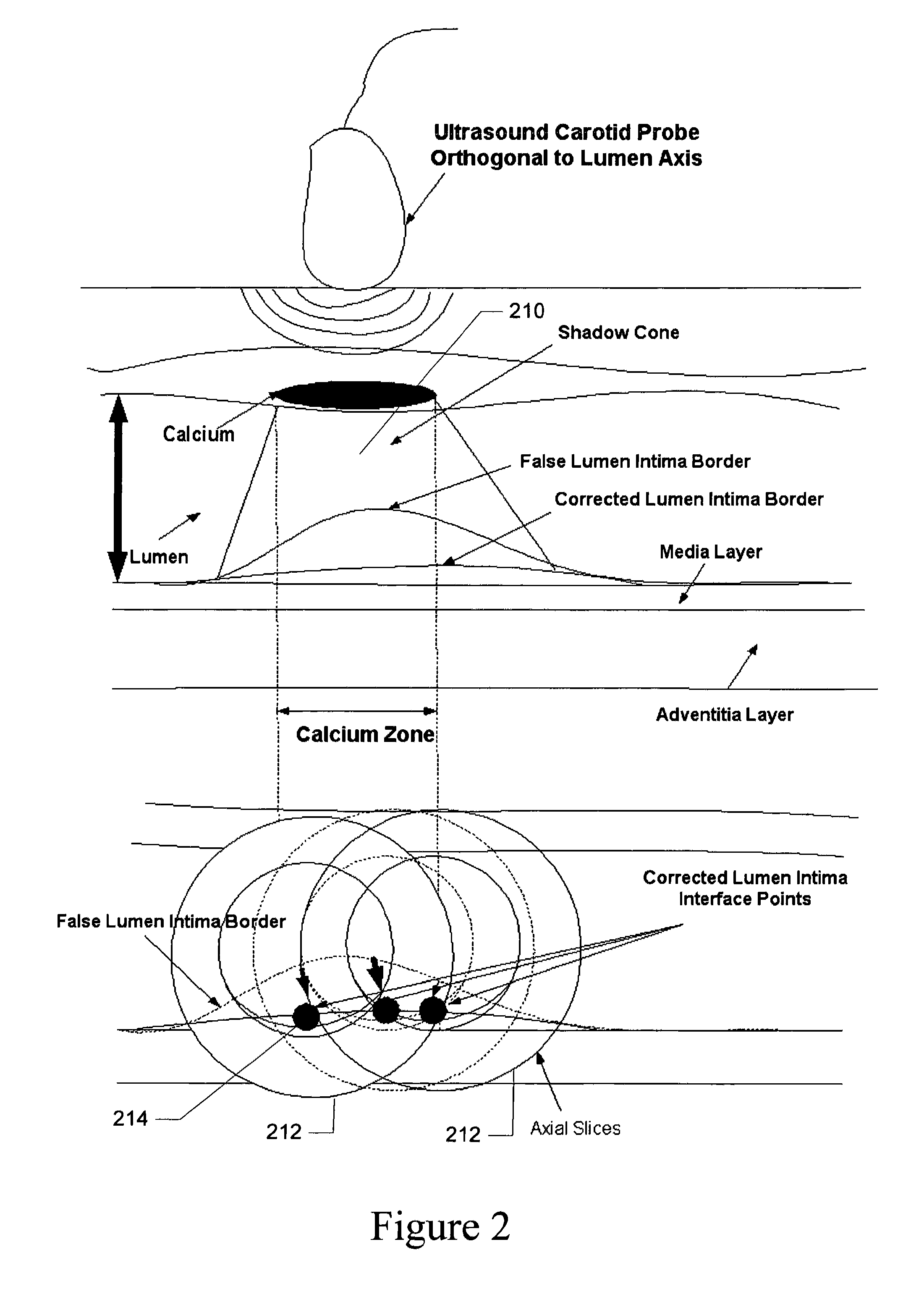
[0039]This patent application discloses various embodiments of a computer-implemented system and method for fast, reliable and automated processing for intima-media thickness (IMT) measurements. In particular, this patent application discloses various embodiments of a computer-implemented system and method for intima-media thickness (IMT) measurements in the presence or absence of calcium at the near (proximal) wall of the arterial vessel. Although the embodiments disclosed herein are described in regard to particular blood vessels (e.g., carotid), the systems and methods disclosed and claimed are also applicable to IMT measurement in any blood vessel in any living org...


PUM


Abstract

A computer-implemented system and method for fast, reliable and automated processing for intima-media thickness (IMT) measurements. Various embodiments include receiving biomedical imaging data and patient demographic data corresponding to a current scan of a patient; checking the biomedical imaging data in real-time to determine if an artery of the patient has a calcium deposit in a proximal wall of the artery; acquiring arterial data of the patient as a combination of longitudinal B-mode and transverse B-mode data; using a data processor to automatically recognize the artery; using the data processor to calibrate a region of interest around the automatically recognized artery; and determining the intima-media thickness (IMT) of an arterial wall of the automatically recognized artery.

Description

COPYRIGHT[0001]A portion of the disclosure of this patent document contains material that is subject to copyright protection. The copyright owner has no objection to the facsimile reproduction of the patent document or the patent disclosure, as it appears in the Patent and Trademark Office patent files or records, but otherwise reserves all copyright rights whatsoever. The following notice applies to the software and data as described below and in the drawings that form a part of this document: Copyright 2009-2010 Jasjit S. Suri, Filippo Molinari, and Biomedical Technologies Inc., All Rights Reserved.TECHNICAL FIELD[0002]This patent application relates to methods and systems for use with data processing, data storage, and imaging systems, according to one embodiment, and more specifically, for ultrasound image processing.BACKGROUND[0003]The state of Atherosclerosis in carotids or other blood vessels can be studied using magnetic resonance imaging (MRI) or Ultrasound imaging. Because...

Claims


Application Information

Patent Timeline
IPC IPC(8): A61B8/00G06K9/00
CPCA61B8/0858A61B8/0891A61B8/5223A61B8/5269G06K9/4609G06T7/0012G06K2209/05G06T2207/10132G06T2207/30101G06K2009/00932G16H50/30G06V40/14G06V10/443G06V2201/03
Inventor SURI, JASJIT S.
Owner ATHEROPOINT
Why Eureka
Try Eureka