Eureka AIR delivers breakthrough ideas for toughest innovation challenges, trusted by R&D personnel around the world.

Preshaped ablation catheter for ablating pulmonary vein ostia within the heart

a pulmonary vein and catheter technology, applied in the field of tissue treatment, can solve the problems of impaired hemodynamics, loss of cardiac efficiency, and disruption of normal activation of the left and right atria, and achieve the effect of maximizing the span of lesions around the ostium

Inactive Publication Date: 2013-04-02
BOSTON SCI SCIMED INC
View PDF107 Cites 32 Cited by
  • Summary
  • Abstract
  • Description
  • Claims
  • Application Information

AI Technical Summary

Benefits of technology

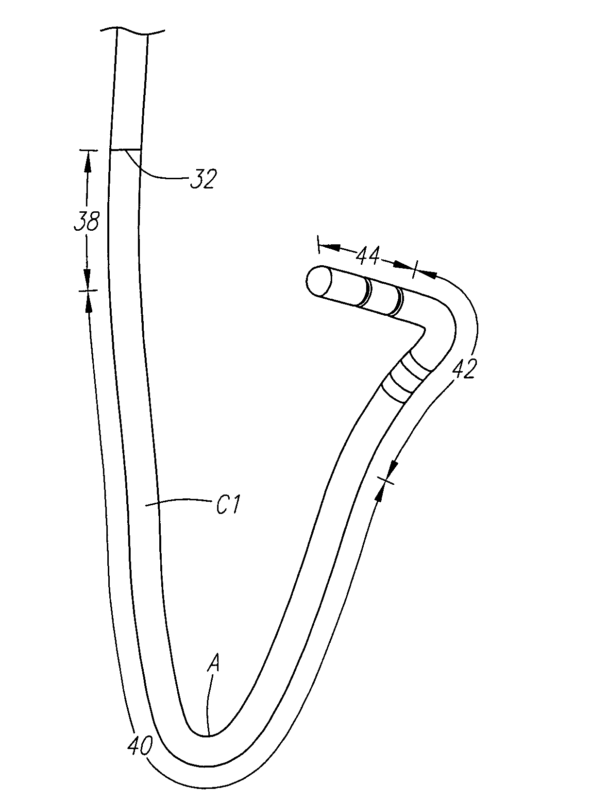
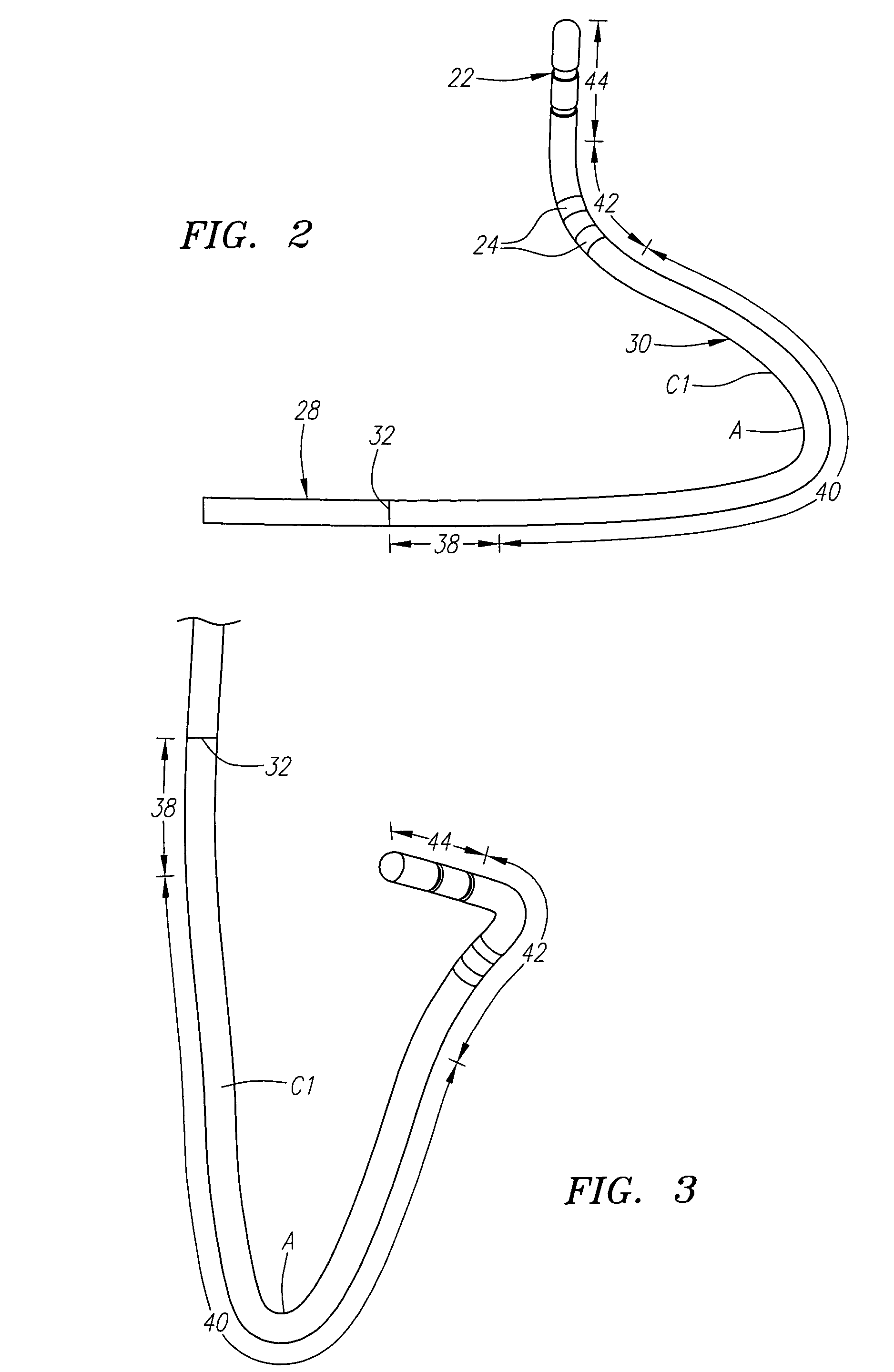
[0009]In either event, the distal shaft portion has a proximal section configured to be internally actuated from a straight geometry to a simple curve (i.e., a curve that lies substantially in a single plane). For example, the catheter can comprise a steering mechanism that is operable to internally actuate the proximal section to form the simple curve, or the proximal section can be pre-shaped to form the simple curve in the absence of an external force, such as gravity or the constraining force apply by a guide sheath. The simple curve bends more than 70 degrees, preferably at least 90 degrees, and more preferably at least 135 degrees, so that, e.g., its apex can be more easily inserted into an ostium of a vessel (e.g., a pulmonary vein ostium). To further facilitate insertion into a vessel ostium, the simple curve may be eccentric, with its apex having the smallest radius curvature of the simple curve. In one embodiment, the proximal section of the distal shaft portion is radio-opaque, so that, e.g., the extent to which the simple curve is inserted into the vessel ostium can be conveniently determined based on the angle formed by the simple curve bending about its apex.
[0011]The distal shaft portion further includes a distal section on which the operative element(s) is mounted, which in one embodiment, forms the distal tip of the catheter body. The distal section may be substantially straight, but can also be pre-shaped to form a simple curve with an apex that points away from the longitudinal axis of the proximal shaft portion. Either configuration lends itself well when linear ablative elements are used, so that a linear ablation can be more efficiently placed around the vessel ostium. The distal shaft portion may optionally include a substantially straight shaft transition section between the proximal shaft portion and the proximal section of the distal shaft portion. Preferably, the proximal shaft portion and shaft transition section are collinear to facilitate the pushability of the catheter (i.e., to minimize the chance that the distal shaft portion will collapse onto the proximal shaft portion in the presence of a resistive axial force, which may otherwise occur if the proximal shaft portion and shaft transition section were angled relative to each other).
[0015]In accordance with a fourth aspect of the present inventions, a method of performing a medical procedure adjacent an anatomical vessel (such as a pulmonary vein) using a catheter is provided. The catheter has a curvable section and an operative element distal to the curved section. The method comprises forming the curvable section into a curve having an apex. This can be accomplished, e.g., by using a steering mechanism or by pre-shaping the curvable section and removing a sheath from the curvable section. The method further comprises inserting the apex within the vessel ostium to place the ablative element in contact with a first tissue site adjacent the vessel ostium, performing the medical procedure on the first tissue site with the operating element, rotating the curve within the vessel ostium about the apex to place the operative element in contact with a second tissue site adjacent the vessel ostium, and then performing a medical procedure on the second tissue site with the operative element. Notably, because the curve is rotated around a fixed point (i.e., the apex), the medical procedure can be performed around the vessel ostium in a controlled and predefined manner.
[0016]This method lends itself well to the medical procedures that involve forming lesions around vessel ostia, and in particular, pulmonary vein ostia, where control of the ablation process is crucial. The lesions can be created for any purpose, but the method lends itself well to therapeutic procedures involving the electrical isolation of arrhythmia causing substrates from the left atrium of the heart. The lesions can either be discrete or can form a continuous lesion, but preferably, are linear or curvilinear, and somewhat tangential, to maximize the span of the lesions about the ostium and the effectiveness of the lesions in blocking the errant electrical pathways from the pulmonary vein. In one method, the lesion formation and rotation steps are performed until a plurality of lesions are circumferentially disposed about the vessel ostium.

Problems solved by technology

These wavelets, called “reentry circuits,” disrupt the normally uniform activation of the left and right atria.
Because of a loss of atrioventricular synchrony, people who suffer from atrial fibrillation and flutter also suffer the consequences of impaired hemodynamics and loss of cardiac efficiency.
They are also at greater risk of stroke and other thromboembolic complications because of loss of effective contraction and atrial stasis.
However, not only is the maze procedure is technically difficult to do, it also requires open heart surgery and is very expensive.
When ablating around the PVs, however, energy is typically applied along the curvilinear path using a free-hand approach, thereby rendering it difficult to accurately move the catheter tip along that path.
Thus, it has proven difficult to form circumferential lesions using conventional devices to isolate the PVs and cure ectopic atrial fibrillation.

Method used


Image

  • Preshaped ablation catheter for ablating pulmonary vein ostia within the heart
  • Preshaped ablation catheter for ablating pulmonary vein ostia within the heart
  • Preshaped ablation catheter for ablating pulmonary vein ostia within the heart

Examples


Embodiment Construction

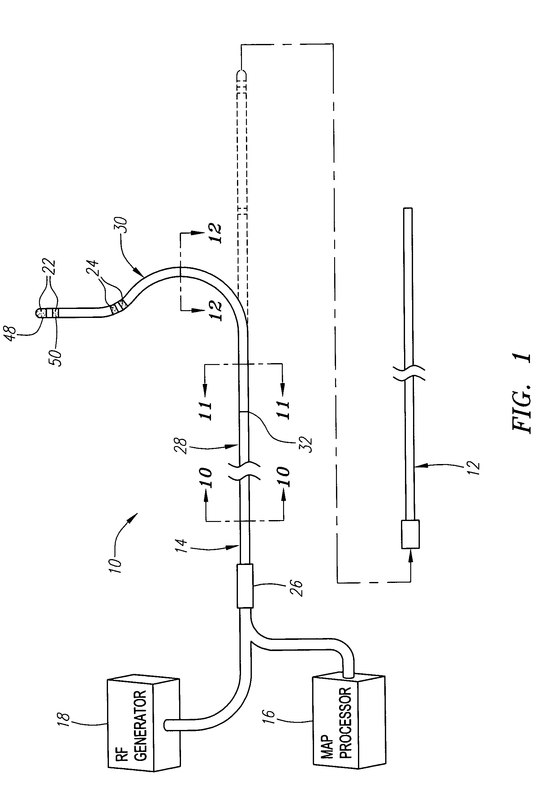
[0035]Referring to FIG. 1, an exemplary tissue ablation system 10 constructed in accordance with the present inventions is shown. The system 10 may be used within body lumens, chambers or cavities for therapeutic and diagnostic purposes in those instances where access to interior bodily regions is obtained through, for example, the vascular system or alimentary canal and without complex invasive surgical procedures. For example, the system 10 has application in the diagnosis and treatment of arrhythmia conditions within the heart. The system 10 also has application in the treatment of ailments of the gastrointestinal tract, prostrate, brain, gall bladder, uterus, and other regions of the body. As an example, the system 10 will be described hereinafter for use in pulmonary veins, and specifically, to electrically isolate one or more arrhythmia causing substrates within the ostium of a pulmonary vein from the left atrium of the heart in order to treat ectopic atrial fibrillation.

[0036...


PUM


Abstract

Catheters and methods are provided for performing medical procedures, such as tissue ablation, adjacent the ostia of anatomical vessels, such as pulmonary veins. The catheter comprises an elongated flexible integrated catheter body having proximal and distal shaft portions and at least one operative element carried by the distal shaft portion. The distal shaft portion has a proximal section configured to be internally actuated (e.g., using a steering mechanism or pre-shaping the proximal section) to form a simple curve with an apex that can be inserted into the vessel ostium, an intermediate section pre-shaped to form a curve that bends opposite the simple curve, and a distal section configured to be placed into a non-radial relationship (tangential or oblique) with the vessel ostium when the apex of the simple curve is inserted into the vessel ostium. By this arrangement, the operative element is configured to be placed firmly in contact with tissue at a predefined radial location relative to the vessel ostium when the apex of the simple curve is inserted into the vessel ostium.

Description

FIELD OF THE INVENTION[0001]The present inventions generally relate to systems and methods for treating tissue, and more particularly to systems and methods for ablating tissue in and around the ostia of vessels, such as pulmonary veins, and other anatomical openings.BACKGROUND OF THE INVENTION[0002]Normal sinus rhythm of the heart begins with the sinoatrial node (or “SA node”) generating an electrical impulse. The impulse usually propagates uniformly across the right and left atria and the atrial septum to the atrioventricular node (or “AV node”). This propagation causes the atria to contract in an organized manner to transport blood from the atria to the ventricles, and to provide timed stimulation of the ventricles. The AV node regulates the propagation delay to the atrioventricular bundle (or “HIS” bundle). This coordination of the electrical activity of the heart causes atrial systole during ventricular diastole. This, in turn, improves the mechanical function of the heart. Atr...

Claims


Application Information

Patent Timeline
Patent Type & Authority Patents(United States)
IPC IPC(8): A61B18/14
CPCA61B18/1492A61M25/0144A61M25/0147A61M25/0152A61B2017/00867A61B2018/00375A61M2025/0161C08L2201/12A61B5/6852A61B2017/00318A61B2017/00323A61B2017/00331A61M25/0041
Inventor AVITALL, BOAZKOBLISH, JOE
Owner BOSTON SCI SCIMED INC
Why Eureka
Eureka Blog
Learn More