Use of multifocal collimators in both organ-specific and non-specific SPECT acquisitions

a multi-focal collimator and organ-specific technology, applied in the field of imaging systems, can solve the problems of increasing costs, insufficient sensitiveness of current planar or spect imaging methods, and inability to achieve simple fan-beam collimators to achieve such requirements, so as to improve confidence and improve the ability to detect small lesions contrast

Active Publication Date: 2013-06-18
SIEMENS MEDICAL SOLUTIONS USA INC
View PDF4 Cites 1 Cited by
  • Summary
  • Abstract
  • Description
  • Claims
  • Application Information

AI Technical Summary

Benefits of technology

[0009]Embodiments of the present invention can perform multiple scans of a subject, iteratively and efficiently, to identify one or more regions of interest and to diagnose pathology in the subject. To obtain optimal results using a non-parallel hole collimator, the region of interest should be within the focus of the collimator. In typical scans, the region of interest would be a predetermined area of the body such as a heart. In full body scans, there is no particular region of interest because the entire body is scanned. The image resulting from a first pass of the scanner yields valuable medical information to assess the overall state and extent of the disease, but it also indicates regions that should be looked at more closely, i.e., regions of interest. These regions can subsequently be examined in more detail by placing them within the focus of the collimator. This will allow for more counts to be collected for a given time compared to parallel hole collimator, resulting in higher statistical power of the tomographic data of this ROI and using a computer resulting in a better detectable and interpretable lesion—in short in a clearer image. After obtaining a clearer image, additional diseased areas may be observed adjacent to the regions of interest, which can then be scanned in another iteration.
[0010]According to one embodiment, a subject is injected with, for example, a radioisotope and is imaged with a gamma camera. Using a variable focal collimator, two or more images can be made of the subject at different locations. The images are then examined to determine regions of interest to be examined further. A second iteration is performed to take additional images having a greater number of counts. Additional iterations can be performed as desired to increase accuracy and complete an examination of the subject. The subsequent iterations allow for increased counts to be obtained from the regions of interest, sensitivity, specificity and more accurate diagnosis. The different iterations can be performed with the same apparatus but at different configurations and orbits using different cameras or collimators.
[0012]Further, in this embodiment, a non-parallel hole collimator can perform non-organ specific scanning, such as in bone SPECT, and can improve the ability to detect smaller lesions contrast than typically possible. The idea can then readily be extended to other types of collimators with similar properties, namely collimators having a non-stationary response to a point source across the detector face, even when the distance is held constant, such as in the most general case: coded apertures, pinholes, slit-slat or any combination thereof. This disclosure focuses on using diagnostic imaging to identify or reject regions of interest (“detection task”) with improved confidence. However, with appropriate data, calibration and reconstruction methods the task of quantification is also possible. This embodiment can be used to diagnose disease, and to plan and monitor treatment.

Problems solved by technology

A simple fan-beam collimator is not suitable to achieve such requirements.
Current methods of Planar or SPECT imaging do not provide sufficiently sensitive results, particularly when scanning over a large area, such as an entire body in a scan of the skeletal system as it is done in oncology imaging.
To obtain sensitive images, multiple scans can be made using the same or different imaging apparatuses, which can require a patient to move from one apparatus to another which causes delay, increases costs, is cumbersome, and has lower specificity and sensitivity.
Additionally, the multiple apparatuses are not optimized and therefore still result in lower image quality.

Method used


Image

  • Use of multifocal collimators in both organ-specific and non-specific SPECT acquisitions
  • Use of multifocal collimators in both organ-specific and non-specific SPECT acquisitions
  • Use of multifocal collimators in both organ-specific and non-specific SPECT acquisitions

Examples


Embodiment Construction

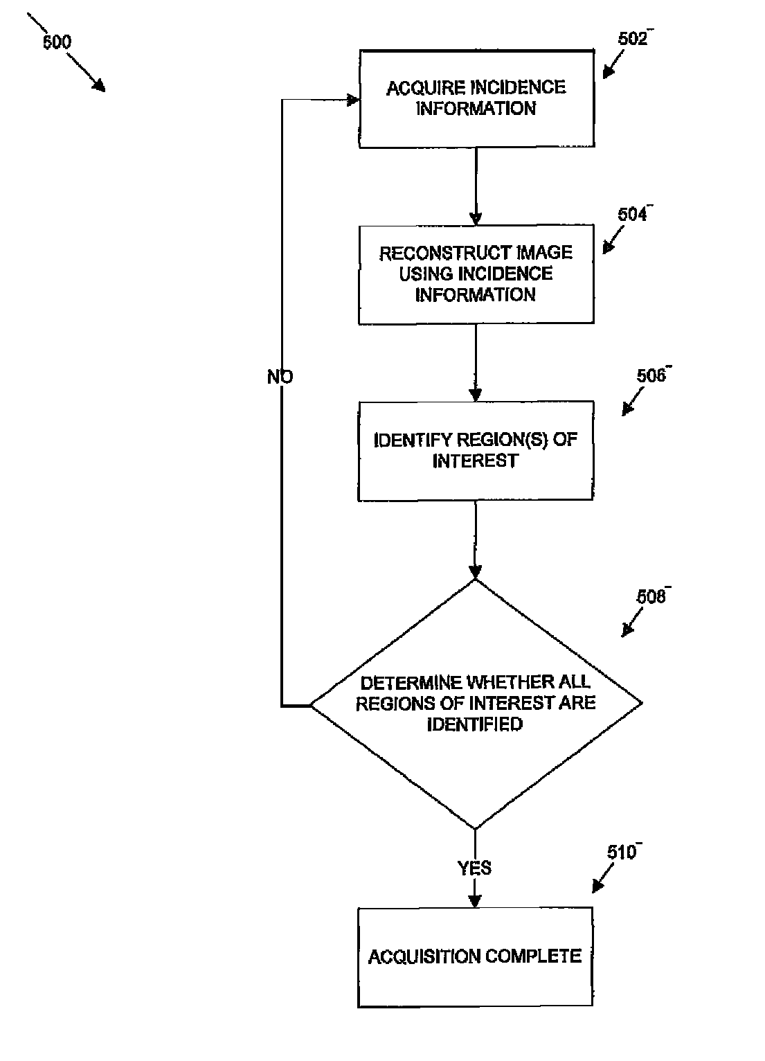
[0018]The following description is presented to enable any person skilled in the art to use a method to efficiently produce superior reconstructed images using, for example, planar imaging or Single Photon Emission Computed Tomography (SPECT). Various modifications to the preferred embodiments will be readily apparent to those skilled in the art, and the generic principles defined herein may be applied to other embodiments and applications without departing from the spirit and scope of the present disclosure. In the following description, numerous details are set forth for the purpose of explanation. However, one of ordinary skill in the art will realize that the present disclosure might be practiced without the use of these specific details. To more efficiently illustrate and describe embodiments of the present disclosure, identical reference numerals are used in the specification and drawings to identify parts that are essentially the same in different stages, versions or instanti...


PUM


Abstract

A method for obtaining nuclear medical images of a patient in which one or more first images are obtained. The one or more first images can be used to identify one or more regions of interest for subsequent images that focus on the regions of interest. The process can occur iteratively until it is determined, either by a physician or a computer program that sufficient images have been obtained to identify lesions or other pathology within a reasonable level of confidence.

Description

RELATED APPLICATIONS[0001]The present patent document claims the benefit of the filing date under 35 U.S.C. §119(e) of Provisional U.S. Patent Application Ser. No. 61 / 254,042, filed Oct. 22, 2009, which is hereby incorporated by reference.BACKGROUND OF THE INVENTION[0002]1. Field[0003]The present disclosure relates generally to imaging systems and more particularly to imaging systems for use in nuclear medicine.[0004]2. Description of Related Art[0005]In a nuclear medicine imaging apparatus, a gamma or scintillation camera can obtain either planar images or Single Photon Emission Computed Tomography (SPECT) images, a collimator is mounted to the face of the imaging apparatus. The collimator is constructed of a dense, high-atomic-number material, such as lead. The material is bored with numerous tiny straight holes that allow radiation (e.g., gamma rays) to pass through. If the radiation is not traveling along the path of the hole, then the material absorbs it and it will not reach a...

Claims


Application Information

Patent Timeline
Patent Type & Authority Patents(United States)
IPC IPC(8): G06K9/00
CPCA61B6/037A61B6/06A61B6/5235A61B8/4416G21K1/025
Inventor VIJA, ALEXANDER HANS
Owner SIEMENS MEDICAL SOLUTIONS USA INC
Why Eureka
Try Eureka