Method and system for stabilizing a series of intravascular ultrasound images and extracting vessel lumen from the images

a technology of intravascular ultrasound and images, applied in image enhancement, instruments, applications, etc., can solve the problems of difficulty in obtaining accurate polar coordinates, and inability to accurately interpret the morphology of blood vessels in ivus dynamic display, etc., to achieve accurate and effective diagnostics and therapeutics.

Inactive Publication Date: 2013-07-09
MEDINOL LTD
View PDF8 Cites 35 Cited by
  • Summary
  • Abstract
  • Description
  • Claims
  • Application Information

AI Technical Summary

Benefits of technology

[0017]The present invention is directed to a method and system for processing and enhancing ultrasonic images from within a tubular environment, such as a blood vessel lumen. In particular, the method according to an embodiment of the invention pertains to generating a stabilized set of intravascular ultrasound (IVUS) images using a probe instrument connected to a processor and a post-processor to obtain a circumferential image depicting a cross-section of a lumen from a series of images, detect the edges of the lumen from the series of images using predictive modeling to generate a binary image of the cross-section, determine the centroid of the binary image, align the centroid with the circumferential image center and, where necessary, correct for rotation and vessel compliance. The method may also involve filtering the series of images to increase the signal to noise ratio before edge detection. The probe instrument, which has a transmitter for transmitting ultrasonic signals and a receiver for receiving reflected ultrasonic signals that contain information about a tubular environment, such as a body lumen, preferably is a catheter. The result is a more accurate and clean image of the tubular environment. The methods further permit extraction of the shape and volume of the inner lumen after the images are stabilized.
[0019]The invention is useful for stabilizing an IVUS image of a blood vessel, where image drift may occur due to heart beat or blood flow, or any tubular environment particularly prone to movements that cause image drift, making image stabilization important for accurate and effective diagnostics and therapeutics.

Problems solved by technology

The data in Polar coordinates are not usually transferred to a display because of the difficulties of interpretation by physicians.
This makes it difficult for the physician to accurately interpret the blood vessel morphology in IVUS dynamic display.
Current IVUS equipment and interpreter algorithms have no stabilization function that compensates for relative movements between catheter and the lumen.
As a result the non-stabilized IVUS images may result in misdiagnosis, inaccurate interpretation of morphology and inaccurate 3D lumen reconstruction, which is widely used by physicians.
When the IVUS video is displayed dynamically, it is not always easy to separate the slowly changing, sometimes subtle morphological features of the blood vessel due to fast image changes.
Currently, software applications allow automatic extraction of the lumen for individual frame or image, but these may be inaccurate and time consuming and therefore may not be feasible for near real-time analysis.
When either manual or automatic methods are applied to rapidly changing images, the analysis of the blood vessel lumen becomes difficult and inaccurate.
Because of image instability, however, such matching is difficult.
Furthermore, the orientation and visualization of the images may be changed, so identification of the anatomy may be time consuming and inaccurate.

Method used


Image

  • Method and system for stabilizing a series of intravascular ultrasound images and extracting vessel lumen from the images
  • Method and system for stabilizing a series of intravascular ultrasound images and extracting vessel lumen from the images
  • Method and system for stabilizing a series of intravascular ultrasound images and extracting vessel lumen from the images

Examples


Embodiment Construction

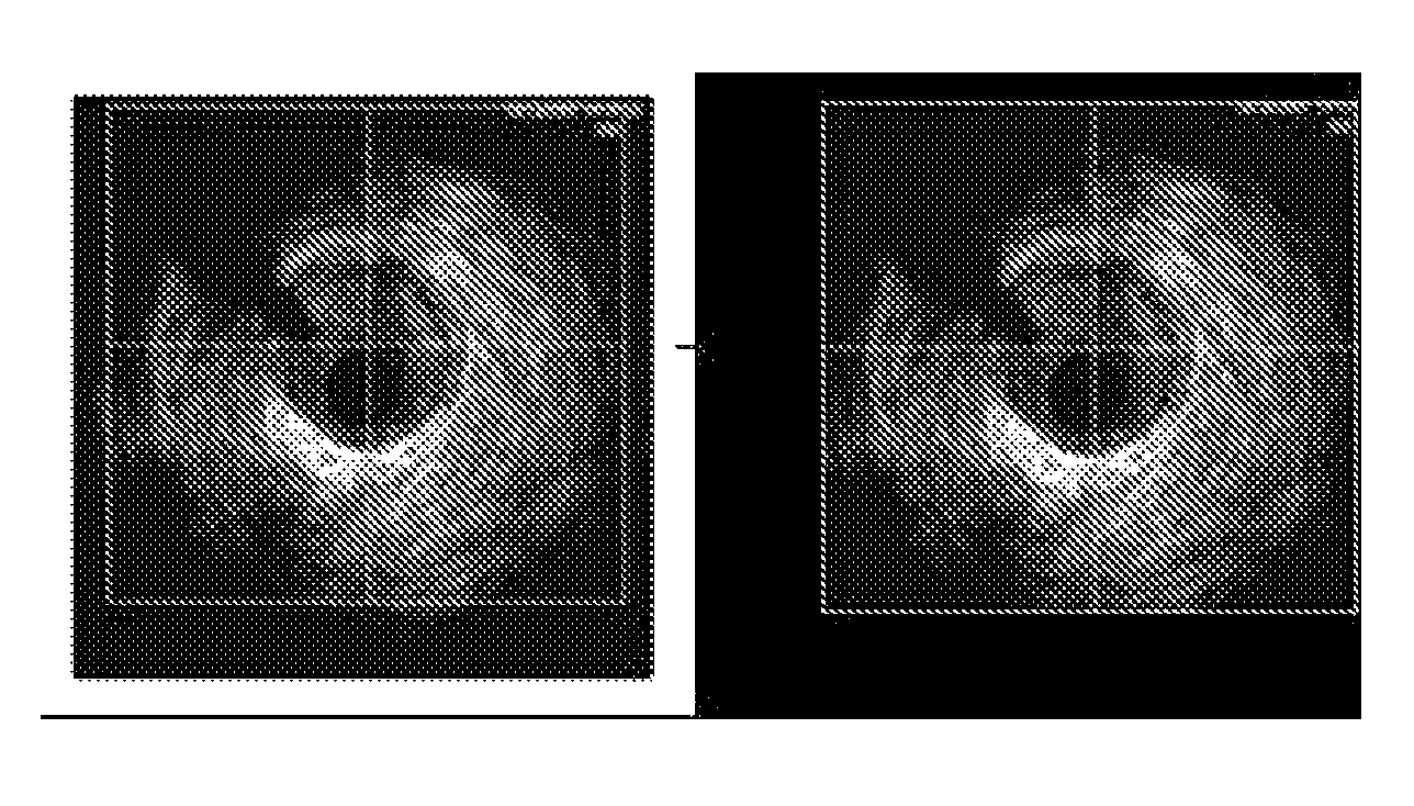
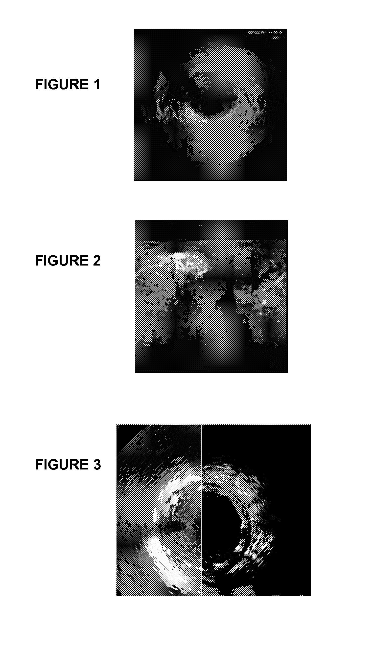
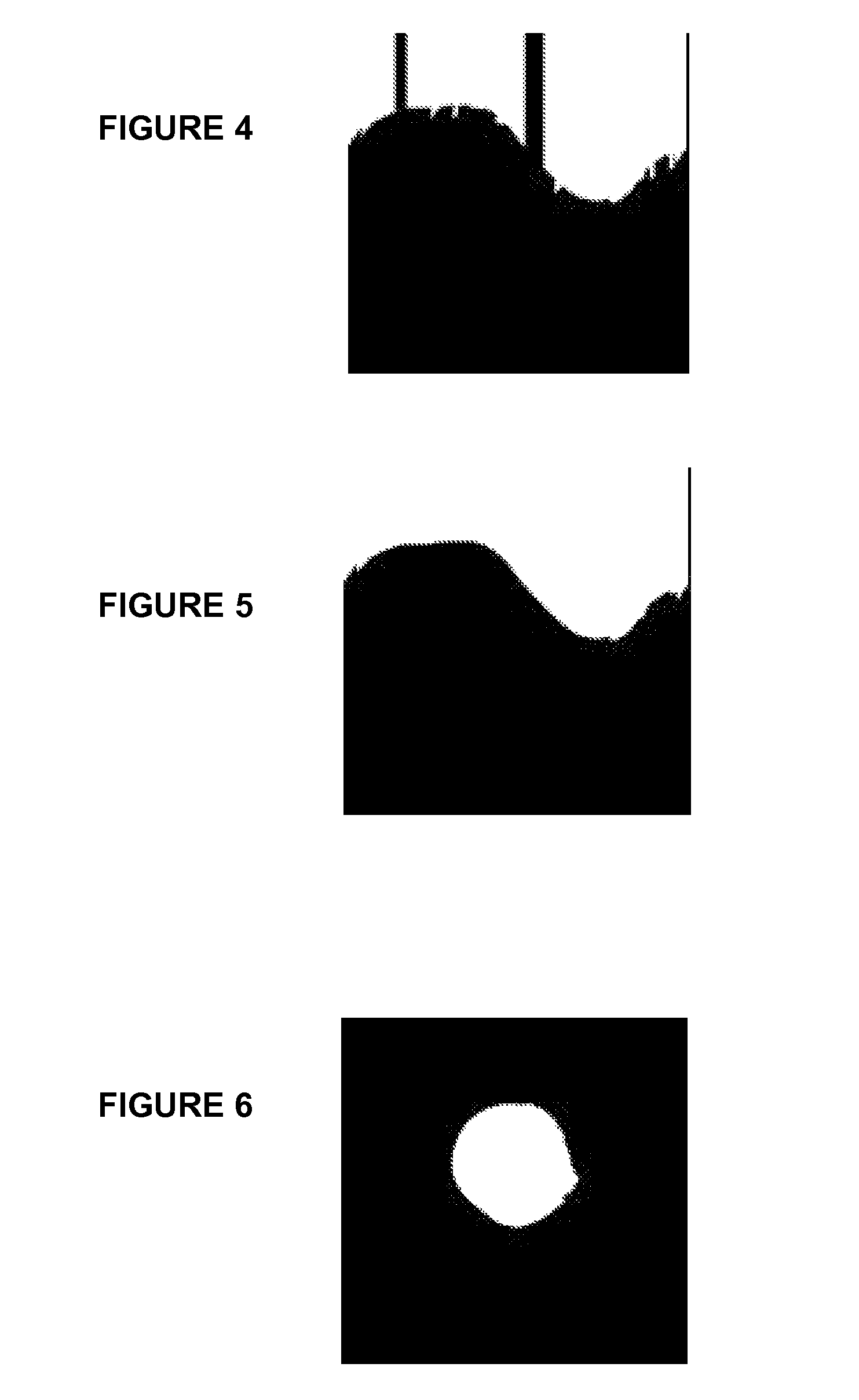
[0031]The invention includes a method and system for stabilizing ultrasonic images of a tubular environment, such as a lumen. “Tubular” means “substantially tubular” herein, because, e.g., in reality a lumen rarely is 100% tubular in a geometrical sense. The method involves receiving ultrasonic signals reflected from the inner surfaces of the tubular environment and converting those signals to an image or images. Preferably the ultrasonic signals have been transmitted in the first instance in a manner that the reflected signals permit generation of a circumferential image of the tubular environment in specific cross-section. Separately, the edges of the tubular environment are detected based on reflected ultrasonic signals, processed into a binary image of the cross-sectional area of the lumen and superimposed over the circumferential image. To stabilize the ultrasonic image, the centroid of the binary image is determined and aligned with the geometric center of the circumferential ...


PUM


Abstract

A method and system for generating stabilized intravascular ultrasonic images are provided. The system includes a probe instrument, having an ultrasonic signal transmitter and a reflected ultrasonic signal receiver, the reflected signals containing information about a tubular environment, and a processor and post-processor, capable of converting inputted signals into one or more, preferably a series of, images. The method for stabilizing images involves the processor and post-processor input and output. The post-processor determines the environment center at each reflection position, detects the tubular environment edges, and aligns the image center with the environment center thereby limiting image drift and stabilizing the images. The processor may also filter images to improve image stabilization and remove motion interference and / or extract the environment's 3D shape. The method and device are of particular use in a vascular lumen, where image drift may occur due to heart beat or blood flow.

Description

FIELD OF THE INVENTION[0001]The present invention relates to a system and method for Intravascular Ultrasound (IVUS) image enhancement. More specifically, a system and method for processing IVUS images in order to enhance quality and provide precise readability of an image or series of images.BACKGROUND[0002]IVUS is used to visualize the interior of blood vessels. Reflected ultrasound signals, usually generated by a transducer or transducer array located at the tip of a catheter that has been inserted into the blood vessel, are received, processed and analyzed to obtain IVUS images. The transducer transmits ultrasound signals, usually within the range of 10-50 MHz, within vessel lumen towards the vessel wall. The ultrasound waves are reflected from the tissue or from the boundaries between two substances with different refraction index, received in the catheter, where the varying pressure waves are converted to electrical signals which are then conducted to an external computer for ...

Claims


Application Information

Patent Timeline
Patent Type & Authority Patents(United States)
IPC IPC(8): G06K9/00A61B8/00G06K9/34G06K9/62G06K9/74G06K9/64A61B8/14
CPCG06T7/0028G06T7/0083G06T2207/10132G06T2207/30101G06T7/12G06T7/33
Inventor RICHTER, JACOBWEIZMAN, OLEGSHIBOLET, ROEE
Owner MEDINOL LTD
Why Eureka
Try Eureka