Use of opioid antagonists

a technology of opioid antagonists and antagonists, which is applied in the direction of drugs, peptides/protein ingredients, instruments, etc., can solve the problems of preventing and/or inhibiting cancer metastasis, and the current methods of treating cancer metastasis are often inadequate, so as to reduce the risk of recurrence

Active Publication Date: 2013-08-27
UNIVERSITY OF CHICAGO
View PDF253 Cites 5 Cited by
  • Summary
  • Abstract
  • Description
  • Claims
  • Application Information

AI Technical Summary

Benefits of technology

[0010]There is a need for improved methods of treating cancer, especially metastatic cancers. It has surprisingly been found that opioid antagonists can inhibit endothelial proliferation and migration associated with angiogenesis, and inhibit growth and metastasis of cancerous tumors.
[0024]In further embodiments, the invention provides methods of treating cancer, wherein a peripheral opioid antagonist and at least one other therapeutic agent that is not an opioid or opioid antagonist are co-administered to the patient. Suitable therapeutic agents include anticancer agents (including chemotherapeutic agents and antineoplastic agents), as well as other antiangiogenesis agents. It has been discovered that opioid antagonists co-administered with various anticancer drugs, radiotherapy or other antiangiogenic drugs can give rise to a significantly enhanced anti-proliferative effect on cancerous cells, thus providing an increased therapeutic effect, e.g., employing peripheral opioid antagonists to certain tumors can potentiate their response to other therapeutic regimens. Specifically, a significantly increased antiproliferative effect, including but not limited to, a significantly increased antiangiogenic effect, is obtained with co-administered combinations as described in more detail below. Not only can an existing regimen be enhanced, but new regimens are possible, resulting, for example, in lower concentrations of the anticancer compound, a lower dosing of radiation, or lower concentration of other antiangiogenic drugs, compared to the treatment regimes in which the drugs or radiation are used alone. There is the potential, therefore, to provide therapy wherein adverse side effects associated with the anticancer or other antiangiogenic drugs or radiotherapy are considerably reduced than normally observed with the anticancer or other antiangiogenic drugs or radiotherapy when used alone.
[0029]In another aspect, the present invention provides a method of inhibiting or reducing angiogenesis, particularly opioid-induced angiogenesis, e.g., of tumor cells, by administering or providing an opioid antagonist, particularly a peripheral opioid antagonist, to cells undergoing angiogenesis. In a further aspect, the invention provides methods of treating opioid-induced angiogenesis in patients receiving opioid treatment or in patients where the angiogenesis is induced by endogenous opioids. The former group is typically cancer patients on opioid-based pain management. The methods comprise administering an opioid antagonist to a patient in an antiangiogenic amount, e.g., an amount sufficient to inhibit or reduce the opioid-induced angiogenesis. In those patients receiving opioid treatment, the opioid and the peripheral opioid antagonist may be co-administered. Peripheral opioid antagonists can, thus, be used to inhibit or reduce the angiogenic effects of opioids on tumor cells, and attenuate the growth of a tumor.
[0035]In yet another aspect, the invention provides a method of reducing the risk of recurrence of a cancer or tumor after medical intervention (such intervention to include but not be limited to surgery, e.g., pulmonary surgery, surgical and endoscopic procedures, e.g., colonoscopy, gastrolaparoscopy, chemotherapy, etc.), comprising co-administering to a cancer patient an opioid antagonist. Thus, the invention contemplates, for example, a method of minimizing the post-operative recurrence of, e.g., breast cancer, in a patient, comprising administering to the patient an effective amount of an opioid antagonist. Peripheral opioid antagonists in accordance with the present invention, e.g., MNTX, can also inhibit VEGF, platelet-derived growth factor (PDGF), or sphingosine 1-phosphate (S1P)-stimulated or induced cell proliferation in the endothelial cells.

Problems solved by technology

The complexity of these processes and the lack of understanding of underlying molecular mechanism have hindered effective therapeutic interventions that prevent and / or inhibit metastasis of cancers.
Current methods of treating metastases of cancers are often inadequate.

Method used


Image

  • Use of opioid antagonists
  • Use of opioid antagonists
  • Use of opioid antagonists

Examples


example 1

Endothelial Cell Migration Assay

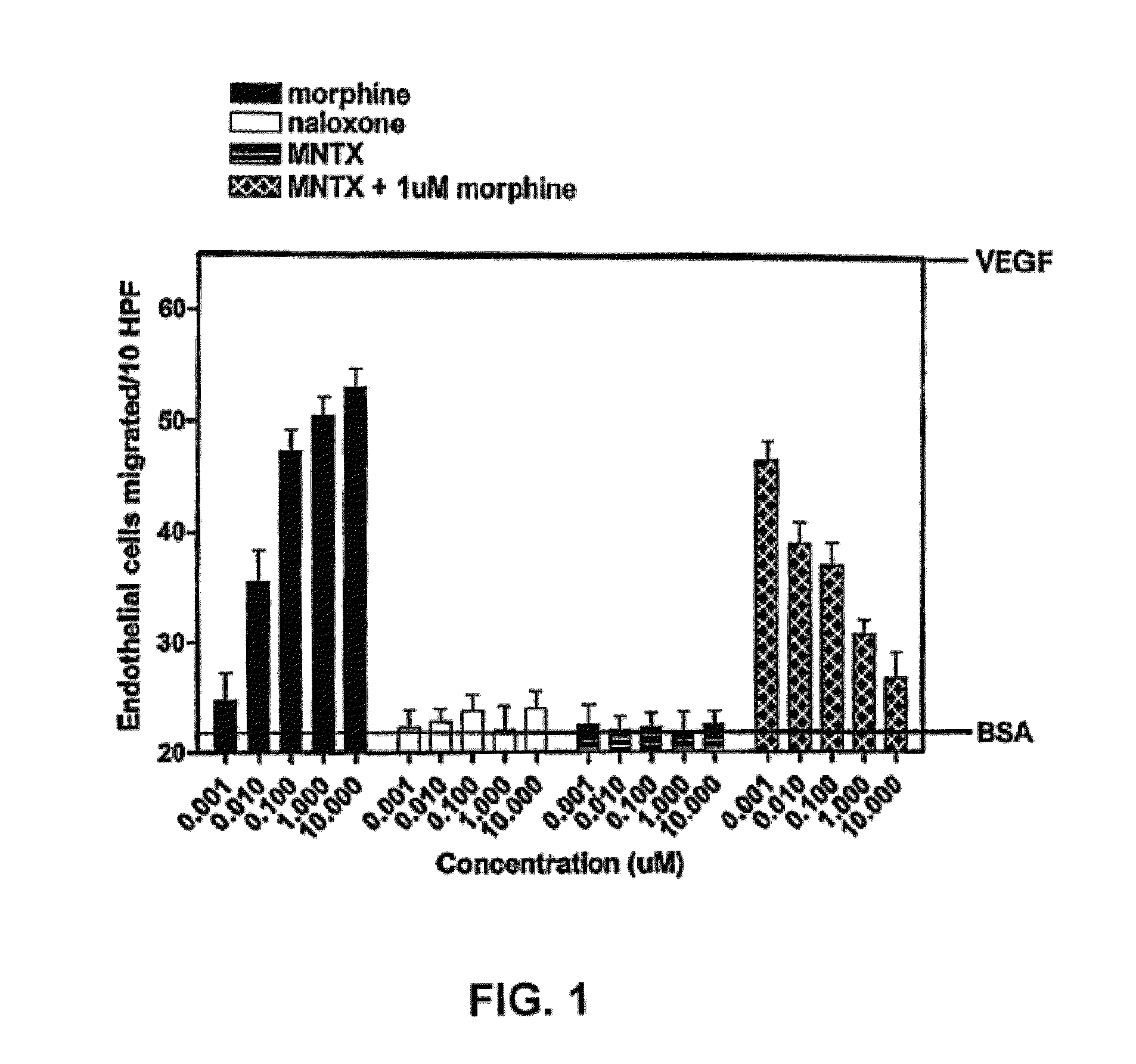
[0193]The antiangiogenic activity of the peripheral opioid antagonists in accordance with the present invention was evaluated in experiments testing the ability of the antagonist to inhibit or modulation capillary endothelial cell migration using a modified Boyden chamber.

[0194]The endothelial cell migration assay was performed as described by Lingen, M. W., Methods in Molecular Medicine, 78: 337-347 (2003), the disclosure of which is incorporated by reference. Briefly, Human Microvascular Endothelial Cells (HMVEC) (Cell Systems, Kirkland, Wash.) were starved overnight in Endothelial Growth Medium (EGM) containing 0.1% bovine serum albumin (BSA). Cells were then trypsinized and resuspended in Dulbecco's Modified Eagle Medium (DME) with 0.1% BSA at a concentration of 1×10⁶ cells / mL. Cells were added to the bottom of a 48-well modified Boyden chamber (NeuroPore Corporation, Pleasanton, Calif.). The chamber was assembled and inverted, and cells were allo...

example 2

Endothelial Cell Migration Assay

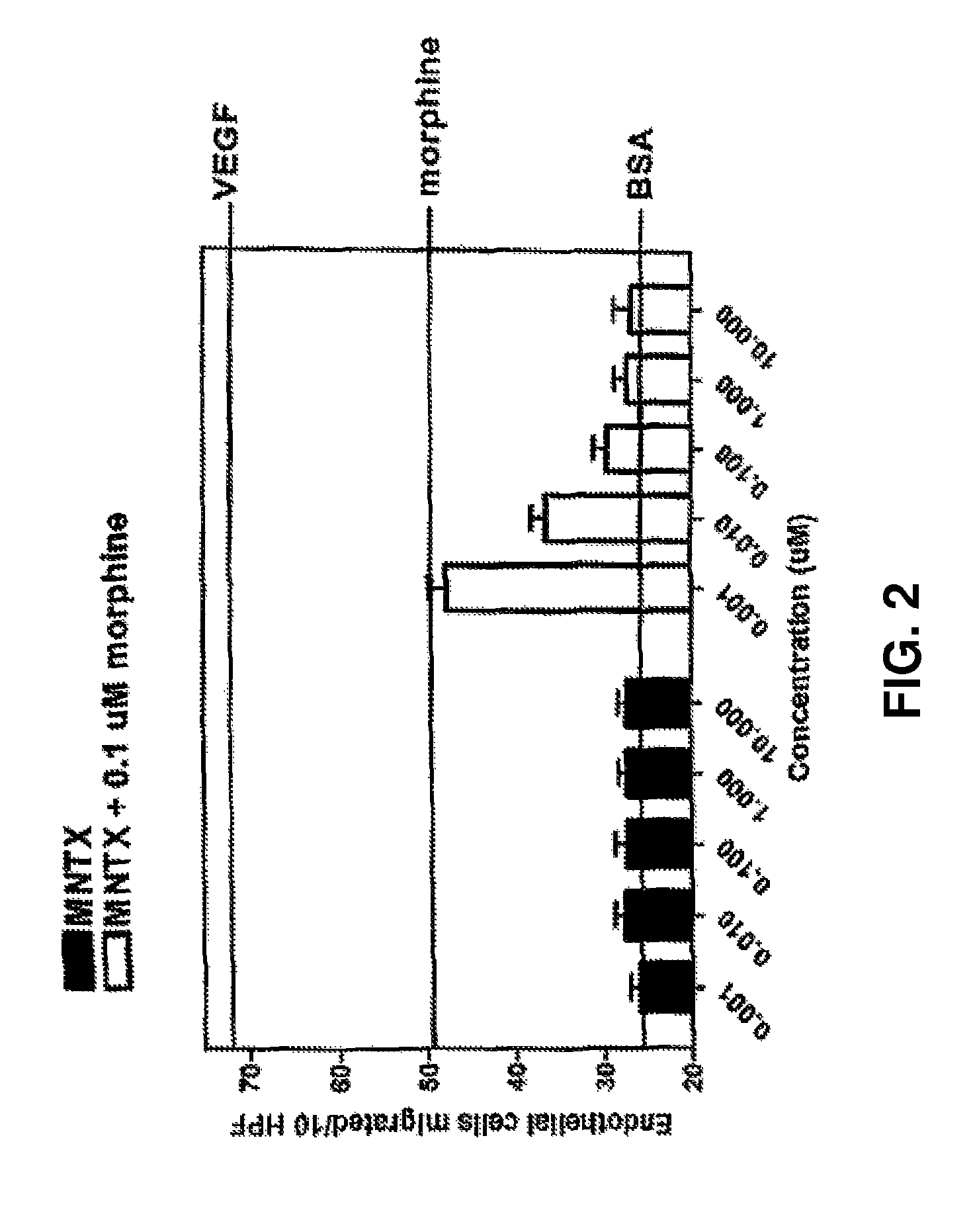
[0196]Another set of experiments was conducted in accordance with the procedure described in Example 1. In this set of experiments, methylnaltrexone and the combination of methylnaltrexone and morphine was again tested for ability to inhibit endothelial cell migration. The methylnaltrexone concentrations when tested alone varied from 0.001 to 10.0 μM. In combination, the concentrations of methylnaltrexone varied from 0.001 to 10.0 μM, while the morphine concentration remained constant at 0.1 μM as described in Example 1. The results are shown in FIG. 2.

[0197]FIG. 2 shows the combination of methylnaltrexone and morphine decreased migration in a concentration-dependent manner, while methylnaltrexone alone did not affect migration.

example 3

Endothelial Cell Migration Induced by DAMGO

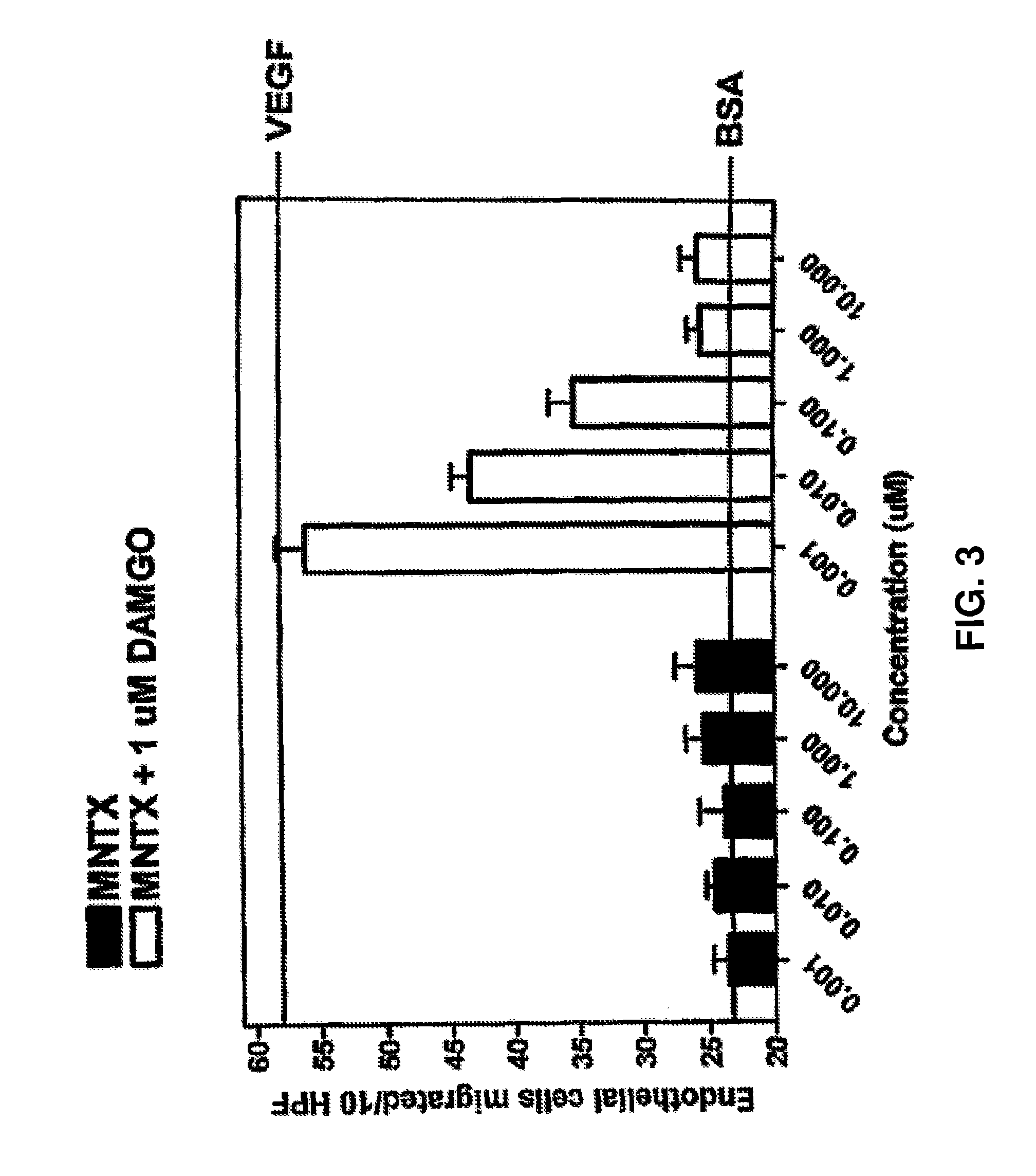
[0198]The drugs used in this study were [D-Ala 2, N-McPhe4, Gly5-ol]enkephalin or DAMGO (Sigma, St. Louis, Mo.); naloxone (Sigma, St. Louis, Mo.); N-methylnaltrexone bromide or methylnaltrexone (Mallinckrodt Specialty Chemicals, Phillipsburg, N.J.). The endothelial cell migration assay was performed as previously described (9). Human dermal microvascular endothelial cells (Cell Systems, Kirkland, W A) were starved overnight in media containing 0.1% bovine serum albumin (BSA), harvested, resuspended into Dulbecco's Modified Eagle's media (DME) with 0.1% BSA, and plated on a semi-porous gelatinized membrane in a modified Boyden chamber (Nucleopore Corporation, Pleasanton, Calif.). Test substances were then added to the wells of the upper chamber and cells were allowed to migrate for four hours at 37° C.

[0199]Membranes were recovered, fixed, and stained and the number of cells that had migrated to the upper chamber per ten high power fields co...


PUM


Abstract

Embodiments of the invention provide methods of attenuating, e.g., inhibiting or reducing, cellular proliferation and migration, particularly endothelial cell proliferation and migration, including that associated with angiogenesis, as well as attenuating cancerous tumor growth and metastasis, using opioid antagonists, including, but not limited to, those that are peripherally restricted antagonists.

Description

RELATED APPLICATIONS[0001]This application is a continuation-in-part of U.S. application Ser. No. 11 / 379,010 filed Apr. 17, 2006, which is a continuation-in-part of International Application No. PCT / US2006 / 07892 filed Mar. 7, 2006, which claims benefit under 35 U.S.C. 119(e) of the filing dates of U.S. Provisional Application Nos. 60 / 659,193 filed Mar. 7, 2005, 60 / 725,703 filed Oct. 12, 2005, 60 / 731,009 filed Oct. 28, 2005, and 60 / 760,851 filed Jan. 20, 2006, the entire disclosures of which are incorporated herein by reference.STATEMENT REGARDING FEDERAL SPONSORED RESEARCH OR DEVELOPMENT[0002]This invention was supported in part by National Institutes of Health grants: DE12322; DE00470; and DE015830. The United States government has certain rights in this invention.FIELD OF INVENTION[0003]The invention relates to methods of attenuating migration and / or proliferation of endothelial cells, especially associated with tumors, as well as attenuating tumor growth and / or metastasis, utiliz...

Claims


Application Information

Patent Timeline
Patent Type & Authority Patents(United States)
IPC IPC(8): A01N43/42A61K31/44C07D221/18
CPCA61K31/485A61K31/452G01N33/5011A61P35/00A61P35/04
Inventor MOSS, JONATHANLINGEN, MARKSINGLETON, PATRICK A.GARCIA, JOE G. N.
Owner UNIVERSITY OF CHICAGO
Why Eureka
Try Eureka