Apparatus and method to carry out image guided radiotherapy with kilo-voltage X-ray beams in the presence of a contrast agent

a technology of contrast agent and kilo-voltage x-ray beam, which is applied in the field of radiotherapy and radio surgery, can solve the problems of inefficient absorbed radiation beam, limited use, and economic cost of apparatus and radiation generation, and achieve the effect of reducing the attenuation of radiation beam

Inactive Publication Date: 2014-03-25
CENT DE INVESTIGACION & DE ESTUDIOS AVANZADOS DEL INST POLITECNICO NACIONAL
View PDF4 Cites 2 Cited by
  • Summary
  • Abstract
  • Description
  • Claims
  • Application Information

AI Technical Summary

Problems solved by technology

It is worth mentioning, however, that as the energy of the radiation beam being used increases, so does the economic cost not only of the apparatus which generates radiation, but also of the installations necessary to accommodate it, given the amount of shielding required to confine the radiation emitted to the treatment room.
However, given the low penetration of this type of radiation, its use has been limited to the treatment of surface tumors, that is, those found at no greater depth than 2 cm in the patient.
The problem is the following: given the low penetration of the X-ray beams with energies in the 20 KeV-500 KeV range, very little radiation reaches the tumor, and that which does reach it, is inefficiently absorbed given the uncertain nature of the interaction of the radiation with the matter.
The concentration of the contrast agent reported in the tumor was that of 10 mg of iodine per gram of tissue (10 mg-l / g), however, this is merely an estimate by the scientists who carried out the study and not a quantitative measurement.
Additionally, given that the bone structures which surround the tumor attenuate in a very pronounced way the X-rays which are directed unto the tumor, the resulting doses which are absorbed by the tumor are very non-uniform.
It has also been shown that the radiation beams must be filtered in a significant manner to remove the very low energy X-rays, as these low energy X-rays interact at a very short distance from the surface, increasing the dosage absorbed by the skin which represents a serious limitation to the clinical application of this treatment embodiment.
The need to filter the used X-ray beams presents a problem to the possible clinical application of this treatment technique, seeing that upon filtering an X-ray beam in a significant manner, the amount of radiation which reaches the tumor is also reduced, resulting in longer treatment times, which represents a serious disadvantage.
Another of the problems with the clinical application of this new treatment embodiment arises from the fact that the absorbed doses imparted to a point within the subject to be irradiated depends on the local concentration of the contrast agent at that specific point, and not quantifying such a concentration in an appropriate manner at each point within the tumor or malformation to be irradiated results in the significant degradation in the quality of the treatment (Garnica-Garza H M Phys. Med. Biol. Vol. 54 pp 5411-5425, 2009).
A significant problem with this patent is that the use of the CAT scanner and the accelerator cannot be simultaneous given the large volume of both apparatuses, it is impossible to take the image during the irradiation of the patient, so first the image is taken, the scanner is moved away from the patient, and the accelerator is placed then proceeding to irradiate the tumor.
A problem with this apparatus is that the diagnostic X-ray equipment used only provides bi-dimensional images of the site to be irradiated, so that even though it is possible to determine lateral tumor movements with respect to the image plane of the tumor, it is impossible to determine movements in the direction of the X-ray beam's incidence.
Another limiting factor of this apparatus is that it only uses one collimated radiation source to produce circular beams with variable diameter, not taking into account the possibility of producing fields in other geometric forms.
A problem with this method is that it assumes the same pattern of deformity of the patient's inner structures occurs both at the time at which the images were taken as at the time of the patient's radiation treatment, which is not necessarily the case, given that physiologic movements are variable.
Additionally, no technique is described to monitor the movement and deformation of the inner structures in real time during treatment.
A problem with this is that it is not possible to use the CAT scanner or NMR in a simultaneous manner with the application of radiation on the tumor or malformation, that is, first the images of the geometry of the site to be treated with the CAT scanner or NMR are obtained, and afterwards the radiation can be applied.
As was previously mentioned, this is not the most convenient given that the tumor or malformation can move during the application of radiation.
In this sense, the optimization of a radio therapeutic plan intended for a high energy radiation beam is not usable for a low energy beam.

Method used


Image

  • Apparatus and method to carry out image guided radiotherapy with kilo-voltage X-ray beams in the presence of a contrast agent
  • Apparatus and method to carry out image guided radiotherapy with kilo-voltage X-ray beams in the presence of a contrast agent
  • Apparatus and method to carry out image guided radiotherapy with kilo-voltage X-ray beams in the presence of a contrast agent

Examples


Embodiment Construction

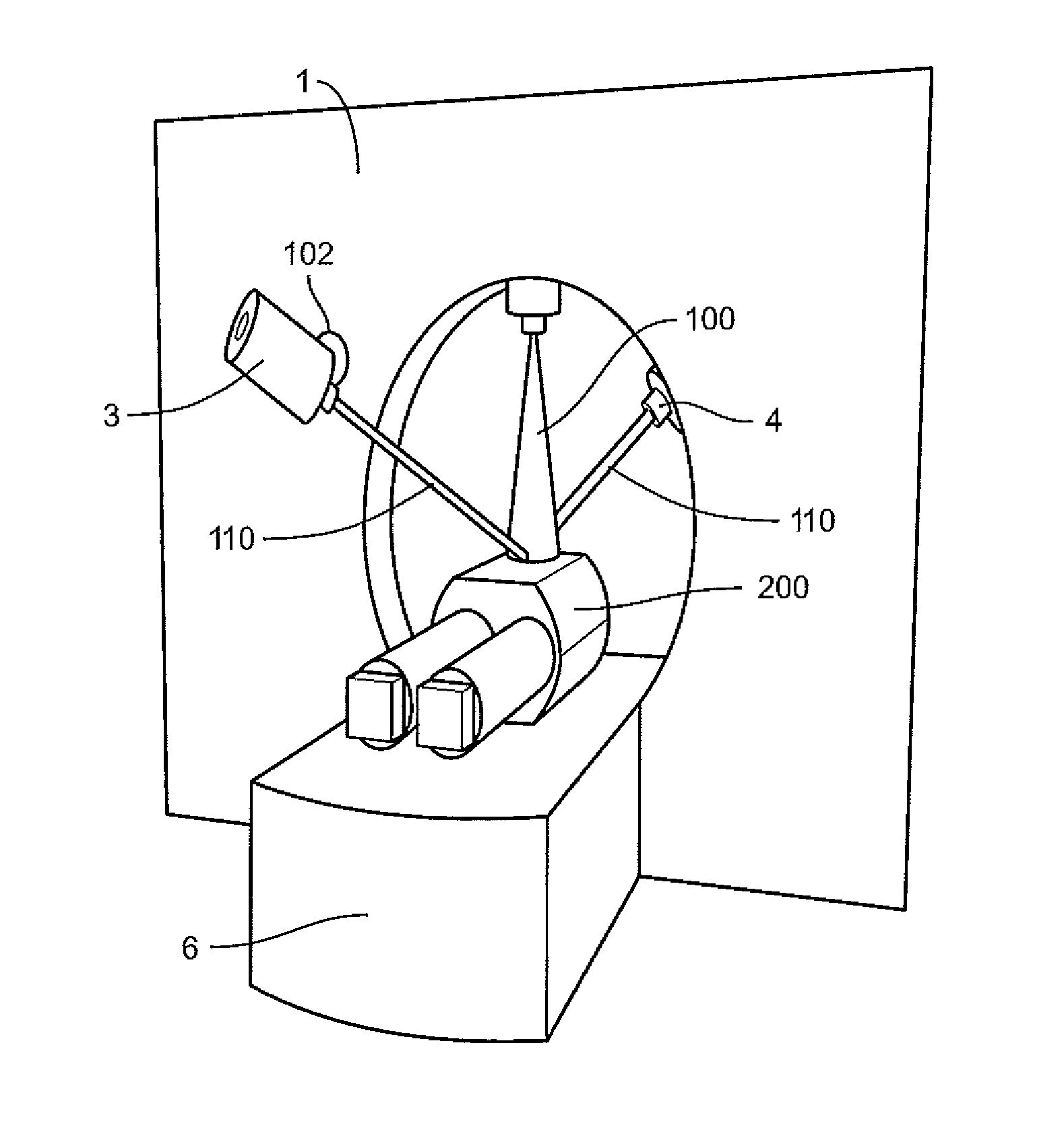
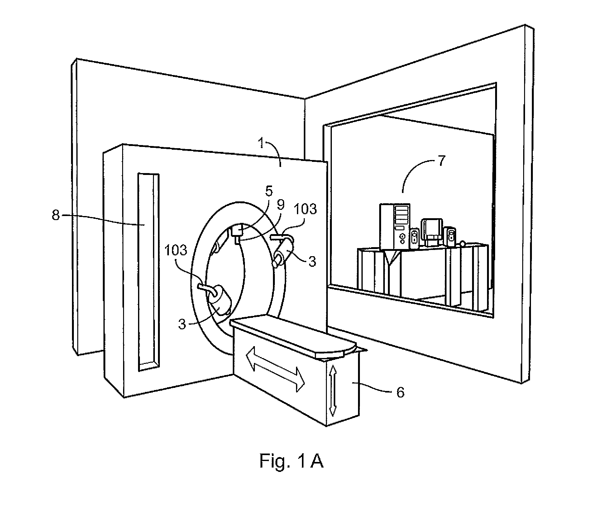
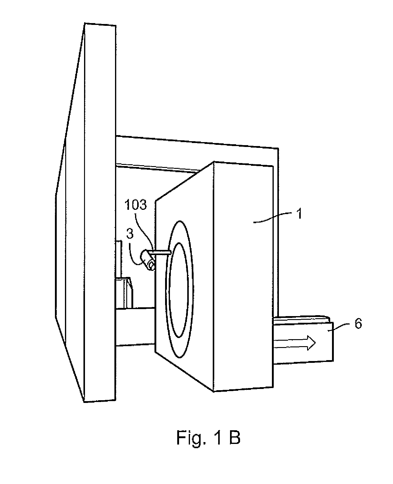
[0047]The present invention provides an apparatus for radiotherapy and / or radiosurgery which includes a system for the obtention of three-dimensional images of the patient (1), a plurality of X-ray generating sources (3), a system to automatically position said X-ray generating sources in such a way that the radiation produced be directed to the site to be irradiated in the patient, and a computer to automatically control the system (8). For the sake of clarity, the present invention is described through an example, without this implying or signaling limitations on the apparatus and revealed techniques.

[0048]The central objective of radiotherapy and / or radiosurgery is to maximize radiation absorbed dose imparted to the tumor while at the same time minimizing the radiation absorbed dose received by the surrounding healthy tissues and structures. To accomplish such in conventional radiotherapy, the following conditions are required:[0049]a) Determine by means of radiological images th...


PUM


Abstract

The present invention is related to an apparatus and method to carry out image-guided radiotherapy or radiosurgical treatments using Kilovoltage X-ray beams; one of the problems with the clinical application of this treatment consists in the absorbed dosage imparted unto a point within the subject to be irradiated depends on the local concentration of the contrast agent at that point, and the lack of properly quantifying the presence of the contrast agent at each point of the tumor or malformation to be irradiated results in a significant decrease in the quality of the treatment; the method and apparatus of the present invention resolve this and other problems. The Method consists of the following steps: a) determining the geometry of the patient and the concentration of the contrast agent at each point of the same, b) planning of treatment, and c) irradiating of the tumor or malformation; the apparatus includes a system to obtain three-dimensional images of the patient, a plurality of X-ray generating sources, a system to automatically position said X-ray generating sources in such a way that the radiation produced is directed towards the site to be irradiated in the patient, and a computer controlled system.

Description

CROSS-REFERENCE TO RELATED APPLICATION[0001]This application claims the benefit of Mexican Patent Application Serial No. MX / a / 2010 / 008470 filed on Jul. 29, 2010.FIELD OF THE INVENTION[0002]The present invention lies in the field of radiotherapy and radio surgery.BACKGROUND OF THE INVENTION[0003]The present invention is related to an apparatus and a method to carry out radiotherapeutic or radiosurgical treatments using kilovoltage X-ray beams, that is, those whose energy spectrum lies within the range varying between 20 keV to 500 keV. The term image guided radiotherapy is used in reference to the combination of an image acquisition system and a radiation generating ionizing equipment (X-rays, electrons, protons, neutrons and others) used to deliver radiation doses to any structure which can be, but is not limited to, a tumor or an arterio-venous malformation (AVM). The system for image acquisition, through the obtaining of a series of radiologic images, is in charge of “guiding” the...

Claims


Application Information

Patent Timeline
Patent Type & Authority Patents(United States)
IPC IPC(8): A61N5/10
CPCA61B6/542A61B6/481A61B6/544H05K999/00A61B6/583
Inventor GARNICA GARZA, HECTOR MAURICIO
Owner CENT DE INVESTIGACION & DE ESTUDIOS AVANZADOS DEL INST POLITECNICO NACIONAL
Why Eureka
Try Eureka