
Method for detecting compounds modulating dimers of VFT domain membrane proteins

a technology of vft domain and dimer, which is applied in the field of compound modulating membrane receptors, can solve the problems of inability to use screening methods on a large scale, poor suitability of existing techniques for demonstrating compounds capable of modulating the activation of dimeric class c gpcrs, and difficult detection of agonist effects

Active Publication Date: 2014-04-15
CIS BIO INT +1
View PDF25 Cites 2 Cited by
  • Summary
  • Abstract
  • Description
  • Claims
  • Application Information

AI Technical Summary

Benefits of technology

This method enables the effective screening of large libraries of compounds for modulating effects on membrane proteins, particularly class C GPCRs, by accurately distinguishing between agonist, antagonist, and allosteric effects, overcoming previous limitations of high background noise and complexity.

Problems solved by technology

These screening methods cannot be used on a large scale.
The existing techniques are poorly suited to demonstrating compounds capable of modulating the activation of dimeric class C GPCRs: agonist effects are very difficult to detect when the receptors have a weak affinity for their natural ligand (this is the case of the mGlu receptors); functional tests based on measuring a secondary message resulting from the signal transduction inside the cell (cAMP, 1P3, IP1) are also not suitable from the viewpoint of the relative difficulty in implementing them and of the complexity of the effector pathways coupled to dimeric receptors.
FRET (Förster resonance energy transfer) techniques based on the expression of GPCRs fused at their intracellular (C-terminal) domains with fluorescent proteins are also not suitable owing to the high background noise that comes from the dimers present in the intracellular compartments.
Moreover, it is essentially illustrated through the use of fluorescent proteins which are not very suitable in the present case owing to the high background noise generated by the dimers present in the various intracellular compartments.
These studies did not make it possible to observe any variation in FRET signal when the receptor is activated by its agonist, GABA, and suggest that this approach is not suitable for studying these dimers.

Method used


Image

  • Method for detecting compounds modulating dimers of VFT domain membrane proteins
  • Method for detecting compounds modulating dimers of VFT domain membrane proteins
  • Method for detecting compounds modulating dimers of VFT domain membrane proteins

Examples


example 1

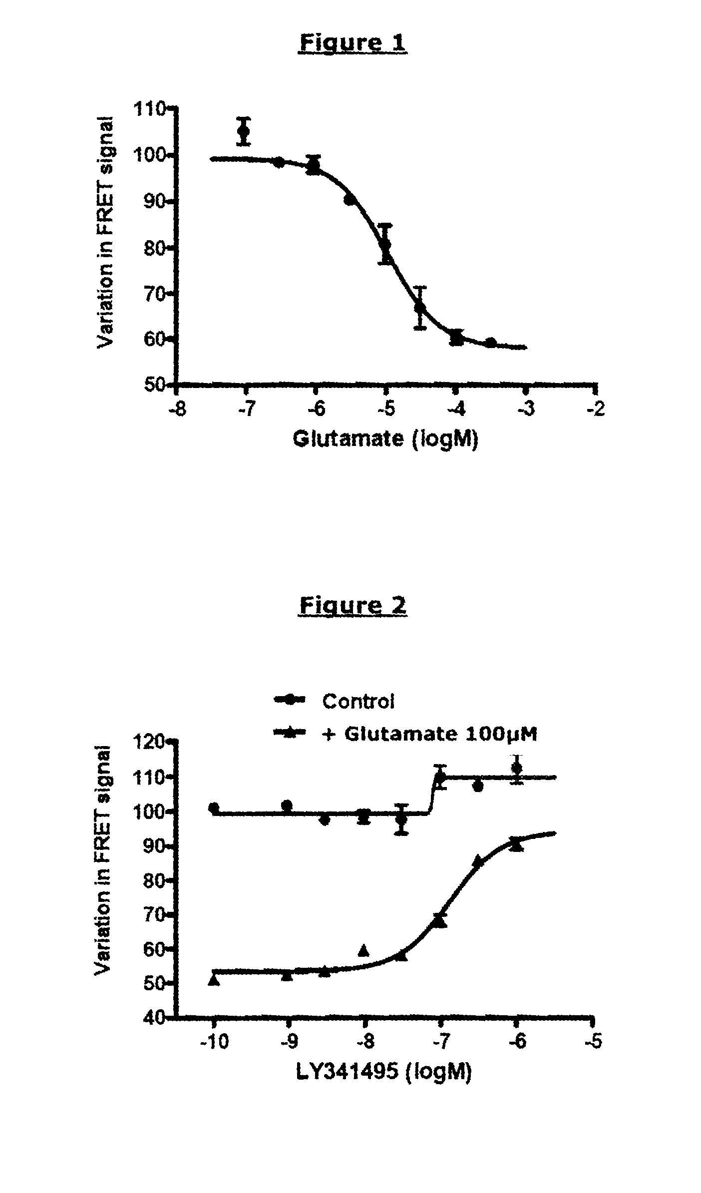
Demonstration of an Activating Compound having an Agonist Effect on an mGluR2-mGluR2 Dimer (R0 of the Pair of FRET Partners Used: 48 Å) Protocol

[0227]Transfection by Electroporation:

[0228]Cos-7 cells were cultured in complete medium at 37° C. and 5% CO2. At confluence, they were washed once with PBS, and detached with a trypsin-EDTA solution for 10 min at 37° C. A suspension of 10 million cells was mixed with 5 μg of total DNA (3.5 μg of prK5 plasmid, 1.2 μg of prK5 Flag-Cliptag-mGluR2 plasmid and 0.3 μg of prK5 HA-Snaptag-mGluR2 plasmid), in a final volume of 300 μl of electroporation buffer, and were then subjected to an electric shock of 280 V, 900 μF using a Gene-Pulser II electroporator (BIO-RAD), in 4 mm electroporation cuvettes (Eurogentec). The cells were then seeded into a 96-well plate (Greiner CellStar 96-well plate), at 150 000 cells per well, in a final volume of 100 μl per well of complete medium, and then incubated for 24 hours.

[0229]Labeling

[0230]24 hours after trans...

example 2

Demonstration of a Deactivating Compound having a Competitive Antagonist Effect on an mGluR2-mGluR2 Dimer (R0 of the Pair of FRET Partners Used: 48 Å)

[0242]Reagents and Plasmids

[0243]The same reagents and plasmids as those described in example 1 were used. The known antagonist compound LY341495 (Tocris Bioscience) was used.

[0244]Protocol

[0245]The same protocol as that described in example 1 was used. The final concentrations were [Glutamate]=100 μM in all the wells and [LY341495]=1000 nM; 316 nM; 100 nM; 31.6 nM; 10 nM; 3.16 nM; or 1 nM.

[0246]Results

[0247]FIG. 2 represents the TRF520 signal as a function of the concentration of LY341495 antagonist and in the presence of a constant concentration of glutamate (control curve: in the absence of glutamate).

[0248]In the presence of glutamate and in the absence of the competitive antagonist LY341495, the receptor is activated, and a variation in TRF520 of 50% is observed. The presence of antagonist prevents this action of glutamate on the ...

example 3

Demonstration of an Activating Compound having a Partial Agonist Effect on an mGluR2-mGluR2 Dimer (R0 of the Pair of FRET Partners Used: 48 Å)

[0250]Reagents and Plasmids

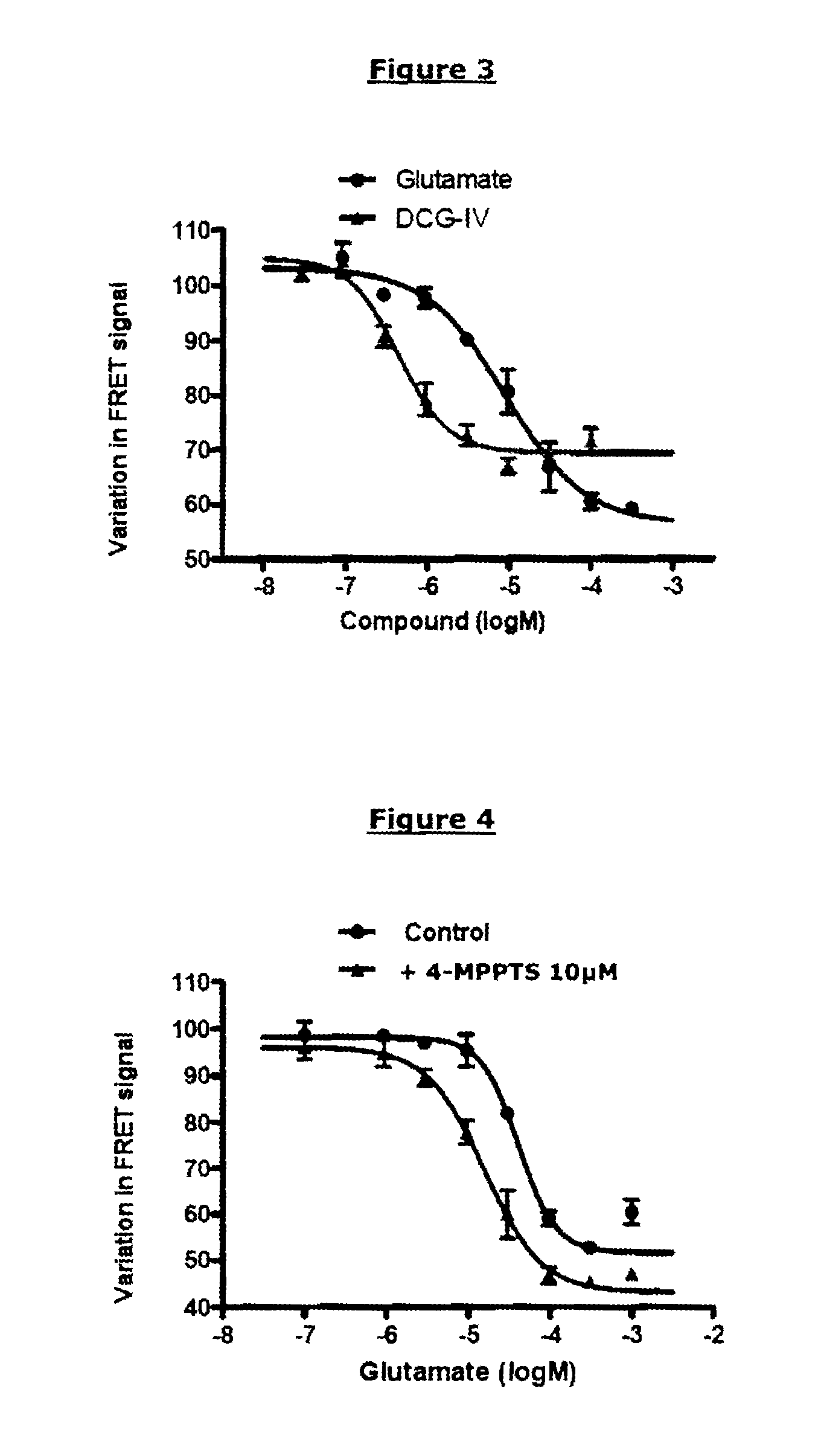
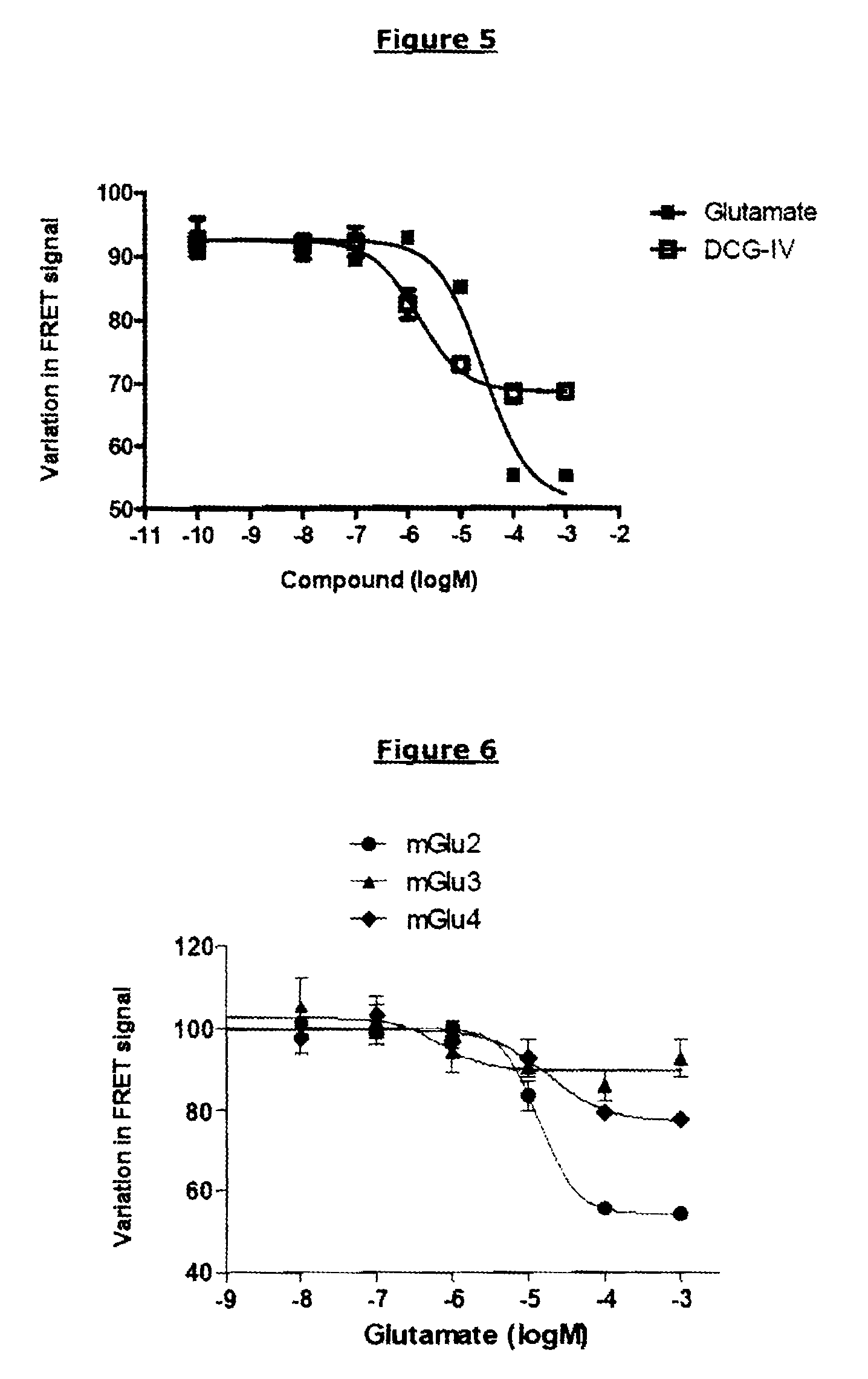
[0251]The same reagents and plasmids as those described in example 1 were used. The known partial agonist compound used was DCG-IV (Tocris Bioscience); it was compared with the reference complete agonist, glutamate.

[0252]Protocol

[0253]The protocol of example 1 was used. The final concentrations of [DCG-IV] were the following: 100 μM; 31.6 μM; 10 μM; 3.16 μM; 1 μM; 0.316 μM; or 0.1 μM.

[0254]Results

[0255]FIG. 3 represents the TRF520 signal as a function of the concentration of glutamate (complete agonist) or of DCG-IV (partial agonist).

[0256]Like glutamate, DCG-IV is capable of inhibiting the TRF520 signal in a dose-dependent manner. The concentration necessary for obtaining a variation equal to half the maximum variation is close to the pharmacological EC50 of DCG-IV (0.4 μM). Nevertheless, the maximum variation obtai...


PUM

PropertyMeasurementUnit
integration timeaaaaaaaaaa

Abstract

The invention relates to a method for selecting compounds having a modulating effect on the activation state of a dimer of VFT-domain proteins expressed in cell membranes present in a measuring medium, said dimer consisting of a first protein and of a second protein, said proteins being identical or different, wherein this method comprises the following steps:(a) labeling the first and second proteins in the N-terminal portion of their VFT domains with the members of a pair of FRET partners, the Förster radius (R0) of said pair being between 20 and 55 Å;(b) measuring the FRET signal in the absence and in the presence of the test compound within a predetermined time window;(c) selecting the test compound as a modulating compound if a difference in FRET signal in the absence and in the presence of test compound is measured in step (b).The invention can be used in the search for new medicaments and new taste modulators.

Description

[0001]This application is a 371 of PCT / FR10 / 50814, filed Apr. 29, 2010, which claims the priority of France 0952908, filed Apr. 30, 2009.PRIOR ART[0002]The invention relates to compounds modulating membrane receptors which are of interest in the search for new medicaments and for new taste modulators and in agriculture.[0003]G-protein coupled receptors (GPCRs) constitute the largest family of mammalian membrane receptors since they represent 3.4% of the genome. The isolation and then the cloning of GPCRs have made it possible to identify, in humans, around 900 genes, of which approximately 500 correspond to olfactory and taste receptors and 400 to receptors capable of binding endogenous ligands. Thus, the heterogeneity of these receptors provides a very broad recognition of both external signals (odors, light, taste molecules) and internal signals (hormones and neurotransmitters).[0004]GPCRs are referenced in three major classes:[0005]Class A, with rhodopsin being the archetype, is ...

Claims


Application Information

Patent Timeline
Patent Type & Authority Patents(United States)
IPC IPC(8): C12Q1/02C12Q1/48C12Q1/34
CPCG01N33/9426G01N2500/04G01N2333/726G01N33/542
Inventor DOUMAZANE, ETIENNEZWIER, JURRIAANTRINQUET, ERICPIN, JEAN-PHILLIPPE
Owner CIS BIO INT
Learn More