Methods and apparatus for displaying enhanced imaging data on a clinical image

a clinical image and imaging data technology, applied in the field of displaying medical images, can solve the problems of reducing the degree of detail of the image, reducing the degree of invasiveness, and and achieving the effect of saving millions of hospital days and millions of dollars annually in hospital residency costs alon

Active Publication Date: 2014-04-22
INTUITIVE SURGICAL OPERATIONS INC
View PDF18 Cites 60 Cited by
  • Summary
  • Abstract
  • Description
  • Claims
  • Application Information

AI Technical Summary

Problems solved by technology

Thus, an increased adoption of minimally invasive techniques could save millions of hospital days, and millions of dollars annually in hospital residency costs alone.
However, the smaller picture of a PIP display may be too small to show a desirable level of detailed information.
Moreover with separate image frames, a surgeon mentally fuses the two separate images or imaging modalities together, which can be fatiguing.
However, switching back and forth between images takes time and can make surgery less efficient as a result.
Moreover, a surgeon may fail to switch back to white light imaging and cut more than necessary or accidentally damage vital tissue while in an alternate imaging mode.

Method used


Image

  • Methods and apparatus for displaying enhanced imaging data on a clinical image
  • Methods and apparatus for displaying enhanced imaging data on a clinical image
  • Methods and apparatus for displaying enhanced imaging data on a clinical image

Examples


Embodiment Construction

[0019]This detailed description describes exemplary implementations that are illustrative of the invention, and so is explanatory and not limiting. The invention is limited only by patented claims. In the drawings, some elements have been omitted to more clearly show the embodiments of the invention.

Introduction

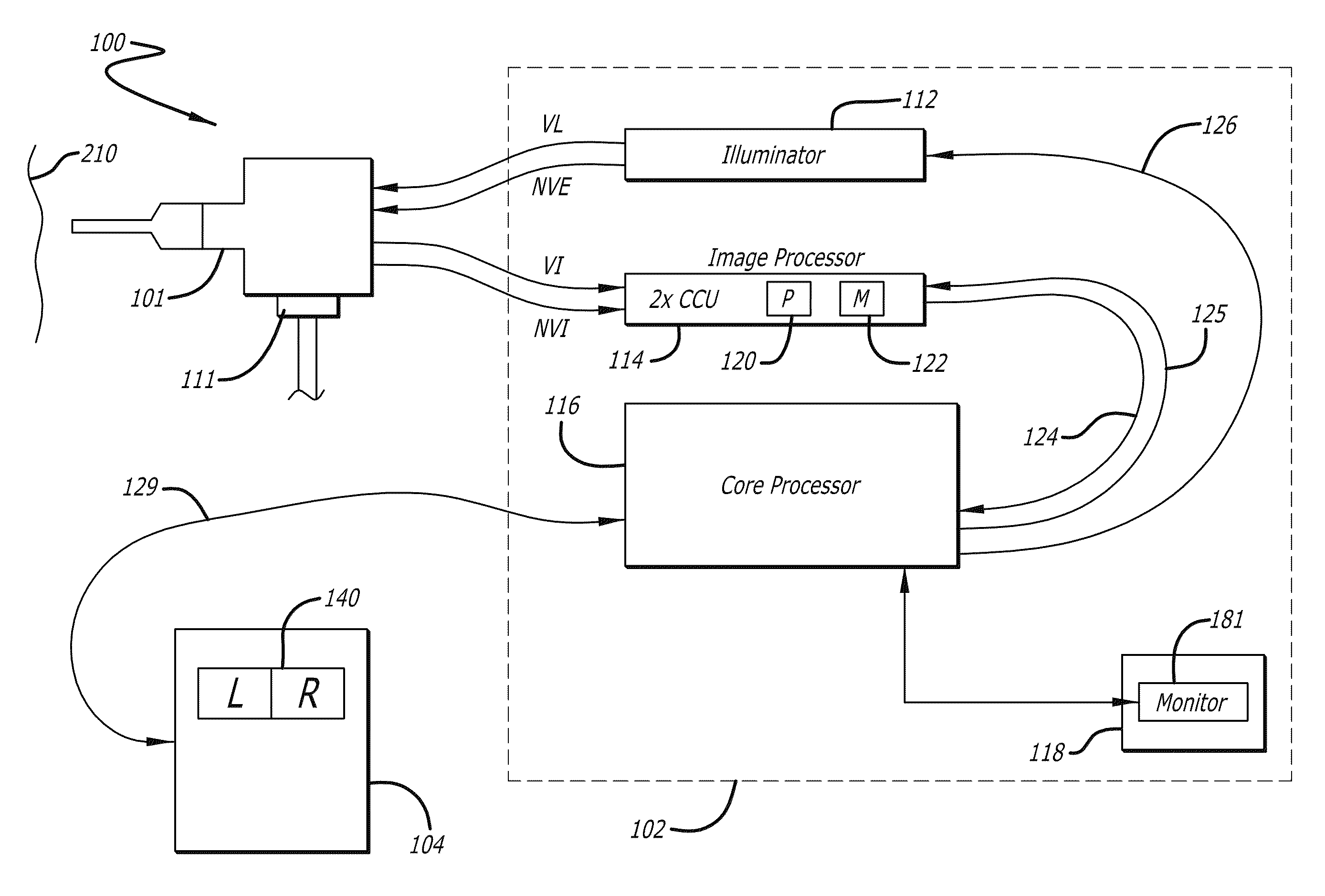
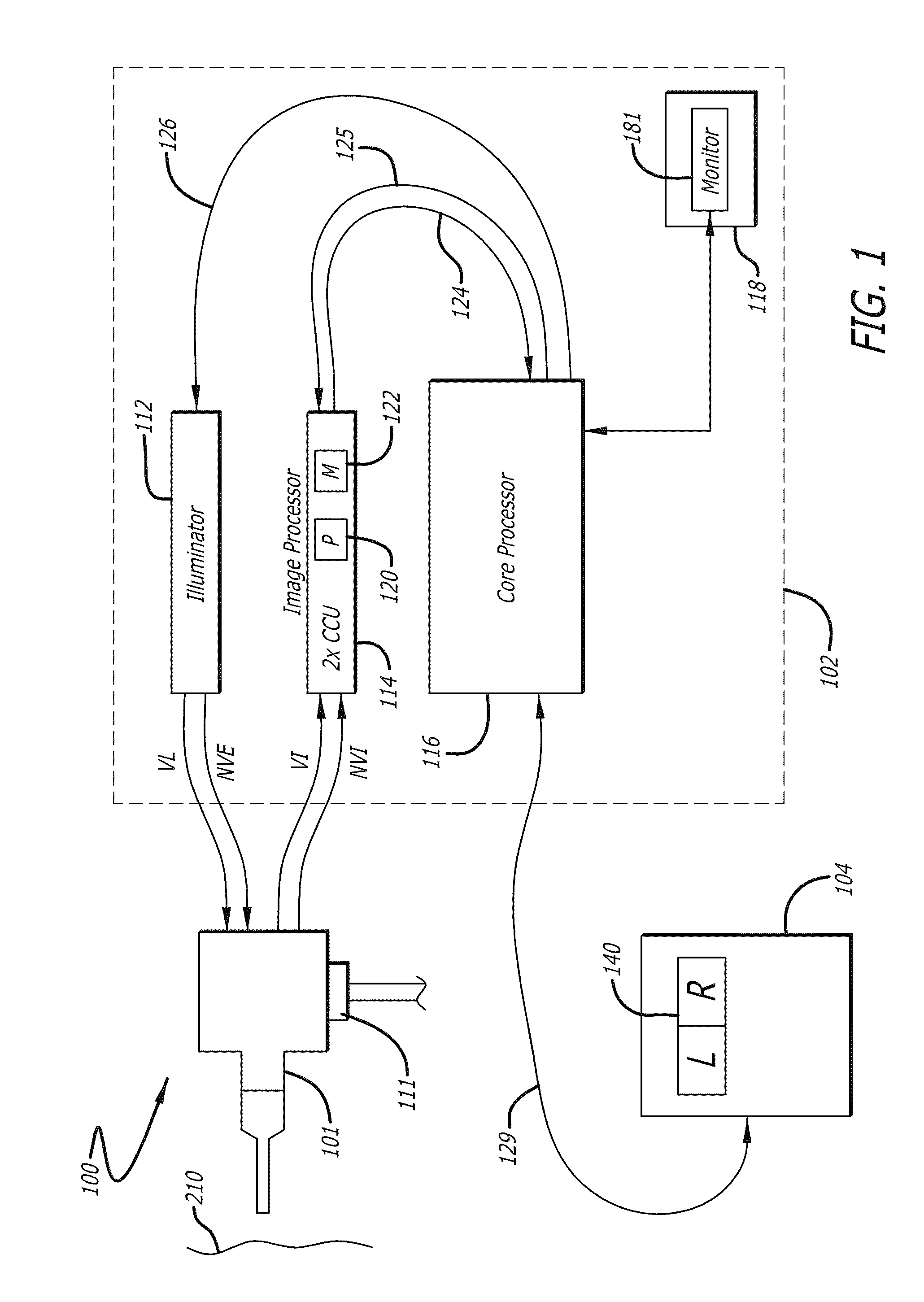
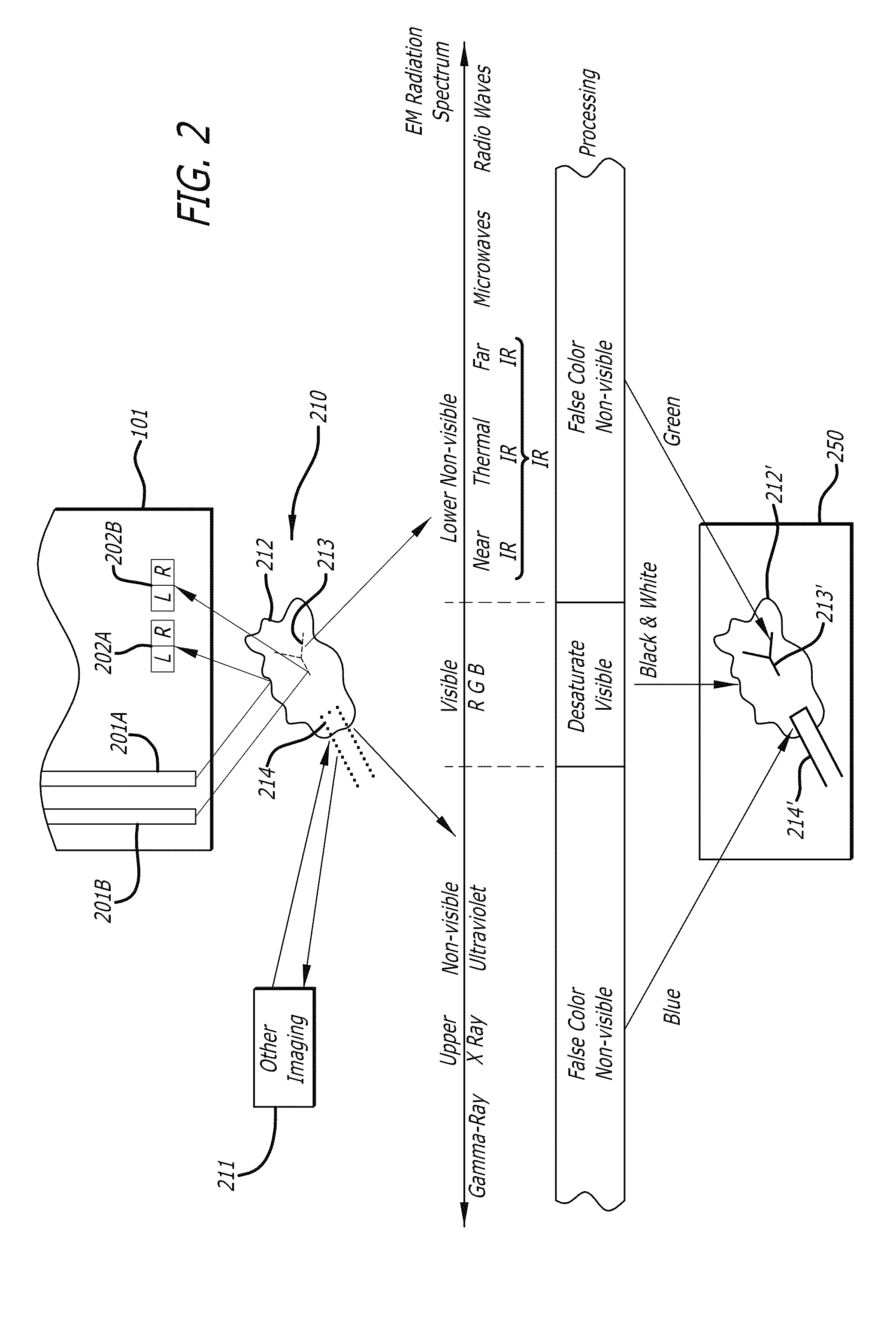
[0020]The embodiments of the invention are aimed at improving the clinical utility of the simultaneous display of a reflected white light image of tissue, and a separately or simultaneously acquired enhanced image of tissue in the same surgical site. The enhanced image of tissue may be captured with technologies such as, but not limited to, near-infrared (NIR) fluorescence, visible light fluorescence, multispectral imaging, fluorescence lifetime imaging, or a raster scan of non-visible light characteristics that contains clinical information with spatial variation. In addition, the enhanced image may be of an image constructed by overlaying point measurements of different typ...


PUM


Abstract

In one embodiment of the invention, an apparatus includes a display device. The display device displays a desaturated image of tissue captured in the visible electro-magnetic (EM) spectrum from a body cavity; and a first color enhanced image combined with the desaturated image. The first color enhanced image represents the first data captured from the body cavity outside the visible electromagnetic spectrum. The relative brightness between the desaturated image and the first color enhanced image is set to emphasize the first data over the tissue captured in the visible electromagnetic spectrum to provide improved information content.

Description

FIELD[0001]The embodiments of the invention generally relate to displaying medical images.BACKGROUND[0002]Minimally invasive medical techniques are aimed at reducing the amount of extraneous tissue that is damaged during diagnostic or surgical procedures, thereby reducing patient recovery time, discomfort, and deleterious side effects. The average length of a hospital stay for a standard surgery may also be shortened significantly using minimally invasive surgical techniques. Thus, an increased adoption of minimally invasive techniques could save millions of hospital days, and millions of dollars annually in hospital residency costs alone. Patient recovery times, patient discomfort, surgical side effects, and time away from work may also be reduced with minimally invasive surgery.[0003]To view a surgical site, an endoscopic camera with an illumination means may be inserted into a patient's body to capture color images of the surgical site. The color images of the surgical site may b...

Claims


Application Information

Patent Timeline
Patent Type & Authority Patents(United States)
IPC IPC(8): A61B5/05
CPCG06T5/50G06T2207/10024G06T2207/30004G06T2207/10048G06T2207/10064G06T2207/10068A61B34/37A61B1/043A61B1/000094A61B5/055G09G5/02G06T2210/41
Inventor MOHR, CATHERINEMCDOWALL, IANMOHR, PAUL
Owner INTUITIVE SURGICAL OPERATIONS INC
Why Eureka
Try Eureka