
Metal nanoshells for biosensing applications

a biosensing and metal nanotechnology, applied in the field of particles, can solve the problems of sensitivity decline, poor reproducibility, and difficulty in substrate preparation of sers enhancement methods of near infrared ft-raman spectroscopy, and achieve the effects of reducing the sensitivity of the ser enhancement method, and improving the reproducibility

Inactive Publication Date: 2005-06-16
RICE UNIV
View PDF43 Cites 63 Cited by
  • Summary
  • Abstract
  • Description
  • Claims
  • Application Information

AI Technical Summary

Benefits of technology

[0015] According to certain embodiments, the in vitro assay method includes selecting one or more optically tuned nanoshells with an absorption or scattering maximum wavelength that substantially matches the wavelength of a desired source of electromagnetic radiation. In some embodiments the chosen nanoshells include one or more conjugated biomolecules. The method also includes associating the nanoshells with one or more molecules of the desired analyte contained in the sample such that an analyte/nanoshell complex is formed. In certain embodiments the method includes associating the nanoshells with a reporter molecule, in which case a reporter/analyte/nanoshell complex is formed. Either complex is capable of producing a Raman signal upon irradiation by the selected source. Preferably the source is in the near-IR range of the electromagnetic spectrum. The method further includes irradiat...

Problems solved by technology

Although infrared excitation eliminates sample fluorescence, it also results in marked decrease in sensitivity, further motivating the need for a sensitization method.
Current methods being used for SERS enhancement of near infrared FT-Raman spectroscopy are frequently plagued by difficult substrate preparation, poor reproducibility, sensitivity to contamination, or limited suitability for in vivo use.
J. Biol. Chem. 227:285-299 (1957)), however, which significantly restricts the use of visible excitation Raman spectroscopy on biological systems.
Although that method was used in a successful sandwich immunoassay, the use of a microscopic silver substrate and the necessity for conjugation of the biomolecules with specific (carcinogenic) chromophores for resonance Raman detection severely limits the adaptability of that approach.
Since near infrared excitation results in a dramatic decrease in sensitivity relative to visible Raman excitation, the most outstanding current limitation to Raman-based glucose monitoring is the lack of sensitivity.

Method used


Image

  • Metal nanoshells for biosensing applications
  • Metal nanoshells for biosensing applications
  • Metal nanoshells for biosensing applications

Examples


example 1

Surface Enhanced Raman Scattering (SERS) Using Metal Nanoshells

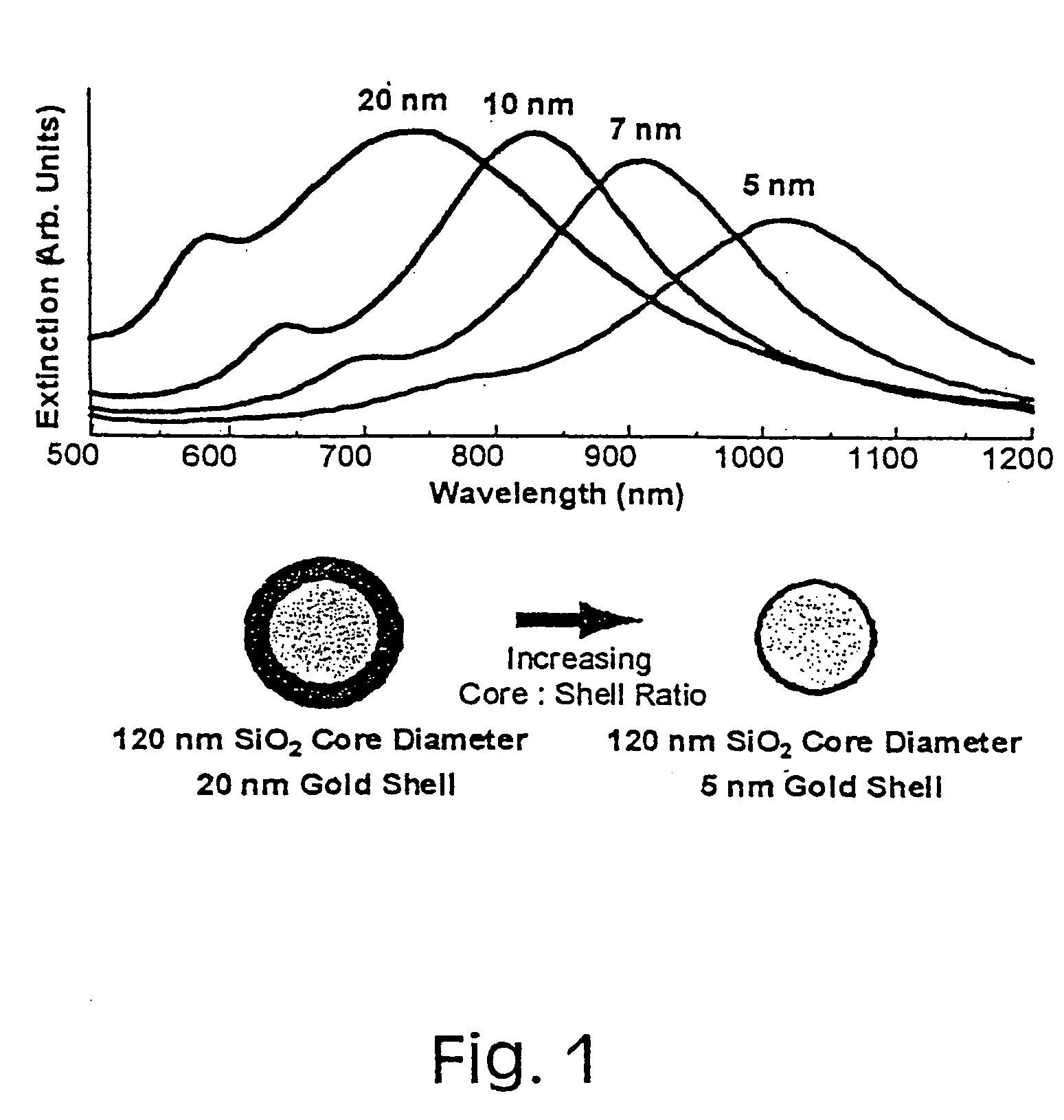
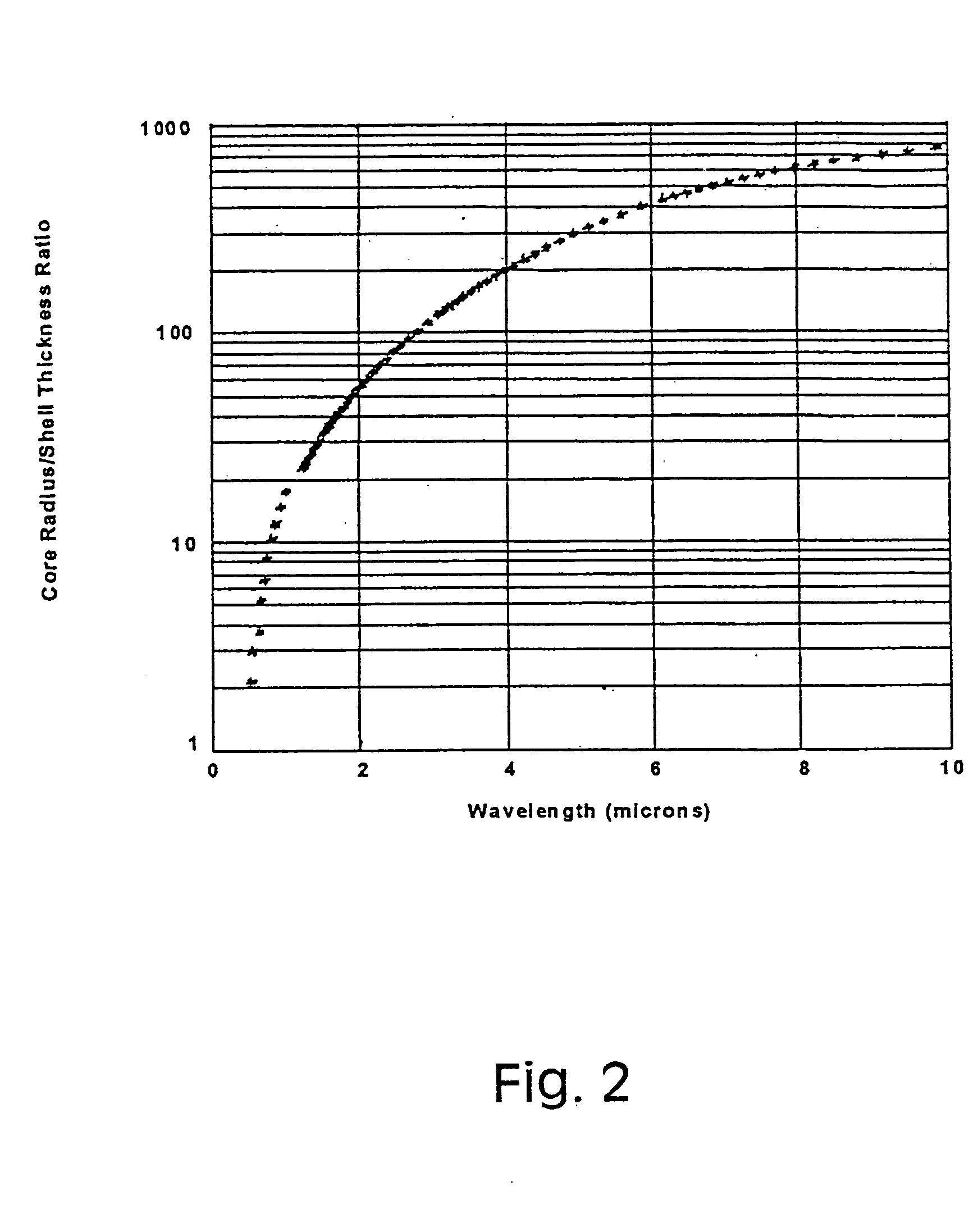
[0042] Since metal nanoshells have a plasmon resonance that is designed into the particle by adjusting the particle core:shell ratio, their plasmon resonance can be shifted during growth of the shell to coincide with the excitation wavelengths of near infrared laser sources, such as the 1064 nm Nd:YAG source used in a FT-Raman laser spectrometer.

[0043] In a series of recent experiments, the SERS enhancement properties of metal nanoshells were investigated (Oldenburg, S. J. et al. J. Chem. Phys. 111:4729-4735 (1999), incorporated in its entirety herein by reference). The nanoshell plasmon resonance was placed at nominally 900 nm, so that the shoulder of the plasmon peak overlapped with the Raman excitation wavelength. FIG. 5 shows the SERS enhancement observed in this study for the molecule mercaptoaniline. An enhancement of 600,000 in the Raman signal was observed. In this case, the strong interaction between mercaptoa...

example 2

Bioconjugation of Gold Nanoparticles / Nanoshells

[0044] Because the reduction of the outer metal layer of gold nanoshells is accomplished using the same chemical reaction as gold colloid synthesis, the surfaces of gold nanoshells are likely to be virtually chemically identical to the surfaces of the gold nanoparticles universally employed in conventional bioconjugate applications. Existing conjugation protocols for the labeling of a broad range of biomolecules with gold colloid (e.g., protein A, avidin, streptavidin, glucose oxidase, horseradish peroxidase and IgG) (M. A. Kerr et al., eds. Immunochemistry Labfax BIOS Scientific Publishers, Ltd., Oxford, U.K. 1994) will be directly repeatable or easily adaptable for use with gold nanoshells. Similar conjugation techniques are also expected to be readily adaptable for conjugation of nanoshells comprising other core materials. In one set of experiments, attachment of glucose oxidase (GO) to 150 nm diameter gold nanoshells was accomplish...

example 3

Gold Nanoshell-Based Biosensors

[0045] A preferred biosensing strategy combines the enormous SERS enhancements provided by metal nanoshells with the facile bioconjugation capabilities of gold nanoshell surfaces. This combination provides a highly sensitive, high information density spectral probe suitable for monitoring specific biochemical processes of physiological importance. Visible light is not suitable for in vivo optical monitoring due to its absorption by hemoglobin. Ultraviolet light is also not suitable due to the potential for photochemical transformation of proteins and DNA. Raman scattering in the near infrared, enhanced by the gold nanoshell plasmon resonance, lacks these disadvantages and is predicted by the inventors to facilitate the same demonstrated SERS sensitivity in regions of high physiological transmissivity, such as the “water windows” of 800-1,300 nm and 1,600-1,850 nm (Anderson, R. R. et al. J. Invest. Dermatol. 77:13-19 (1981); and Duck. F. A., Physical P...


PUM

PropertyMeasurementUnit
Thicknessaaaaaaaaaa
Thicknessaaaaaaaaaa
Thicknessaaaaaaaaaa

Abstract

The present invention provides nanoshell particles (“nanoshells”) for use in biosensing applications, along with their manner of making and methods of using the nanoshells for in vitro and in vivo detection of chemical and biological analytes, preferably by surface enhanced Raman light scattering. The preferred particles have a non-conducting core and a metal shell surrounding the core. For given core and shell materials, the ratio of the thickness (i.e., radius) of the core to the thickness of the metal shell is determinative of the wavelength of maximum absorbance of the particle. By controlling the relative core and shell thicknesses, biosensing metal nanoshells are fabricated which absorb light at any desired wavelength across the ultraviolet to infrared range of the electromagnetic spectrum. The surface of the particles are capable of inducing an enhanced SERS signal that is characteristic of an analyte of interest. In certain embodiments a biomolecule is conjugated to the metal shell and the SERS signal of a conformational change or a reaction product is detected.

Description

CROSS-REFERENCE TO RELATED APPLICATIONS [0001] This application is a divisional application of U.S. Patent Application No. 09,616,154 filed Jul. 14, 2000 entitled “Metal Nanoshells for Biosensing Applications” which is a continuation-in-part of co-pending U.S. patent application Ser. No. 09 / 038,377 filed Mar. 11, 1998, and also claims the benefit of U.S. Provisional Application No. 60 / 144,136 filed Jul. 16, 1999. The disclosures of those applications are incorporated herein by reference.STATEMENT REGARDING FEDERALLY SPONSORED RESEARCH OR DEVELOPMENT [0002] This invention was made with government support under Grant No. N00014-97-1-0217 awarded by the Office of Naval Research and under Grant No. ECS-9258118 awarded by the National Science Foundation. The United States government has certain rights in the invention.BACKGROUND OF THE INVENTION [0003] 1. Field of the Invention [0004] The present invention generally relates to particles composed of a nonconducting core coated with a very...

Claims


Application Information

Patent Timeline
IPC IPC(8): A61K41/00B01J13/02B22F1/054B22F1/18C08K9/02C09C1/30C09K3/00G01N33/543G01N33/58G02F1/355G21K1/10
CPCA61K41/0042Y10T428/2991B22F1/0018B22F1/02B82Y5/00B82Y15/00B82Y30/00C01P2002/82C01P2002/84C01P2004/04C01P2004/12C01P2004/32C01P2004/38C01P2004/50C01P2004/54C01P2004/61C01P2004/62C01P2004/64C01P2004/80C01P2006/22C01P2006/33C01P2006/40C01P2006/60C08K9/02C09C1/3081C09C1/309C09K3/00G01N33/54373G01N33/587G01N33/588G02F1/355B01J13/02B22F1/054B22F1/056B22F1/18
Inventor WEST, JENNIFER L.HALAS, NANCY J.OLDENBURG, STEVEN J.AVERITT, RICHARD D.
Owner RICE UNIV
Learn More