

Methods for the detection of glycated hemoglobin
a technology of human hemoglobin and detection method, which is applied in the field of detection methods of glycated human hemoglobin, can solve problems such as up to a 40% elevation
- Summary
- Abstract
- Description
- Claims
- Application Information
AI Technical Summary
Benefits of technology
Problems solved by technology
Method used
Image

Examples
example 1
Digestion of Human Whole Blood Samples with Pepsin and Proline-Specific Endopeptidase
[0047]7.5 μL of DCA N (Normal control, DCA® 2000 Analyzer reagent kit (Siemens Medical Solutions Diagnostics)) or DCA AB (Abnormal control, DCA® 2000 Analyzer reagent kit (Siemens Medical Solutions Diagnostics)) were incubated at room temperature for 20 minutes with 150 μL Sample Denaturant Reagent (ADVIA® HbA1c reagent kit (Siemens Medical Solutions Diagnostics)) containing 250 U / mL of pepsin. 150 μL proline-specific endopeptidase (30 U / mL) in 0.1 mol / L phosphate buffer, pH 7.0, was added to half the pepsin-treated samples, and the proline-specific endopeptidase-treated samples were incubated for 30 minutes at room temperature. The remaining pepsin-treated samples were not treated with proline-specific endopeptidase, and 150 μL 0.1 mol / L phosphate buffer, pH 7.0, was added to those samples.
[0048]All samples were then tested in an agglutination inhibition immunoassay on an ADVIA® 1650 instrument by ...
example 2
Formulation of a Proline-Specific Edopeptidase-Containing Agglutinator Reagent
[0050]Conditions for the stable formulation of a reagent containing the agglutinator used in Example 1 (β-chain amino-terminal glycated sexapeptide of human hemoglobin conjugated to polyaspartamide) and proline-specific endopeptidase at pH 7.0 were determined.
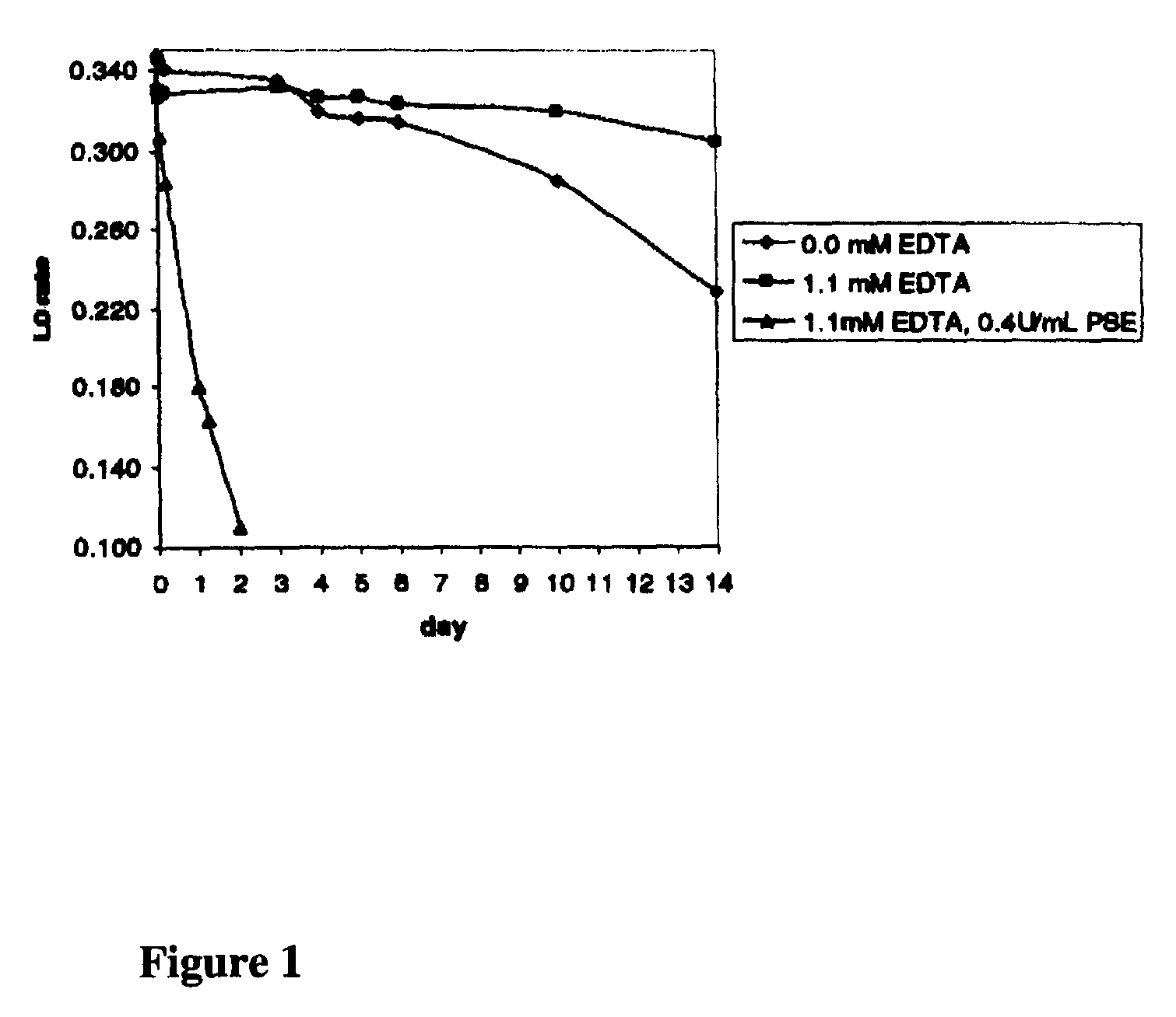
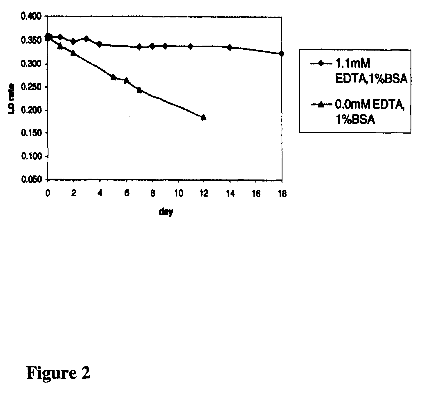
[0051]Solutions of the agglutinator (approximately 1.5.mu.g / mL) containing 0.02 mol / L morpholino-propane-sulfonic acid (MOPS), 0.05% NaN.sub.3, and 0.2% TWEEN®-detergent, pH 7 were prepared either without ethylenediamine tetraacetic acid (EDTA) and 0.4 U / mL proline-specific endopeptidase, with 1.1 mmol / L EDTA, or with both 1.1 mmol / L EDTA and 0.4 U / mL proline-specific endopeptidase. The ability of the agglutinator in the various formulations to cause agglutination of latex coated with mouse monoclonal anti-HbA1c antibody over a period of 14 days was then determined to assess the stability of the agglutinator over time. Samples were tested in an agglut...
example 3
Determination of Pepsin Digestion Conditions
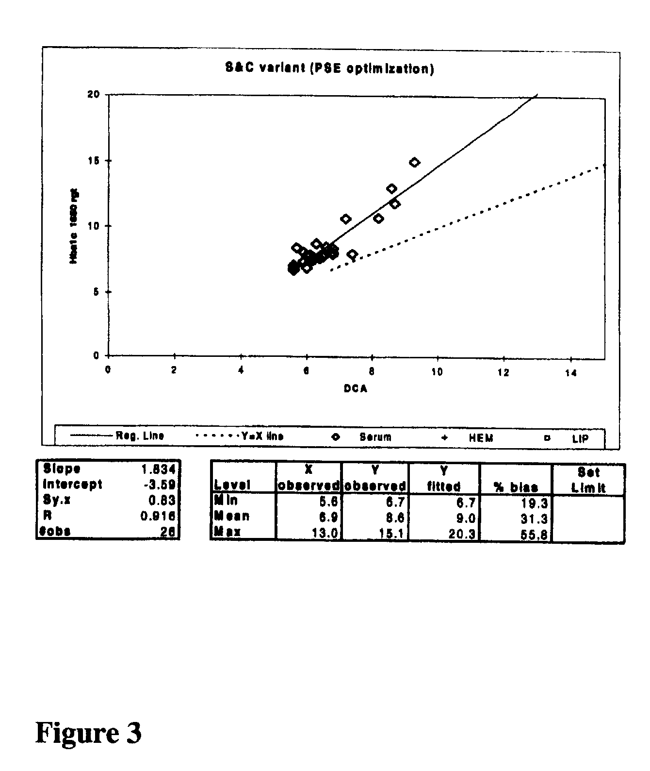
[0053]2 μL samples of lyophilized human whole blood containing various concentrations of HbA1c (Whole Blood HbA1c Calibrators, Aalto Scientific) were manually digested with 148 μL of 250 U / mL pepsin (Sample Denaturant Reagent (ADVIA® HbA1c reagent kit (Siemens Medical Solutions Diagnostics)) for 10 minutes, were digested for nine seconds with approximately 6000 U / mL pepsin in a 0.02 mmol / L citrate, 0.2% Triton X100 pH 2.4 solution in an automated fashion, or were digested for ten minutes with approximately 6000 U / mL pepsin in a 0.02 mmol / L citrate, 0.2% Triton X100 pH 2.4 solution in an automated fashion. The samples were then assessed in an agglutination inhibition immunoassay on an ADVIA® 1650 instrument as described in Example 2 where R1 was the agglutinator reagent containing 0.4 U / mL proline-specific endopeptidase. The lack of change among the agglutination rates listed for the different denaturing conditions for each respective sampl...
PUM
| Property | Measurement | Unit |
|---|---|---|
| pH | aaaaa | aaaaa |
| pH | aaaaa | aaaaa |
| pH | aaaaa | aaaaa |
Abstract
Description
Claims
Application Information