
Electrical bioimpedance analysis as a biomarker of breast density and/or breast cancer risk

a biomarker and breast density technology, applied in the field of electric bioimpedance analysis as a biomarker of breast density and/or breast cancer risk, can solve the problems of high mortality rate, difficult tissue detection, and inability to detect the presence of proliferative cells

Inactive Publication Date: 2014-05-27
EPI SCI LLC
View PDF45 Cites 5 Cited by
  • Summary
  • Abstract
  • Description
  • Claims
  • Application Information

AI Technical Summary

Benefits of technology

The patent describes a method for estimating the percentage of breast tissue that is made up of mammographic density. This is important for assessing a woman's risk of breast cancer or other breast abnormalities. The method involves measuring the electrical properties of the breast tissue using a special electrode configuration and algorithm. The method can be used in conjunction with X-ray images, ultrasound, or magnetic resonance imaging to provide a more accurate estimate of breast density. The method can also be adjusted for the distance between the electrodes and the individual's age and body mass index. Overall, the method provides a reliable and non-invasive way to estimate breast density and assess breast cancer risk.

Problems solved by technology

Difficulty in detecting proliferative, abnormal pre-cancerous or cancerous tissue before treatment options become non-viable is one of the reasons for the high mortality rate.
Detecting of the presence of proliferative, abnormal or cancerous tissues is difficult, in part, because such tissues are largely located deep within the body, thus requiring expensive, complex, invasive, and / or uncomfortable procedures.
For this reason, the use of detection procedures is often restricted until a patient is experiencing symptoms related to the abnormal tissue.
Many forms of cancers or tumors, however, require extended periods of time to attain a detectable size (and thus to produce significant symptoms or signs in the patient).
It is often too late for effective treatment by the time the detection is performed with currently available diagnostic modalities.
Both are fraught with problems of inaccuracy.
Clinical breast examinations are limited because lesions less than one cm are usually undetectable and larger lesions may be obscured by diffuse nodularity, fibrocystic change, or may be too deep in the breast to enable clinical detection.
Accordingly, mammography and clinical breast examination have relatively poor specificity in diagnosing breast cancer.
Therefore many positive mammographic findings or lesions detected on clinical breast examination ultimately prove to be false positives resulting in physical and emotional trauma for patients.
Another disadvantage of the above referenced system is that the frequency range of the electrical signal is not defined.
Another disadvantage with the above referenced system is that the topography of altered impedance is not examined.
A further disadvantage of the above referenced methods is that they do not probe the specific conductive pathways that are altered during the malignant process.
This approach relies on the differences in conductivity and impedivity between different tissue types and relies on data acquisition and image reconstruction algorithms which are difficult to apply clinically.
Another disadvantage with using EIT to diagnose breast cancer is the inhomogeneity of breast tissue.
Single nucleotide polymorphisms of genes whose protein products are involved in carcinogen and hormone metabolism and / or DNA repair are associated with relative risks of 1.4-2.0, however combined polymorphisms may be associated with significantly higher relative risks.
However none have been established to definitively identify high-risk women.
Assessing density typically requires the use of radiation-based methods and is subject to inter-observer variability.

Method used


Image

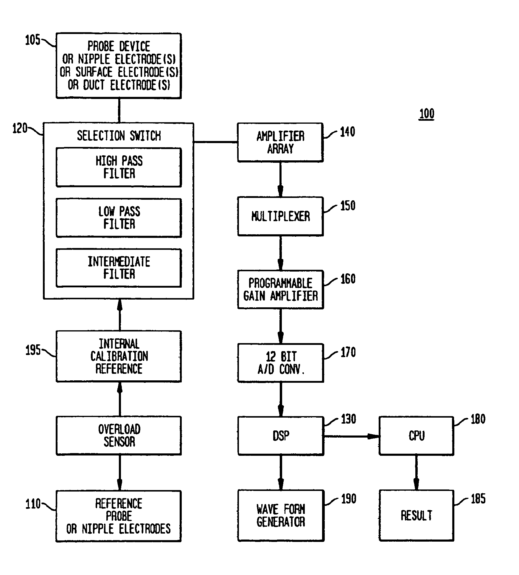
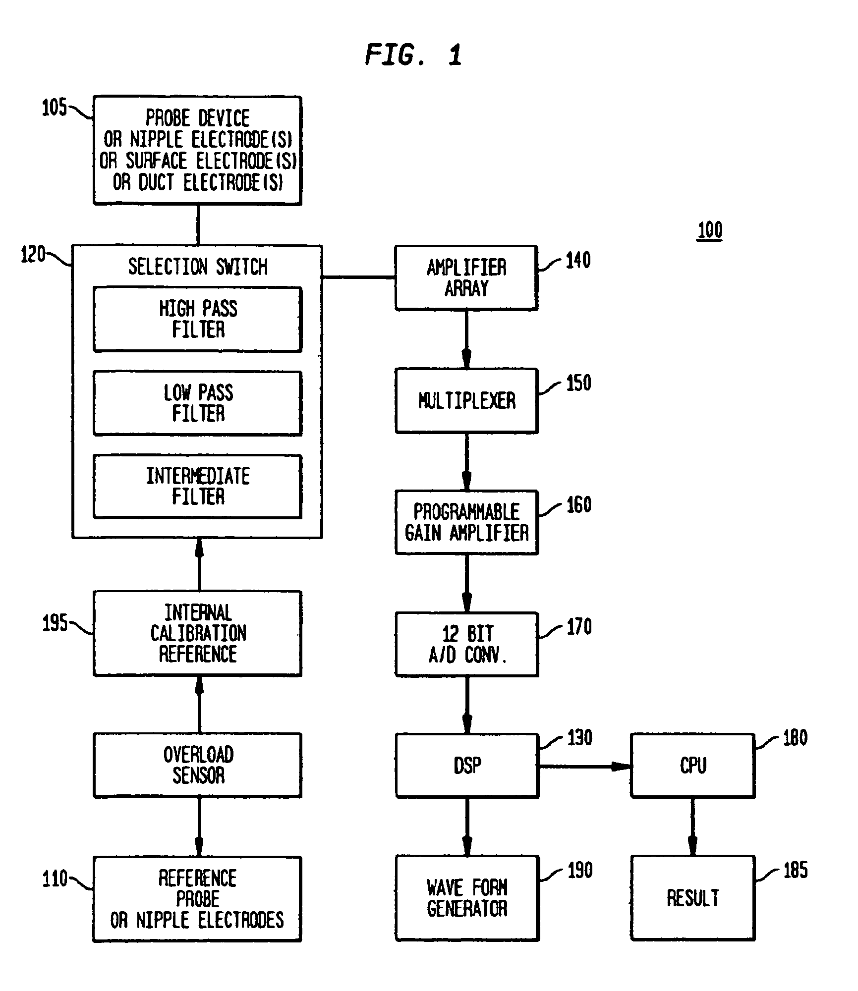
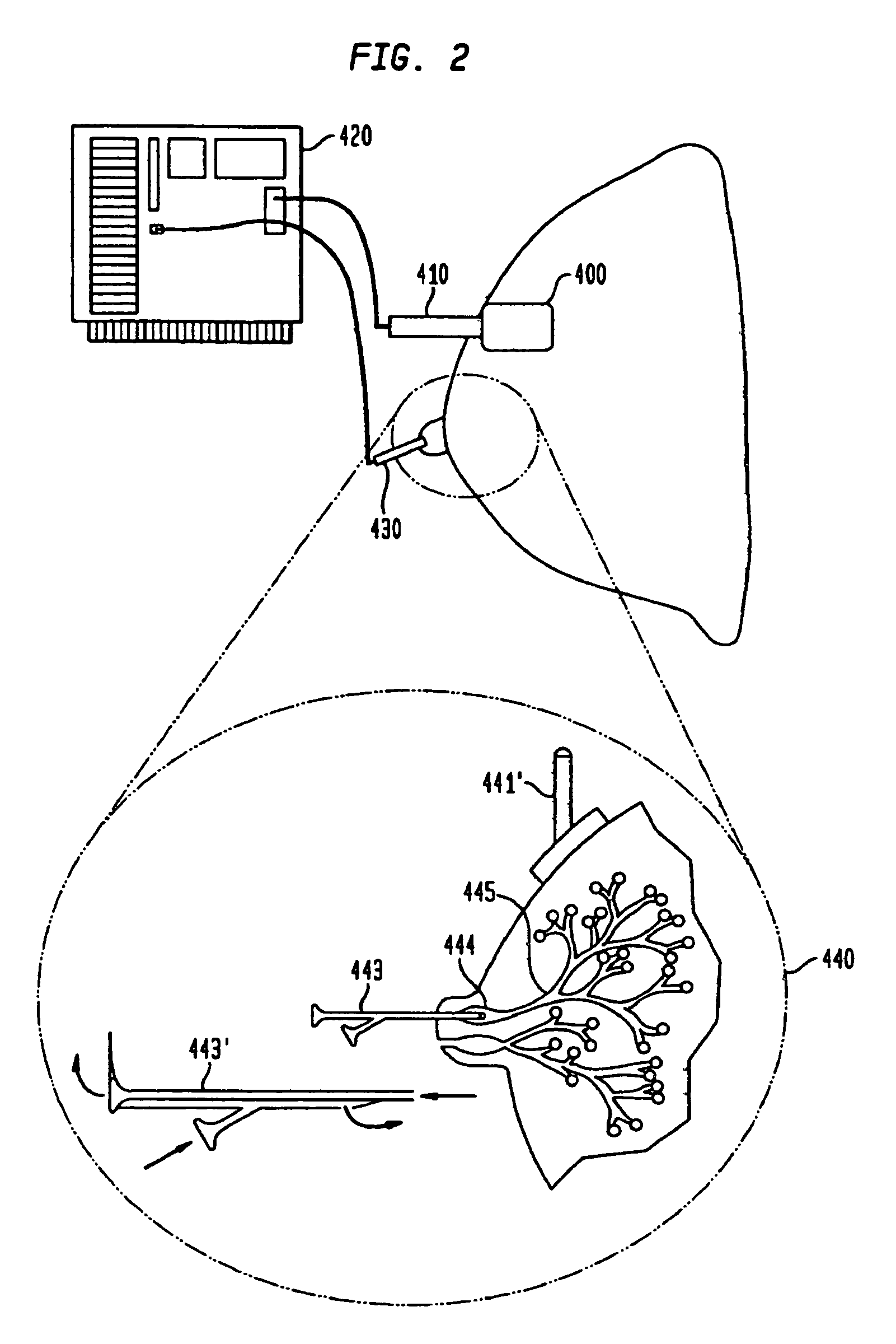
  • Electrical bioimpedance analysis as a biomarker of breast density and/or breast cancer risk
  • Electrical bioimpedance analysis as a biomarker of breast density and/or breast cancer risk
  • Electrical bioimpedance analysis as a biomarker of breast density and/or breast cancer risk

Examples


Embodiment Construction

[0109]For purposes of the present invention the following terms have the indicated meanings:

[0110]Sub-epithelial impedance, referred to herein as Zsub, means the impedance of the breast tissue that is underneath (sub) or beyond the ducts of the breast, i.e., the stroma or mesenchymal tissue of the breast (including fat, fibroglandular tissue, etc.). At high frequencies, such as about 50 KHz to about 60 KHz and higher, the epithelia (ducts and ductal-alveolar units of the breast) and overlying skin of the breast contribute very little to the total measured impedance, so the remainder is the sub-epithelial impedance (Zsub). Technically, Zsub is defined as the impedance at infinite frequency or the highest frequency tested, provided that the frequency is sufficiently high that the dielectric properties of the epithelial breast cells and the overlying skin break down and thus do not cause the test result to differ significantly from the true value at infinite frequency or at a significa...


PUM


Abstract

Methods and systems are provided for the noninvasive measurement of the subepithelial impedance of the breast and for assessing the risk that a substantially asymptomatic female patient will develop or be at substantially increased risk of developing proliferative or pre-cancerous changes in the breast, or may be at subsequent risk for the development of pre-cancerous or cancerous changes. A plurality of electrodes are used to measure subepithelial impedance of parenchymal breast tissue of a patient at one or more locations and at least one frequency, particularly moderately high frequencies. The risk of developing breast cancer is assessed according to measured and expected or estimated values of subepithelial impedance for the patient and according to one or more experienced-based algorithms. Devices for practicing the disclosed methods are also provided.

Description

CROSS-REFERENCE TO RELATED APPLICATIONS[0001]This application claims the benefit of the filing date of U.S. Provisional Patent Application No. 61 / 007,128 filed Dec. 11, 2007, the disclosure of which is hereby incorporated herein by reference.BACKGROUND OF THE INVENTION[0002]The ideal or preferred biomarker of breast density and / or cancer risk includes the following characteristics: (1) biologic plausibility; (2) a higher rate of expression in high-risk compared to low-risk populations; (3) an association with cancer in prospective studies; (4) expression minimally influenced by normal physiologic processes, or the ability to control for the influences of physiology; (5) the ability to obtain the biomarker using minimally invasive techniques at low costs; and (6) reproducibility. For purposes of the present invention the generally accepted National Institutes of Health definition of a “biomarker” is adopted: “a characteristic that is objectively measured and evaluated as an indicator...

Claims


Application Information

Patent Timeline
Patent Type & Authority Patents(United States)
IPC IPC(8): A61B5/05
CPCA61B6/482A61B5/0537A61B8/00A61B5/418A61B6/00A61B5/415A61B5/055
Inventor DAVIES, RICHARD, J.
Owner EPI SCI LLC
Learn More