Method of rapidly visualizing essential neural pathways

a neural pathway and rapid technology, applied in the field of computer assisted surgery, can solve the problems of long time taken to use software in preparation for surgery, its routine use in the cranial cavity, etc., and achieve the effect of improving the safety, efficiency and precision of brain tumor surgery

Inactive Publication Date: 2014-07-22
UNIV OF SOUTH FLORIDA +1
View PDF4 Cites 0 Cited by
  • Summary
  • Abstract
  • Description
  • Claims
  • Application Information

AI Technical Summary

Benefits of technology

The patent is about a new method for brain tumor surgery that improves safety, efficiency, and precision. It uses advanced imaging software to quickly and accurately identify important fiber bundles in the brain. This saves time and reduces the risk of complications during surgery. The method also helps to maximize functional outcomes and quality of life for patients with brain tumors. Overall, the patent presents a valuable contribution to the field of neurosurgery, and the use of DTI-FT software is a key advantage in the process.

Problems solved by technology

However, the prolonged amount of time taken to use the software in preparation for surgery is well known.
This drawback discourages its routine use in the cranial surgery despite its obvious advantage and application in improving the surgical outcome.

Method used


Image

  • Method of rapidly visualizing essential neural pathways
  • Method of rapidly visualizing essential neural pathways
  • Method of rapidly visualizing essential neural pathways

Examples


example 1

(FIGS. 1-3)

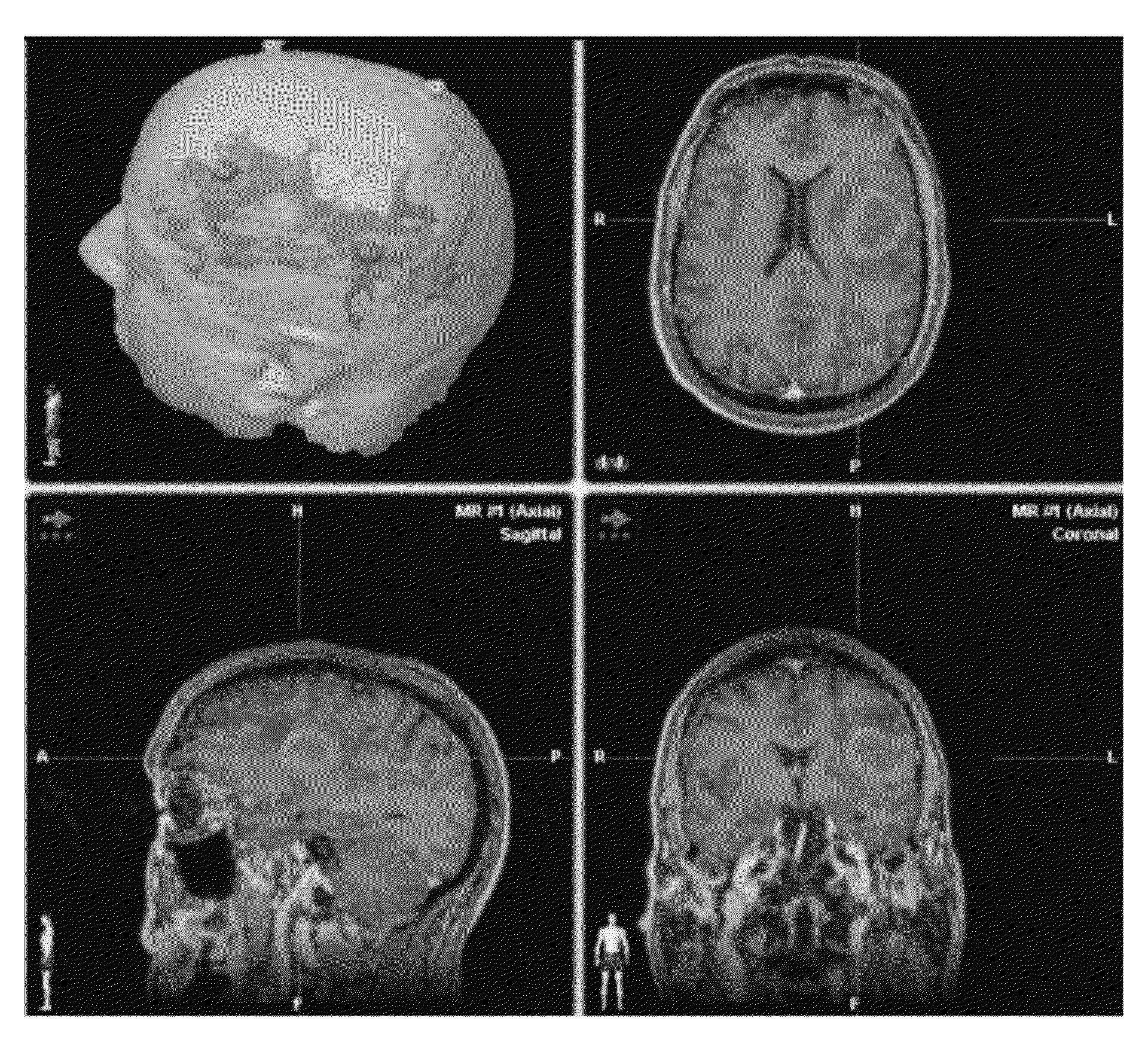
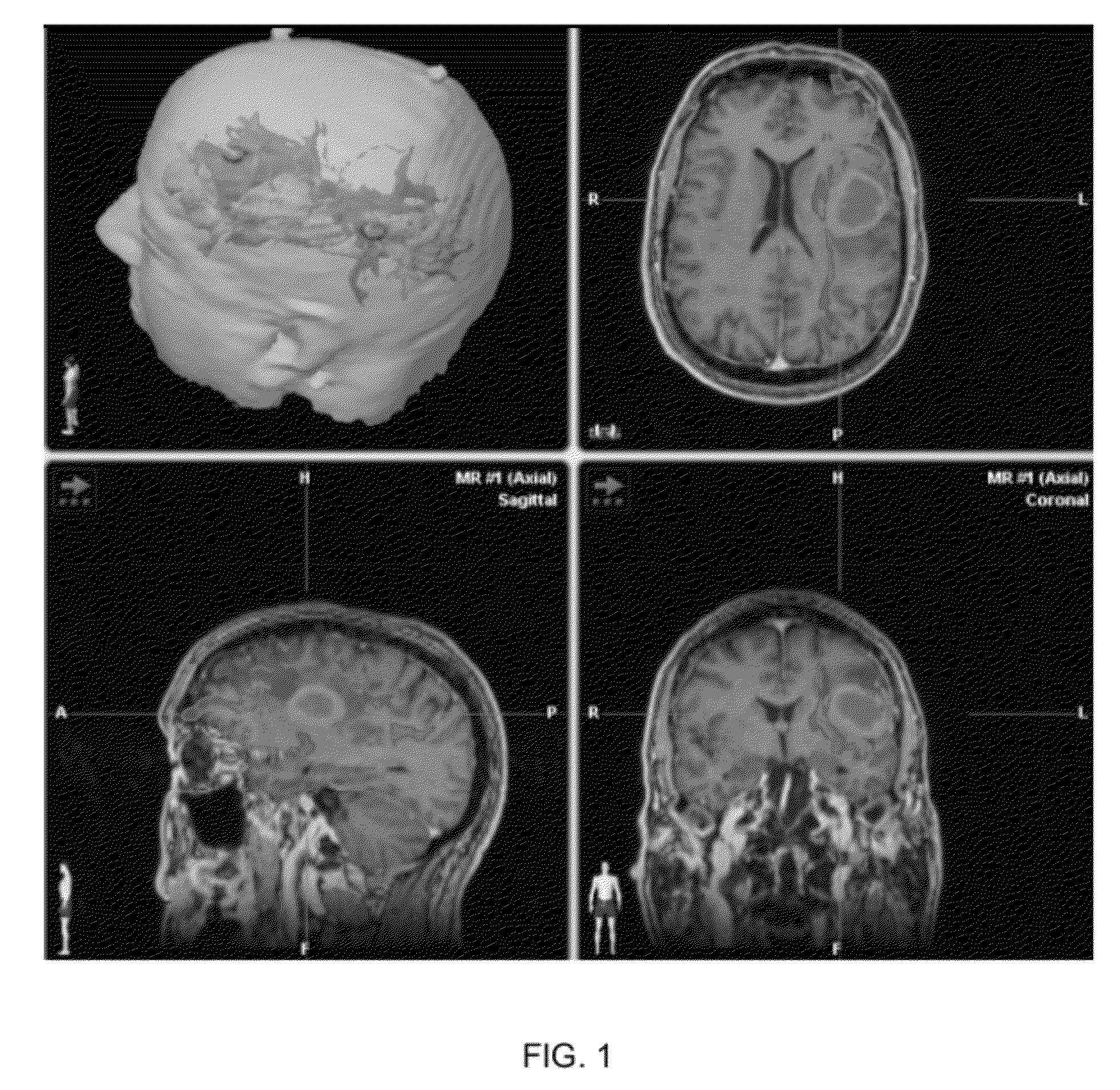
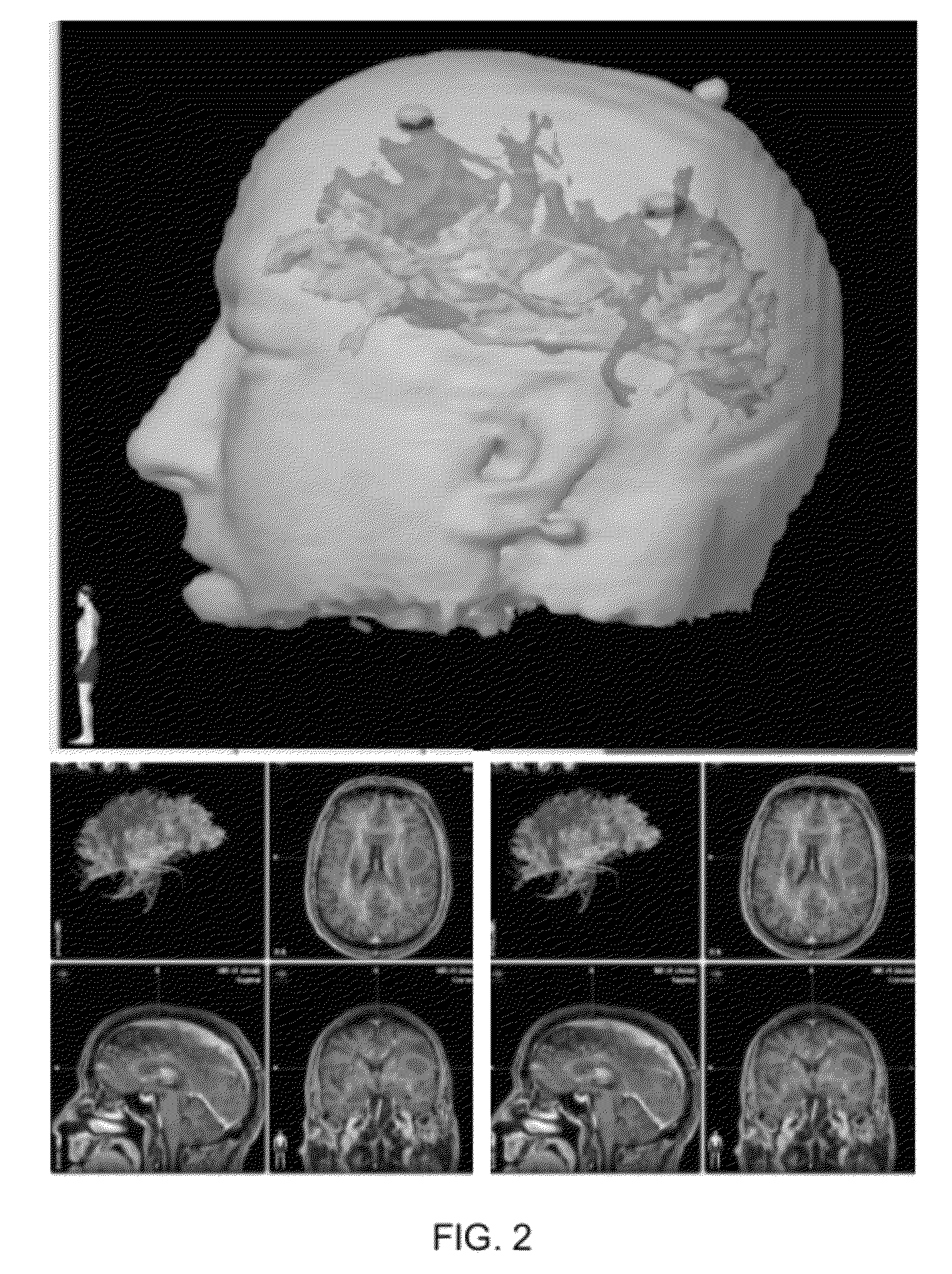
[0054]Patient: 45-year-old male, right-handed with Broca's area type ‘A’ cortical lesion.[0055]Preoperative Status: Seizure disorder with intermittent speech arrest and word finding difficulties.[0056]Procedure: DTI-FT, f-MRI BOLD SPPECH Mapping, intra-operative somatosensory evoked potential (SSEP) studies.[0057]Alternative approach: Awake craniotomy. Patient refused to undergo awake procedure.[0058]DTI Data: superior longitudinal fasiculus (SLF), inferior longitudinal fasiculus (ILF) uncinate fasiculus (UNC) reconstructed. Infiltration of speech fibers along medial wall of tumor. Type ‘ A’ subcortical tumor presentation. Fiber tracts displayed along ¾ wall of the tumor except a 5 mm area in the wall superiorly where fibers were not present.[0059]BOLD Data: Speech cortex infiltrating the tumor.[0060]Outcome: 5 mm opening made after initial transsulcal approach. Subtotal resection achieved. Significant improvement in speech after surgery. No new postoperative deficits. Se...

example 2

(FIGS. 4-6)

[0062]Patient: 81-year-old male, right-handed with frontal PS type ‘B’ motor cortex and type ‘B’ subcortical newly diagnosed GBM.[0063]Preoperative Status: No motor weakness from lesion.[0064]Procedure: DTI-FT, f-MRI BOLD MOTOR mapping, and intraoperative electrophysiological motor mapping studies integrated with MR based neuronavigation.[0065]DTI-Results: Type ‘B’ subcortical CST presentation. Fiber tracts displaced by tumor bulk without associated disruption. All fiber tracts appear posterior, inferior, and lateral to tumor in 3D plane.[0066]BOLD Data: Motor cortex displaced laterally.[0067]Outcome: DTI data helped in surgical planning and a gross total resection achieved with postoperative deficits.[0068]Impression: Postoperative DTI study is helpful to analyze and quantify change in fiber tracts characteristics in response to tumor resection and thereby, predict prognosis.

EXAMPLE 3

(FIGS. 7-11)

[0069]Patient: 31-year-old, newly diagnosed, Rt. Frontal non-enhancing type ...

example 3

(FIGS. 12-15)

[0076]Patient: 78-year-old female with metastatic brain tumors: type ‘C’ cortical, type ‘B’ subcortical tumor.[0077]Preoperative Status: Headaches. No deficits. KPS 90.[0078]Procedure: DTI-FT, f MRI BOLD SPEECH-Motor mapping, Intraoperative electrophysiological motor mapping studies.[0079]DTI results: Type ‘B’ subcortical tumor. Fiber tracts displaced posteriorly by the tumor.[0080]BOLD results: Type ‘C’ cortical tumor. Speech reflected to the left brain.[0081]Outcome: Gross total resection of tumor achieved. No postoperative deficits.[0082]Impression: Lack of neurological deficits suggests displacement of motor fibers in the subcortical plane rather than compression by the tumor. Postoperative DTI is helpful in validating this finding.


PUM


Abstract

A method of optimizing pre-operative procedures in the field of neurosurgery. The method facilitates the identification of essential fiber tract pathways that must be avoided when planning and performing an operation, thereby increasing surgical safety and avoiding complications. The method includes the combined use of four variables in Diffusion Tensor Imaging (DTI)-Fiber Tracking (FT) of projection fibers in the brain. These variables include Jain-Brem™ fractional anisotropy constant for tractography (JB-FACT), Jain-Brem™ fiber length (JB-FL), subcentimeter size placement of regions of interest (ROI), and the placement of ROIs to cover the anatomical pathway of projection fibers. JB-FACT and JB-FL provide rapid, reproducible tracking for projection fibers in the brain.

Description

CROSS REFERENCE TO RELATED APPLICATIONS[0001]This application is a continuation of currently PCT application PCT / US2010 / 044572 filed Aug. 5, 2010, which claims priority to U.S. Provisional Patent Application No. 61 / 231,378, filed Aug. 5, 2009 which are hereby incorporated by reference.FIELD OF INVENTION[0002]This invention relates to computer-assisted surgery in the field of neurosurgery.BACKGROUND[0003]Existing navigation software systems in the field of neurosurgery related, such as[0004]BrainLab's VectorVision®, have a steep learning curve. There is a long-felt need for surgical planning software that reduces complexity and likelihood of individual error.[0005]BrainLab's iPLAN® is diffusion tensor imaging (DTI)—fiber tracking (FT) software that identifies and tracks white matter fibers of the brain and recognizes different fiber bundles, such as commissural / association and projection fibers, and their location in relation to a brain tumor to assist a surgeon with surgical plannin...

Claims


Application Information

Patent Timeline
IPC IPC(8): A61B5/055
CPCG01R33/56341
Inventor JAIN, SURBHIBREM, STEVEN
Owner UNIV OF SOUTH FLORIDA
Why Eureka
Try Eureka