
Methods, tools and devices for percutaneous access in minimally invasive spinal surgeries

a technology for spinal surgery and percutaneous access, applied in the field of methods, tools and devices for providing percutaneous access in minimally invasive spinal surgery, access cannula, etc., can solve the problems of prolonged recovery time, increased risk of infections, and extensive tissue trauma

Inactive Publication Date: 2014-09-16
KIC VENTURES LLC
View PDF53 Cites 7 Cited by
  • Summary
  • Abstract
  • Description
  • Claims
  • Application Information

AI Technical Summary

Problems solved by technology

These procedures result in extensive tissue traumatization and consequently in prolonged recovery time, risk of infections, high hospitalization costs, pain that can be more severe than the pain due to the initial ailment, and in some cases permanent scarring.
The avoidance of general anesthesia reduces post-operative recovery time and the risk of complications.
Minimally invasive surgical procedures are especially desirable for spine surgeries because spine pathologies are located deep within the body without clear muscle planes and there is danger of damaging the adjacent neural and vascular tissues.
The destruction to the spinal structures is even more extensive during fusion procedures, which require more lateral tissue dissection and exposure to access the transverse processes and pedicles for placement of pedicle screws, rod constructs for stability, and bone graft under direct vision.
Unfortunately, the effectiveness and safety of the enzyme, chymopapain used for chemonucleolysis, have been complicated by severe spasms, post-operative pain, and sensitivity reactions including anaphylactic shock.
Loupe magnification and microscopes are helpful for improving visualization but are not effective without retractor systems and specialized instruments and devices to make minimal access surgery effective.
A problem that occurs frequently in minimally invasive surgical procedures is related to the fact that it is not always known how deep the pathology is located.
Placement of these spacers or connecting elements requires open surgery, which is currently one of the major limitations of other percutaneous cannula access methodologies.

Method used


Image

  • Methods, tools and devices for percutaneous access in minimally invasive spinal surgeries
  • Methods, tools and devices for percutaneous access in minimally invasive spinal surgeries
  • Methods, tools and devices for percutaneous access in minimally invasive spinal surgeries

Examples


Embodiment Construction

[0113]The present invention relates to improved methods, tools and devices for providing percutaneous access in minimally invasive spinal surgeries, and more particularly to cannula system that includes multi-stage dilators and multi-stage cannulae.

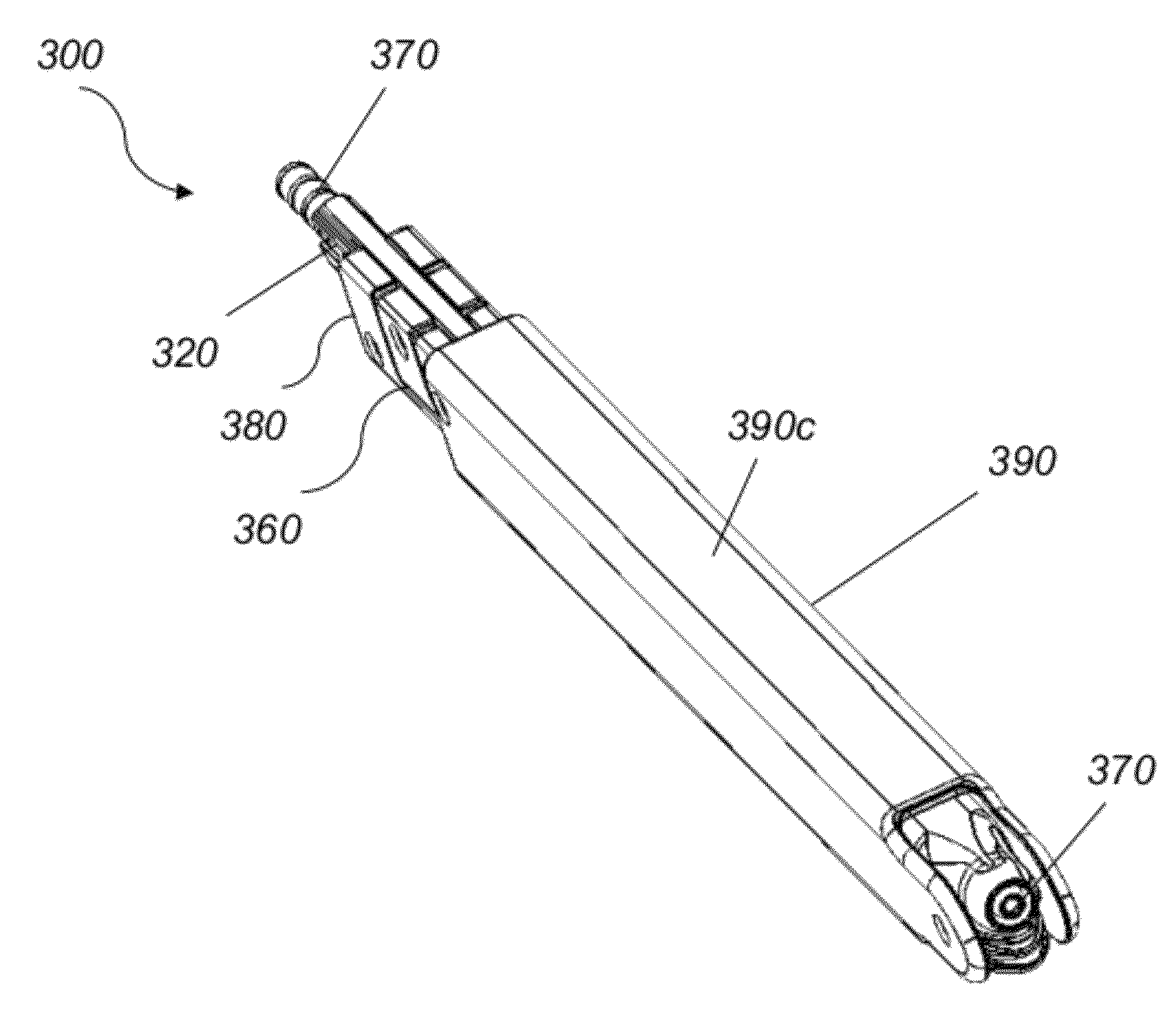
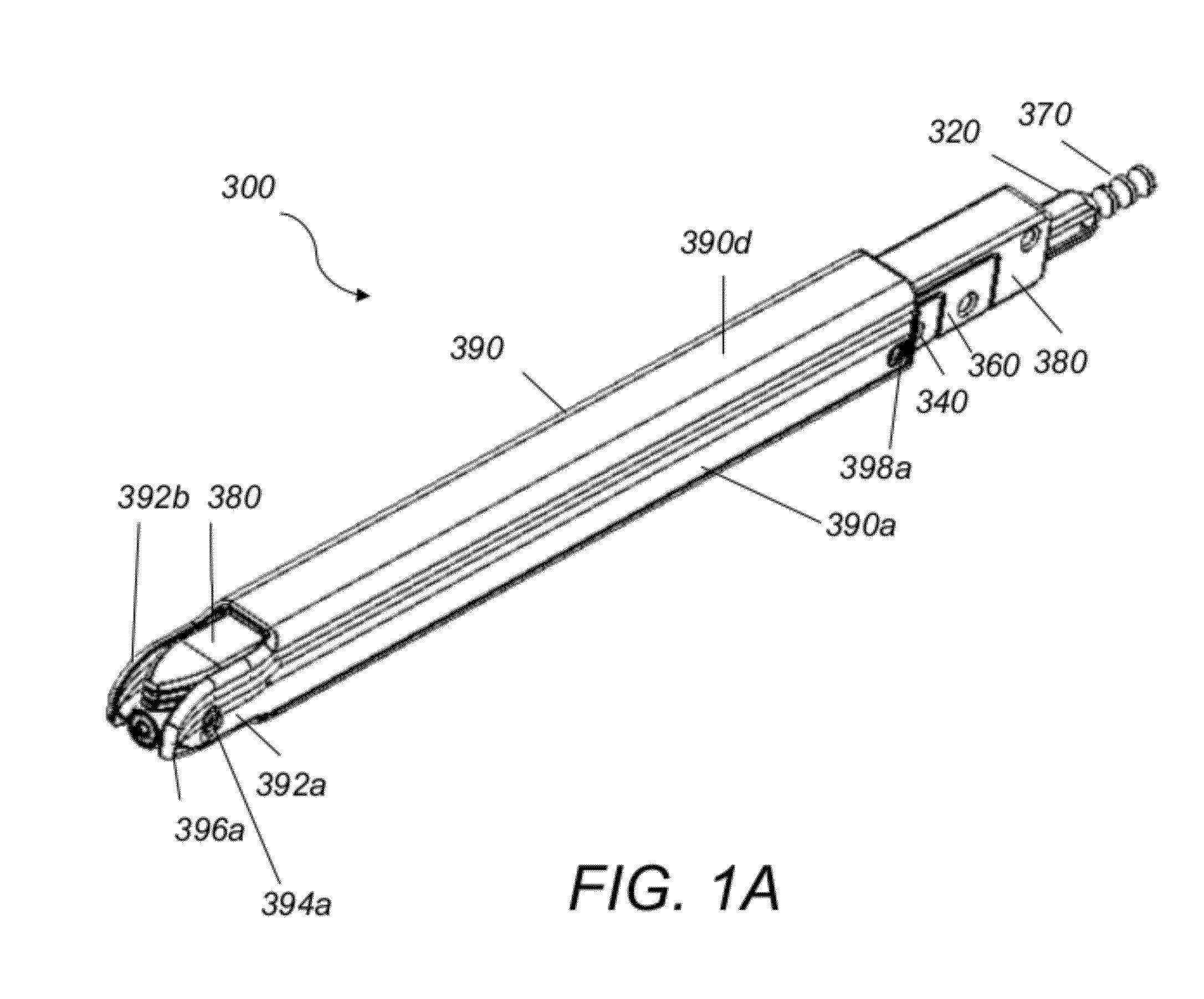
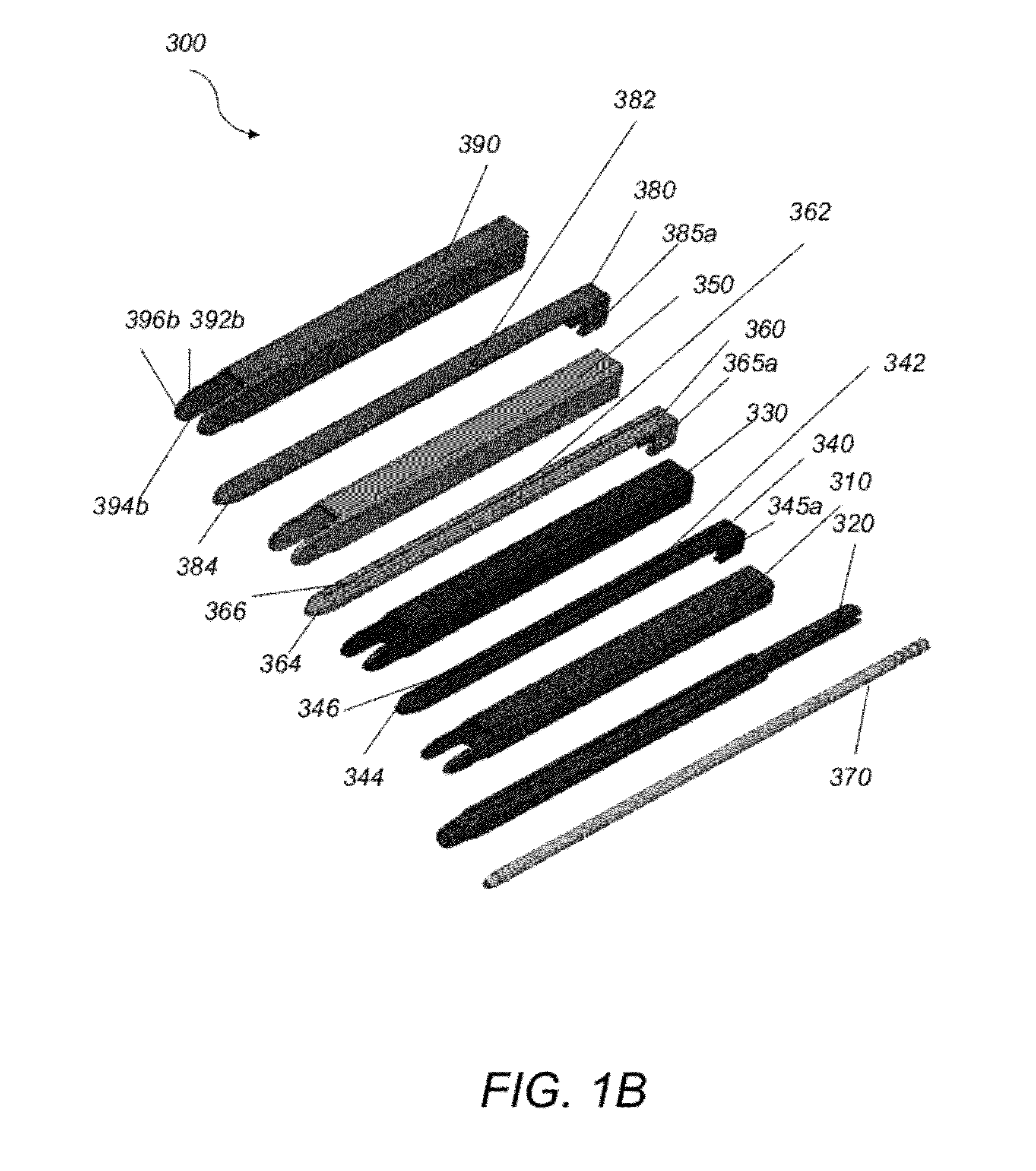
[0114]Referring to FIG. 1A, and FIG. 1B, access cannula system 300 includes a 14 mm working cannula 390 surrounding sequentially a 14 mm dilator 380, a 12 mm cannula 350, a 12 mm dilator 360, a 10 mm cannula 330, a 10 mm dilator 340, an 8 mm cannula 310, an 8 mm dilator 320 and a nerve probe dilator 370. Working cannula 390 includes an elongate tube 390 having a rectangular cross section and four side surfaces 390a, 390b (shown in FIG. 3B), 390c (shown in FIG. 4A), and 390d. Side surfaces 390a and 390b are opposite and parallel to each other and their distal ends terminate in parallel fork extensions 392a, 392b, respectively, that are tapered. Fork extensions 392a, 392b are rigid, are used for distraction purposes, and are dimensioned to ...


PUM


Abstract

A cannula assembly for providing percutaneous access in minimally invasive spinal surgeries, includes an outer cannula, a nerve probe dilator and a multistage dilator system comprising a first dilator, a second dilator, a third dilator and a fourth dilator. The outer cannula and the dilators are slidable relative to each other and are arranged sequentially so that the fourth dilator surrounds the nerve probe dilator, the third dilator slides over a surface of the fourth dilator, the second dilator slides over a surface of the third dilator, the first dilator slides over a surface of the second dilator, and the outer cannula surrounds the first dilator.

Description

CROSS REFERENCE TO RELATED CO-PENDING APPLICATIONS[0001]This application claims the benefit of U.S. provisional application Ser. No. 61 / 490,655 filed May 27, 2011 and entitled “IMPROVED METHODS, TOOLS AND DEVICES FOR PERCUTANEOUS ACCESS IN MINIMALLY INVASIVE SPINAL SURGERIES”, the contents of which are expressly incorporated herein by reference.FIELD OF THE INVENTION[0002]The present invention relates to improved methods, tools and devices for providing percutaneous access in minimally invasive spinal surgeries, and more particularly to an access cannula that includes multi-stage dilators and multi-stage cannulae.BACKGROUND OF THE INVENTION[0003]It is well known that traditional surgical procedures in locations deep within a patient's body require a long incision, extensive muscle stripping, prolonged retraction of muscles for visualization, and denervation and devascularization of the adjacent tissue. These procedures result in extensive tissue traumatization and consequently in pr...

Claims


Application Information

Patent Timeline
Patent Type & Authority Patents(United States)
IPC IPC(8): A61M29/00A61B17/00A61B17/02A61B17/16A61B17/34A61B17/70
CPCA61M29/00A61B17/70A61B17/3421A61B17/0206A61B17/7092A61B17/025A61B17/7077A61B17/1671A61B2017/0256A61B2017/0262A61B2017/0046A61B17/1626
Inventor CHIN, KINGSLEY R.HENSHAW, CRAIGIBARRA, MATTHEWLORE, VITOCROSSGROVE, JEREMY
Owner KIC VENTURES LLC
Learn More