Labeled molecular imaging agents, methods of making and methods of use
a molecular imaging agent and labeling technology, applied in the field of labeling molecular imaging agents, can solve the problems of effective changes in treatment regimens, and achieve the effect of low toxicity/immune respons
- Summary
- Abstract
- Description
- Claims
- Application Information
AI Technical Summary
Benefits of technology
Problems solved by technology
Method used
Image
Examples
example 1
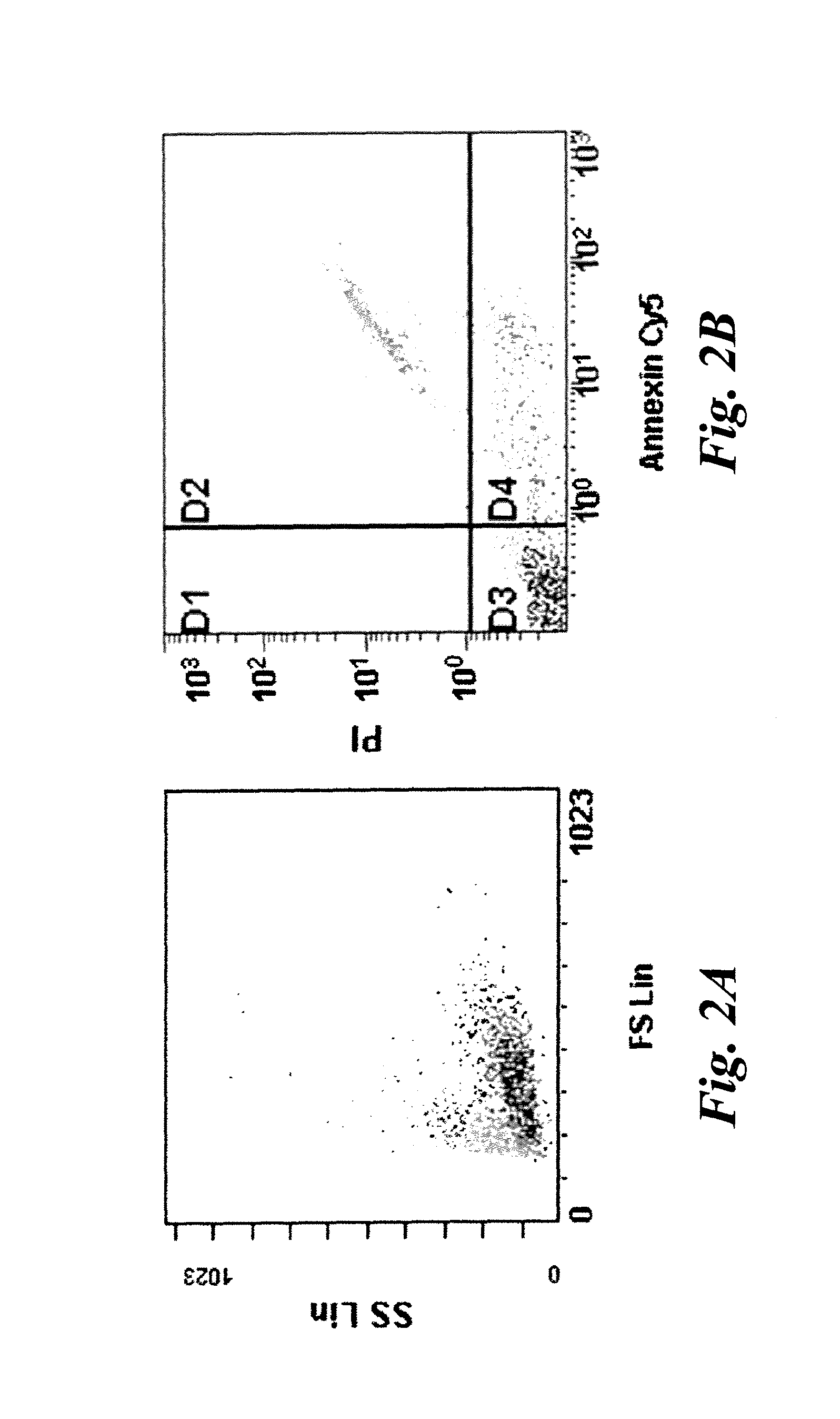
[0049]Human Jurkat cells were cultured with or without 1 uM staurosporine for 16 hours in an in vitro assay for apoptotic cells and stained with propidium iodide(PI) and Cy5-Annexin V (Annexin). Typically ˜30-50% of cells were found to be in some stage of apoptosis or necrosis. Flow cytometry was used to identify population of cells defined as normal (PI and Annexin negative), early apoptotic (PI negative, Annexin positive), and late apoptosis and necrosis (PI and Annexin positive). The results are shown in FIGS. 2A and 2B. FIG. 2A is a forward scatter / side scatter plot showing some late apoptotic cells (light gray) with a different granularity than non-apoptotic cells (darker gray). In FIG. 2B, Annexin V and PI negative cells are shown in quadrant D3 and represent non-apoptotic cells. Cells in quadrant D4 indicate early apoptotic cells that are positive for Annexin V staining, but negative for PI staining. Cells in quadrant D2 indicate late apoptotic and necrotic cells that are pos...
example 2
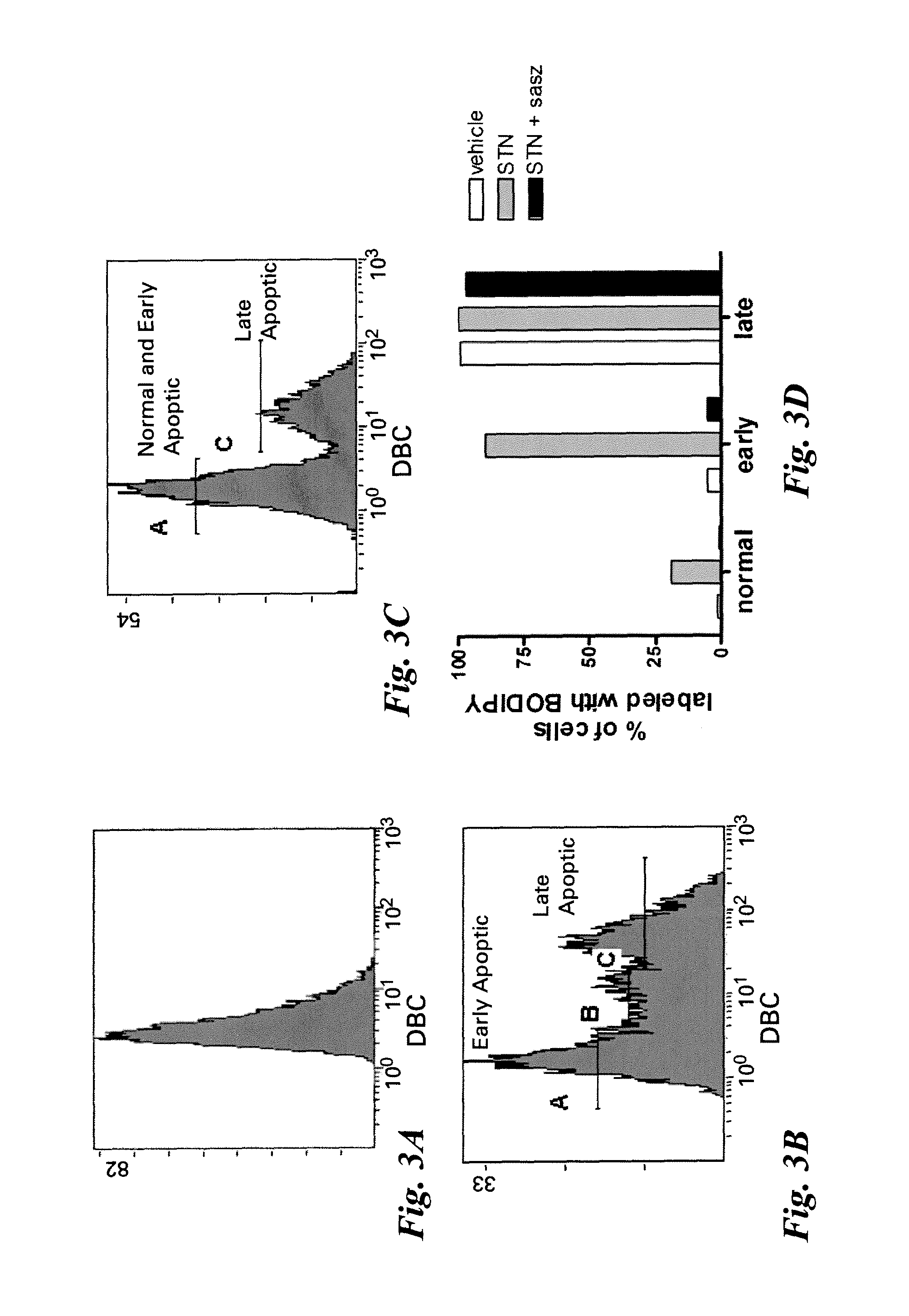
[0050]Jurkat cells were incubated with (FIG. 3B) and without (FIG. 3A) 1 μM staurosprine (STN) for 16-18 hours, stained with Annexin V-Cy5 and propidium iodide, and incubated with DBC for 30 minutes. DBC is a commercially available product from Invitrogen (catalog #B20340), which is sold for the purpose of reversible thiol labeling of nucleotides, proteins and cells via a disulfide exchange reaction at acidic conditions. DBC has the structure shown below.
[0051]
[0052]In FIG. 3A, untreated cells show some low intensity fluorescence corresponding to uptake or nonspecific binding of the DBC molecule. In FIG. 3B, staurosporine induced cells have a subpopulation of cells with high intensity DBC staining and this correlated primarily to the late apoptotic / necrotic cells (+Annexin V, +PI). A population of cells with intermediate DBC staining also appeared, which correlates well with the early apoptotic cells (+Annexin V, −PI). To more specifically address the role of the the xc- transporter...
example 3
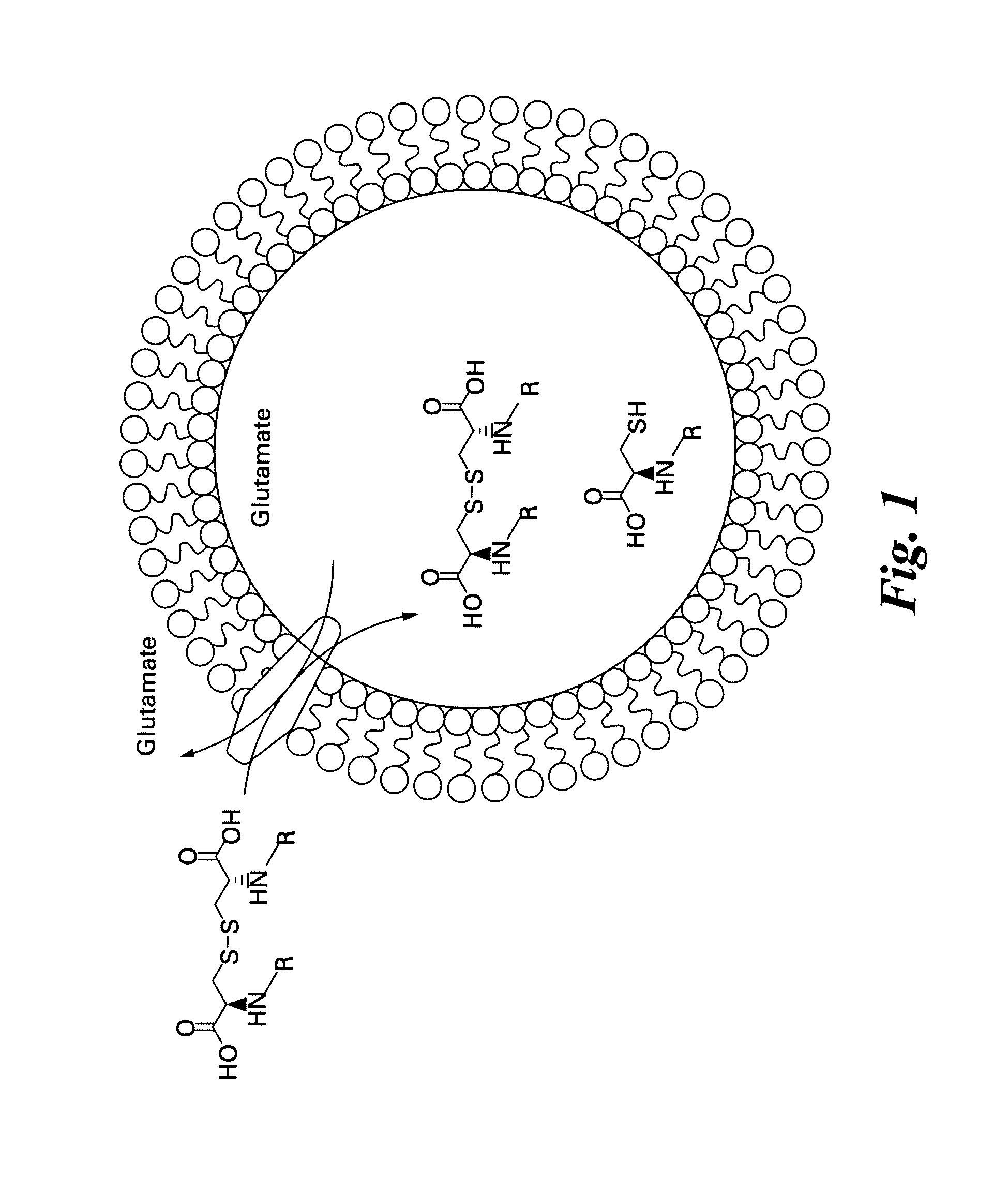
[0053]The apoptotic Jurkat cell uptake of DBC shows not only that the xc- transporter is activated in cells undergoing oxidative stress (and in this case apoptosis), but also that the xc- transporter is promiscuous enough to allow this substrate (cystine) with amine appended green fluorophores (bodipyFL) into the cell. A labeled cystine derivative was synthesized with a red fluorophore (bodipy650) to further determine the promiscuity of the transporter. In a side-by-side comparison to the bodipyFL labeled cystine, DBC-650 did not appear to label apoptotic cells any more than normal cells, as shown in FIGS. 4A and 4B, indicating that some labels are not sufficient for maintaining xc- transporter substrate status. In this example, the bodipy-650 fluorophore and associated linker are larger than that of DBC (FIG. 4A), and therefore size represents one limitation of what groups are appended to the amines while retaining the ability to pass through the transporter.
PUM
| Property | Measurement | Unit |
|---|---|---|
| time | aaaaa | aaaaa |
| time | aaaaa | aaaaa |
| time | aaaaa | aaaaa |
Abstract
Description
Claims
Application Information
 - R&D
- Intellectual Property
- Life Sciences
- Materials
- Tech Scout
- Unparalleled Data Quality
- Higher Quality Content
- 60% Fewer Hallucinations
Browse by: Latest US Patents, China's latest patents, Technical Efficacy Thesaurus, Application Domain, Technology Topic, Popular Technical Reports.
© 2025 PatSnap. All rights reserved.Legal|Privacy policy|Modern Slavery Act Transparency Statement|Sitemap|About US| Contact US: help@patsnap.com


