2D/3D image registration method

a technology of image registration and 3d, applied in the field of image processing, can solve the problems of difficult fluorometric data, automatic registration of the heart or other structures of the body, and difficulty in registering the different images, so as to achieve the effect of reducing the image dissimilarity metri

Active Publication Date: 2015-01-27
SIEMENS HEALTHCARE GMBH
View PDF5 Cites 13 Cited by
  • Summary
  • Abstract
  • Description
  • Claims
  • Application Information

AI Technical Summary

Benefits of technology

[0015]An embodiment of the present invention may also provide a system for providing multi-modal 2D / 3D image registration during a medical procedure, comprising an imager that acquires 2D image data of an anatomical region of a patient and a processor that manipulates the acquired image data and stored 3D image data of the anatomical region to align a stored 3D image to a 2D image and to automatically track the anatomical region position during the medical procedure, said processor manipulating the 3D image data, in part, to render the stored 3D image in two dimensions for a positional comparison between the 3D image and the 2D image. The processor may have a graphics processing element that accelerates the manipulation of the 3D image data to render the stored 3D image in two dimensions. The processor may apply a cost function to evaluate and optimize a Chan-Vese-like functional used as an image dissimilarity metric and an optimization method to minimize the image dissimilarity metric.

Problems solved by technology

While image guidance systems and techniques can provide visualization of the catheter tip, and sometimes localization of the tip within some coordinate space, the challenge is often in relating that tip information to the actual location of the anatomy of interest.
Sometimes this might be accomplished by using optimal imaging planes that clearly show both the anatomy and the device, although this can be difficult in a complex anatomy such as the heart.
In the case of the heart, this is further complicated by the heart beating motion, patient breathing motion and catheter motion.
However, this catheter 8 is not always available during different types of clinical procedures.
The automatic registration of the heart or other structures of the body, to fluoro data, however, can be very challenging due to the low contrast of most tissues in fluoro images and possibly the missing-angle projection geometry (e.g., bi-plane geometry) used in clinical settings.
Currently, there is no automatic method available to 1) correct the patient's setup error between the pre-op 3D image (e.g., CT or MR image) and the ob-board fluoroscopy or 2) track the intra-treatment heart, for example, position under respiratory motion.
Moreover, registrations between pre-op MR images and fluoroscopic images present additional difficulties due to the substantial imaging gap between two different modalities (MR and X-ray).
For example, when generating DRRs (Digitally Reconstructed Radiographs) from an MR image to perform a registration with the fluoro images, there is no density information available to allow the generation of a realistic X-ray image.

Method used


Image

  • 2D/3D image registration method
  • 2D/3D image registration method
  • 2D/3D image registration method

Examples


Embodiment Construction

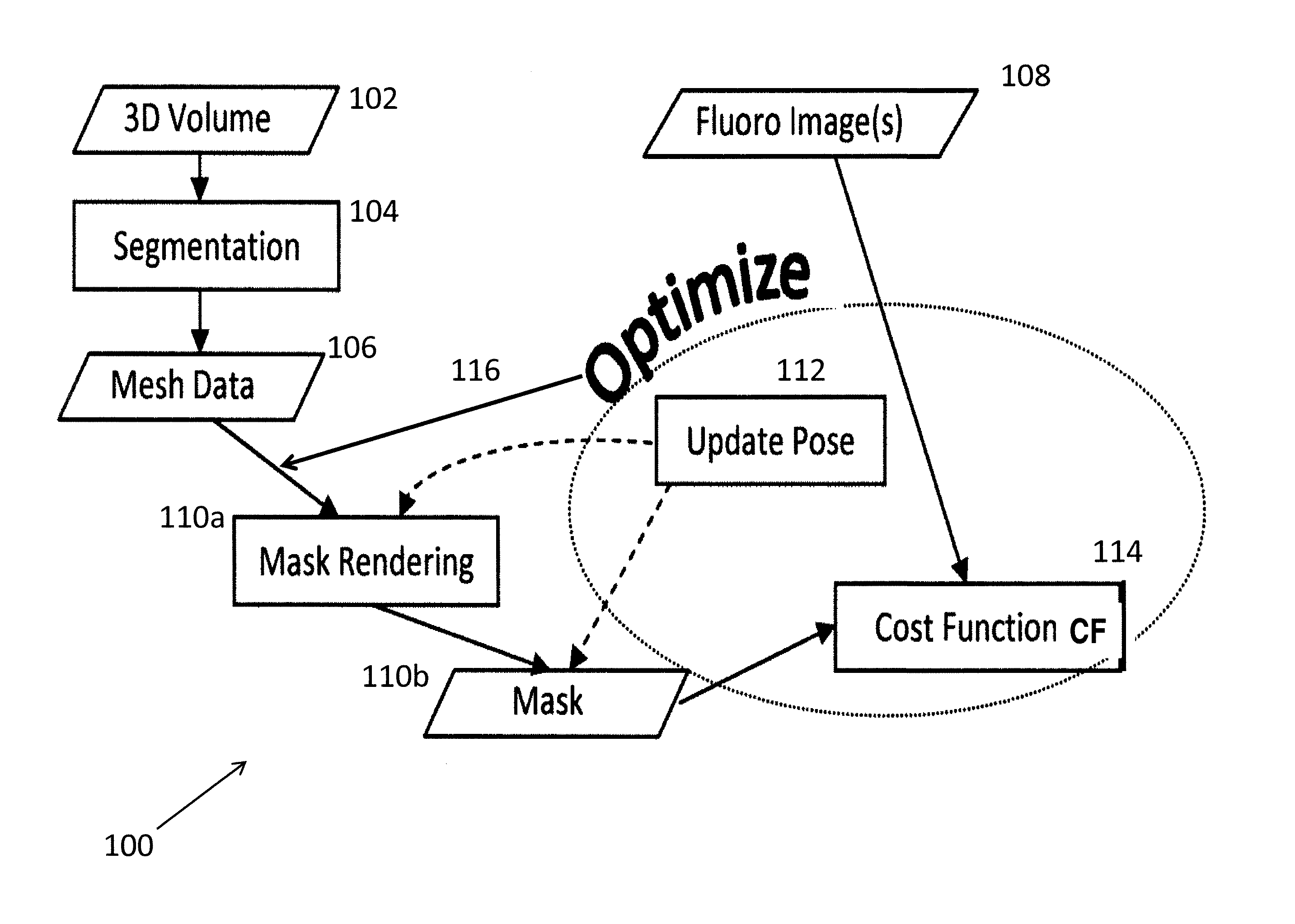
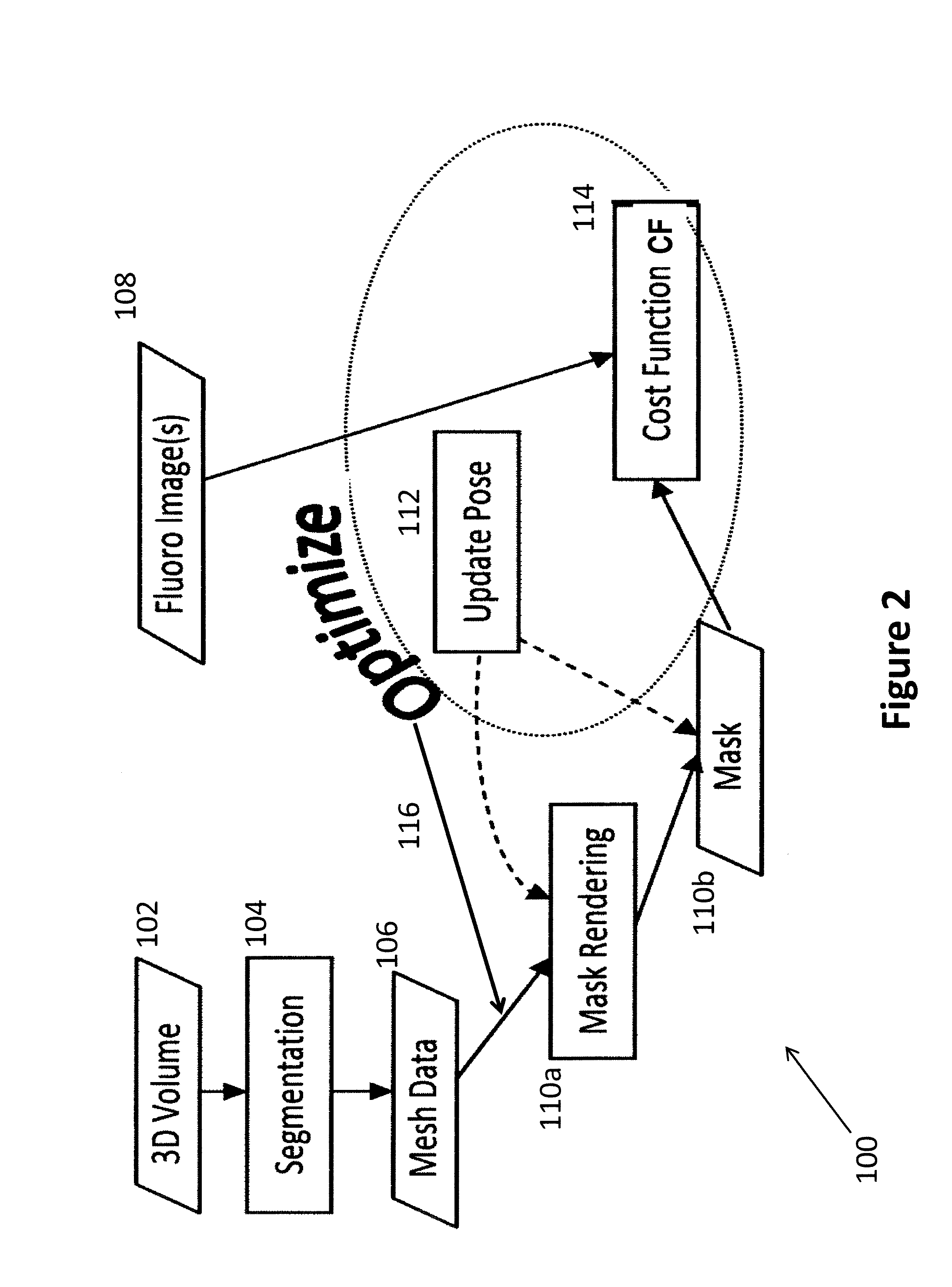
[0026]FIG. 2 is a schematic view of a method 100 carried out in accordance with an embodiment of the present invention. Generally, volumetric imaging data of the patient and, more particularly, of the anatomical region of interest (for example, the heart) is acquired (Step 102). Normally, this 3D data is acquired prior to the medical procedure to be performed and, as noted above, the data may be acquired using CT or MR imaging. As an alternative, the data may be obtained from a 3D model of the anatomical region of interest. The method 100 performs automatic or manual segmentation of the 3D volume (Step 104) and obtains a 3D mesh from the pre-acquired 3D volume (Step 106). A 3D mesh is a 3D object representation consisting of a collection of vertices and polygons that define the shape of an object in 3D. This 3D mesh is then used as a basis for registration with real-time fluoro data, which is acquired during the medical procedure (Step 108).

[0027]Specifically, the method 100 compute...


PUM


Abstract

A method (100) that registers a 3D heart volume (112, 114) obtained from either a pre-operative MR image or CT image (102) to an intra-operative fluoroscopic image using a mesh of the heart structure (106) as the basis for the registration.

Description

CROSS-REFERENCE TO RELATED APPLICATIONS[0001]This application claims the benefit of both Provisional U.S. Patent Application Ser. No. 61 / 528,834, entitled, “Novel Multi-modal 2D / 3D Registration Method for Fluoro Data”, filed in the name of Chen-Rui Chou, Atilla Peter Kiraly, and Christophe Chefd'hotel, on Aug. 30, 2011, and Provisional U.S. Patent Application Ser. No. 61 / 585,690, entitled, “3D-2D Mesh-Based Registration”, filed in the name of David Thivierge-Gaulin, Atilla Peter Kiraly, Christophe Chefd'hotel, and Chen-Rui Chou, on Jan. 12, 2012, the disclosure of each is also hereby incorporated herein by reference.FIELD OF THE INVENTION[0002]This invention generally relates to image processing and, more particularly to, the alignment or registration of different images.BACKGROUND OF THE INVENTION[0003]Cardiac electrophysiology (EP) studies that are used to diagnose and treat heart arrhythmia are conducted using catheters in minimally invasive medical procedures. Specifically, the ...

Claims


Application Information

Patent Timeline
Patent Type & Authority Patents(United States)
IPC IPC(8): G06T7/00
CPCG06T7/0032G06K2209/057G06T2200/04G06T2207/10081G06T2207/10088G06T2207/10121G06T2207/10124G06T2207/30048G06T7/344G06V2201/034
Inventor CHOU, CHEN-RUIKIRALY, ATILLA PETERCHEFD'HOTEL, CHRISTOPHETHIVIERGE-GAULIN, DAVID
Owner SIEMENS HEALTHCARE GMBH
Why Eureka
Try Eureka