

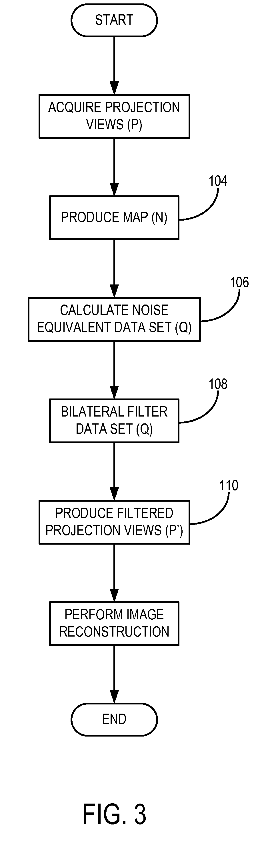
Projection-space denoising with bilateral filtering in computed tomography
a computed tomography and projection space technology, applied in image enhancement, image analysis, instruments, etc., can solve the problems of increasing the noise level of the measured projection data and subsequent reconstructed images, the risk of cancer or other diseases associated with the radiation exposure of ct scans, and the diagnostic value of ct examinations is severely degraded, so as to achieve the effect of ct image filtering
- Summary
- Abstract
- Description
- Claims
- Application Information
AI Technical Summary
Benefits of technology
Problems solved by technology
Method used
Image


Examples
Embodiment Construction
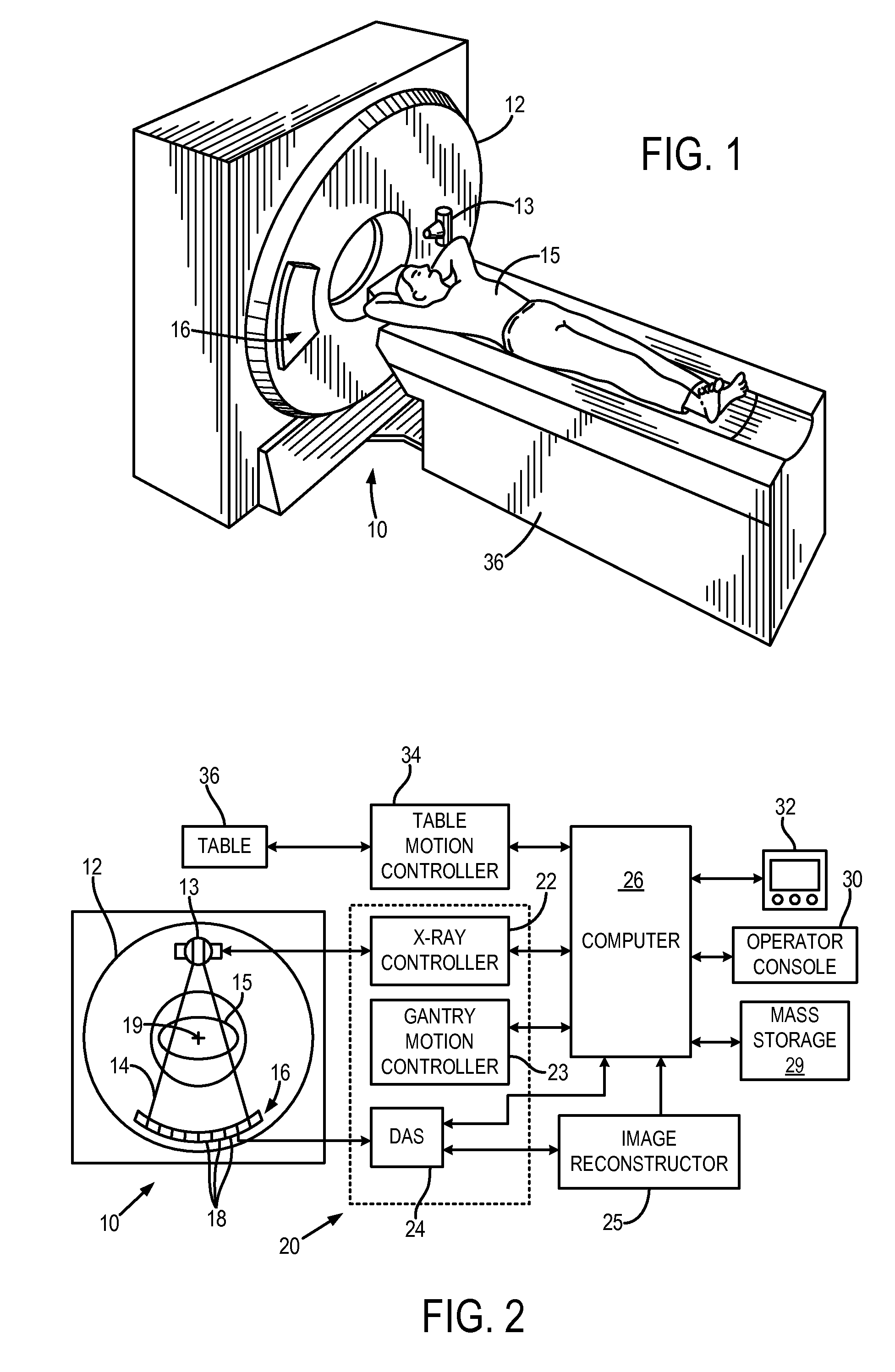
[0018]With initial reference to FIGS. 1 and 2, a computed tomography (CT) imaging system 10 includes a gantry 12 representative of a “third generation” CT scanner. Gantry 12 has an x-ray source 13 that projects a beam such as a fan or cone beam of x-rays 14 toward a detector array 16 on the opposite side of the gantry. The detector array 16 is formed by a number of detector elements 18 which together sense the projected x-rays that pass through a medical patient 15. Each detector element 18 produces an electrical signal that represents the intensity of an impinging x-ray beam and hence the attenuation of the beam as it passes through the patient. During a scan to acquire x-ray projection data, the gantry 12 and the components mounted thereon rotate about a center of rotation 19 located within the patient 15.
[0019]The rotation of the gantry and the operation of the x-ray source 13 are governed by a control mechanism 20 of the CT system. The control mechanism 20 includes an x-ray cont...
PUM
 Abstract
Description
Claims
Application Information