
Method of expanding double negative T cells

a double negative t cell and ex vivo technology, applied in the field of ex vivo expansion of double negative t cells, can solve the problems of severe limited treatment, severe autoimmune disease in treated patients, cancer relapse, etc., and achieve the effect of reducing tumor growth and reducing tumor growth

Active Publication Date: 2015-04-28
UNIV HEALTH NETWORK
View PDF9 Cites 1 Cited by
  • Summary
  • Abstract
  • Description
  • Claims
  • Application Information

AI Technical Summary

Benefits of technology

The patent text describes a way to purify and expand human T cells that can effectively inhibit the growth of tumors. These cells, known as double negative (DN) T cells, were found to have a better anti-tumor effect than other types of T cells in the laboratory. When transferred into mice with immunodeficiencies, DN T cells were more effective at fighting tumors than other types of T cells. This suggests that DN T cells may have potential as a therapeutic treatment for cancer.

Problems solved by technology

However, this treatment is severely limited by the need to identify relevant tumor antigens.
Moreover, cancer relapse occurs even after repeated injections (Meidenbauer et al.
This approach can also result in severe autoimmune disease in treated patients (Dudley and Rosenberg 2003).
Because identifying a single class I, particularly a dominant MiHA that is present in patients but not in donors, is a difficult task, its clinical application to patients is complex.
However, the role of human DN T cells in tumor immunity has not been investigated previously due to the limited number of DN T cells that can be obtained.

Method used


Image

  • Method of expanding double negative T cells
  • Method of expanding double negative T cells
  • Method of expanding double negative T cells

Examples


example 1

Murine DN Cells

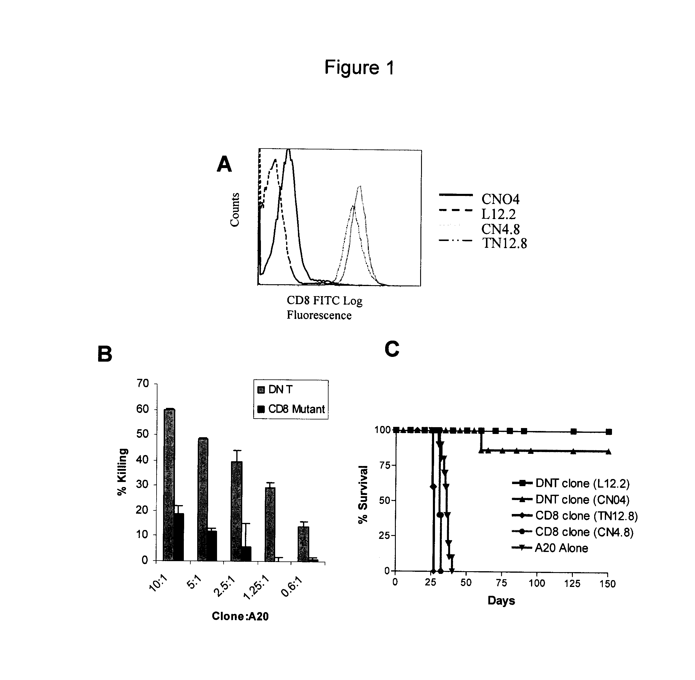
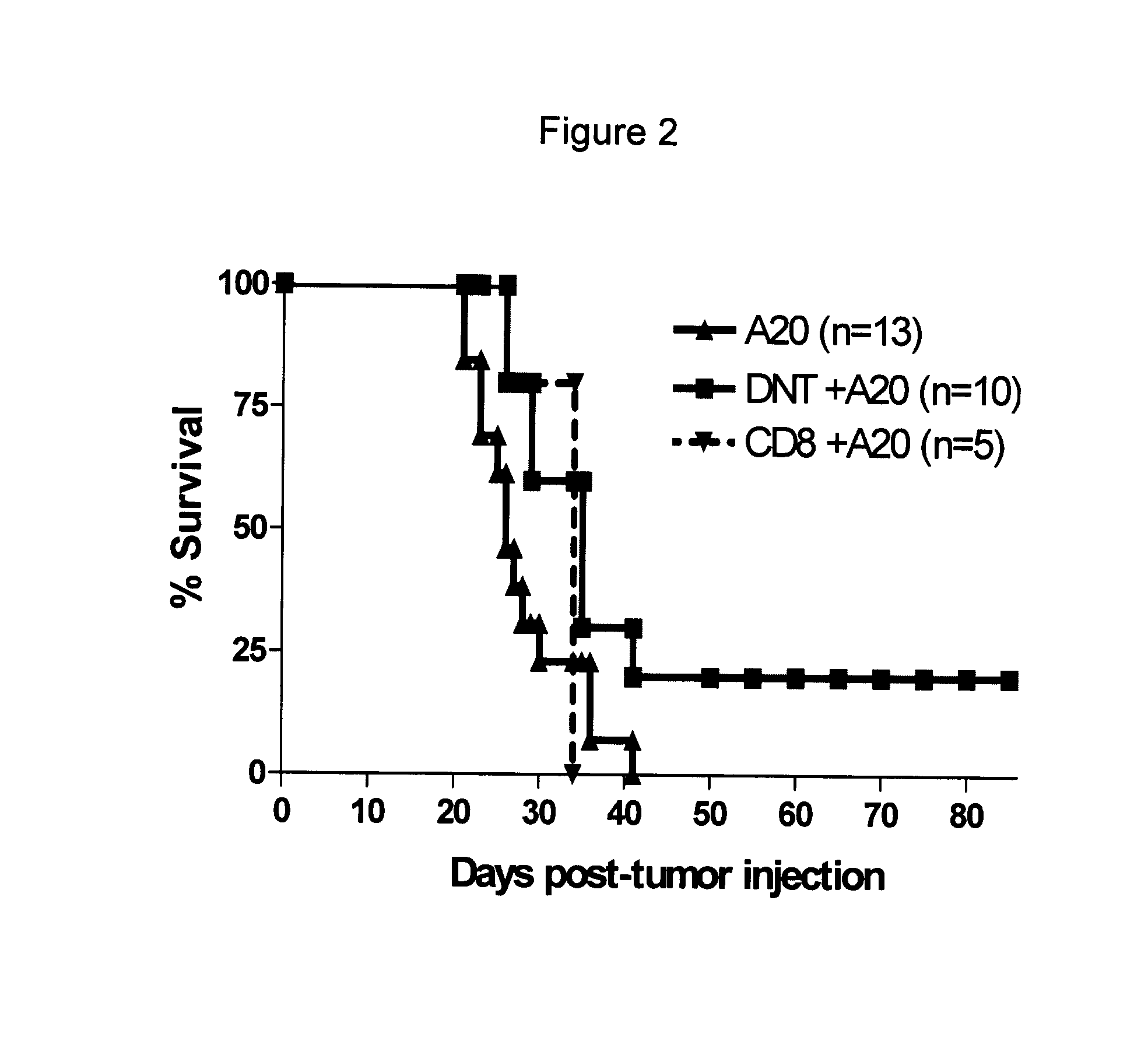
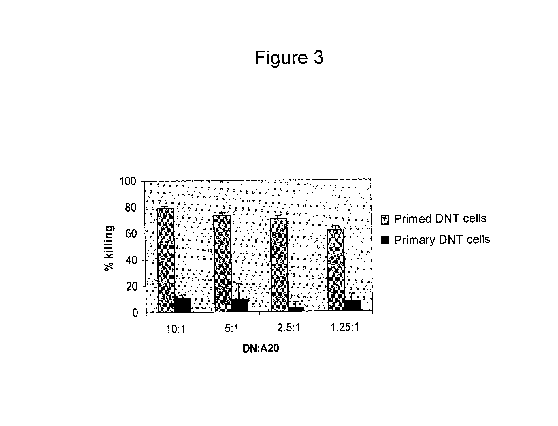
[0064]The inventors have generated a panel of mouse DN T cell clones. During long-term cultivation, several natural mutant T cell clones arose which express CD8 (FIG. 1A). In FIG. 1A, Ld− DN T cell clones CN04 and L12.2 and their natural mutants CN4.8 and TN12.8 were stained with CD8-FITC, and analyzed by flow cytotometry. Both mutant clones are CD8+. The mutant clones have reduced cytotoxicity to A20 tumor in vitro (FIG. 1B). In FIG. 1B, DN T cell clone CN04 and its CD8 mutant clone CN48 were used as effector cells at ratios as indicated. Ld+ A20 tumour cells were used as targets. Specific lysis of the target cells was measured in a cytotoxic assay. While a single infusion of DN T cell clones can prevent recipient tumor development from a lethal injection of lymphoma cells, adoptive transfer of the same number of CD8+ mutant clones have no protective effect (FIG. 1C). In FIG. 1C, (B6×BALB / c)F1 mice (Ld+) were infused with 105 / mouse A20 tumor cells either alone or tog...

example 2

Human DN Cells

[0071]The population of DN T cells in human peripheral blood is very low (FIG. 4A). To determine whether human DN T cells can be used as a novel cancer adoptive immunotherapy, the inventors developed a protocol by which human DN T cells can be expanded ex vivo. Peripheral blood samples were collected from healthy individuals and the red blood cells were lysed. The remaining PBMC were stained with CD3-Cy-Chrone, CD4-FITC and CD8-FITC. Percentage of CD3+CD4−CD8− T cells in PBMC is shown in G4 region of FIG. 4A. Erythrocytes and CD4, CD8 T cells were depleted using the Human CD4 / CD8 depletion cocktail kit (Stem Cell Technologies). The residual DN T cell enriched population was then cultured in anti-CD3 mAb pre-coated 24-well plates for 3 days in the presence of recombinant human interleukin-2 (rhIL-2, 50 U / mL) and rhIL-4 (30 U / mL). Activated T cells were washed and cultured for another 4 days in the presence rhIL-2 and rhIL-4. On day 7, viable cells were split and culture...


PUM

PropertyMeasurementUnit
timeaaaaaaaaaa
timeaaaaaaaaaa
volumeaaaaaaaaaa

Abstract

A method of expanding double negative T cells in culture is described. The method comprises (a) providing a starting sample comprising DN T cells or precursors thereof; (b) substantially depleting CD8+ and CD4+ T cells from the starting sample; (c) culturing the sample from step (b) with an immobilized T cell mitogen in a culture medium comprising an agent that can stimulate DN T cell growth; (d) washing the cells obtained in step (c) and resuspending in a culture medium comprising the agent without the T cell mitogen; and (e) washing the cells obtained in step (d) and resuspending in a culture medium comprising the agent and a soluble T cell mitogen. The DN T cells obtained by the method are useful in a variety of applications including the treatment of cancer, infectious diseases, graft versus host disease and autoimmune disease.

Description

FIELD OF THE INVENTION[0001]The present invention relates to a method of expanding double negative T cells ex vivo.BACKGROUND OF THE INVENTION[0002]Adoptive cancer immunotherapy involves isolation, ex vivo activation and expansion of immune cells, and subsequent injection into patients with cancer. Since Rosenberg et al first introduced adoptive immunotherapy for the treatment of human cancers (Rosenberg et al. 1985; Rosenberg et al. 1988), several types of immune cells, including lymphokine activated killer cells (LAK), CD8+ cytotoxic T lymphocytes (CTL), natural killer (NK) cells, NK T cells and tumor infiltrating lymphocytes (TIL) have been used in clinical trials (Verneris et al. 2002; Toh et al. 2005; Leemhuis et al. 2005; Yee et al. 2002; Dudley and Rosenberg 2003; Dudley et al. 2005). Currently the transfer of ex vivo activated and expanded autologous tumor specific CD8+ CTLs may induce objective clinical responses in significant number of patients (Gattinoni et al. 2005; Ros...

Claims


Application Information

Patent Timeline
Patent Type & Authority Patents(United States)
IPC IPC(8): C12N5/0783
CPCC12N5/0636C12N2501/23A61P31/00A61P35/00A61P37/02A61K39/4611A61K2239/31A61K2239/55A61K39/4644A61K2239/26A61K2239/38C12N2501/2302C12N2501/2304C12N2501/515
Inventor ZHANG, LIHAN, MEIDOKOUHAKI, POUNEH
Owner UNIV HEALTH NETWORK
Learn More