
Apical puncture access and closure system

a puncture and access system technology, applied in the field of apical puncture access and closure system, can solve the problems of high invasive operation, achieve the effects of facilitating sealing fabric securing, and reducing the risk of puncture damag

Active Publication Date: 2015-06-16
EDWARDS LIFESCIENCES CORP
View PDF64 Cites 80 Cited by
  • Summary
  • Abstract
  • Description
  • Claims
  • Application Information

AI Technical Summary

Benefits of technology

Enables secure access and closure of heart tissue punctures during procedures like transapical heart valve replacement, reducing blood loss and facilitating minimally invasive surgeries by providing a reliable seal and minimizing recovery time.

Problems solved by technology

Conventional heart valve surgery is an open-heart procedure conducted under general anesthesia, and is a highly invasive operation.

Method used


Image

  • Apical puncture access and closure system
  • Apical puncture access and closure system
  • Apical puncture access and closure system

Examples


Embodiment Construction

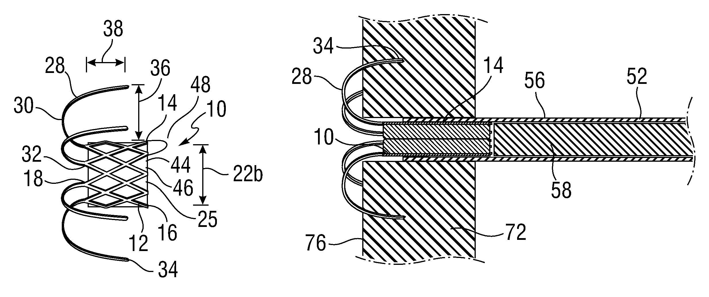
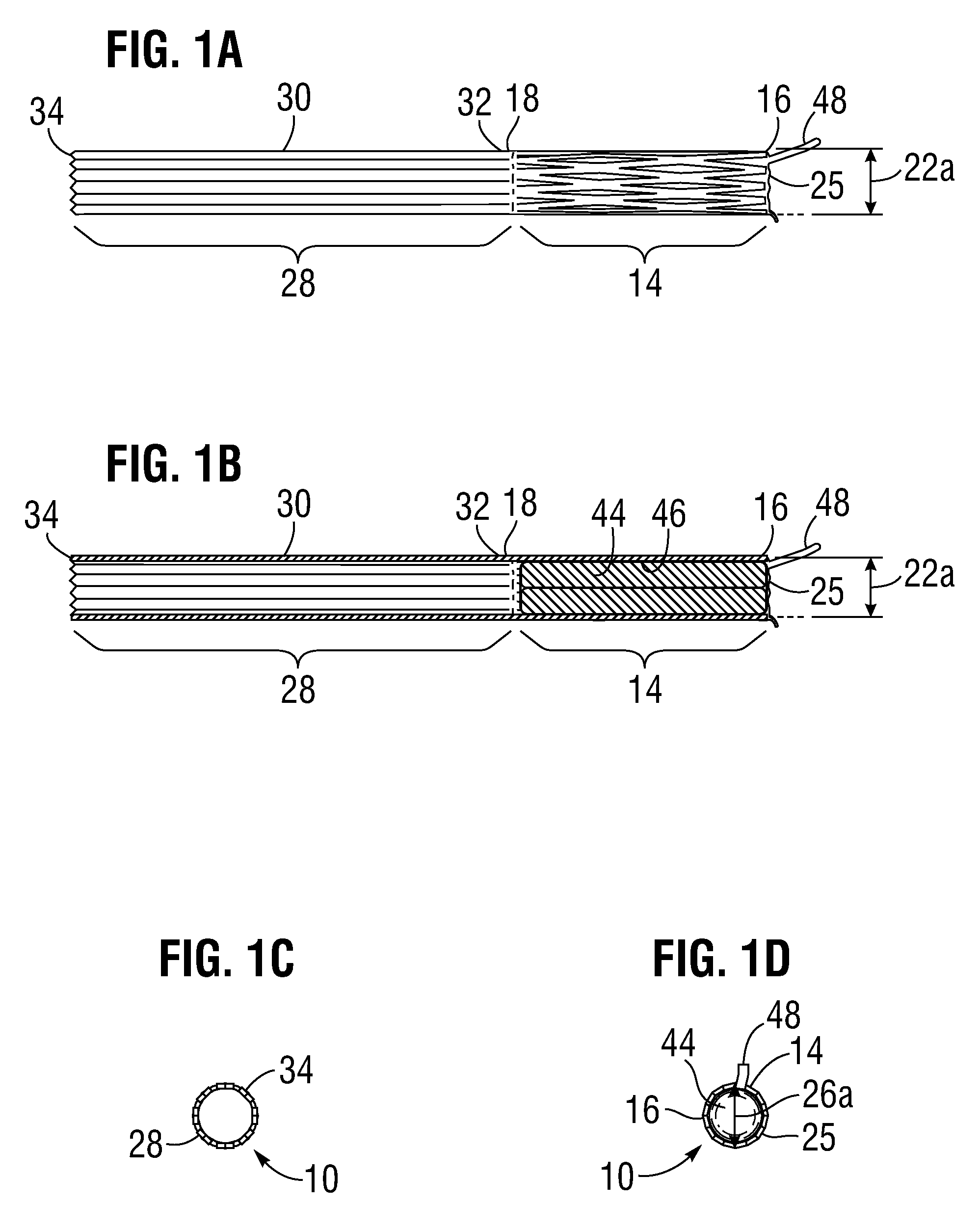
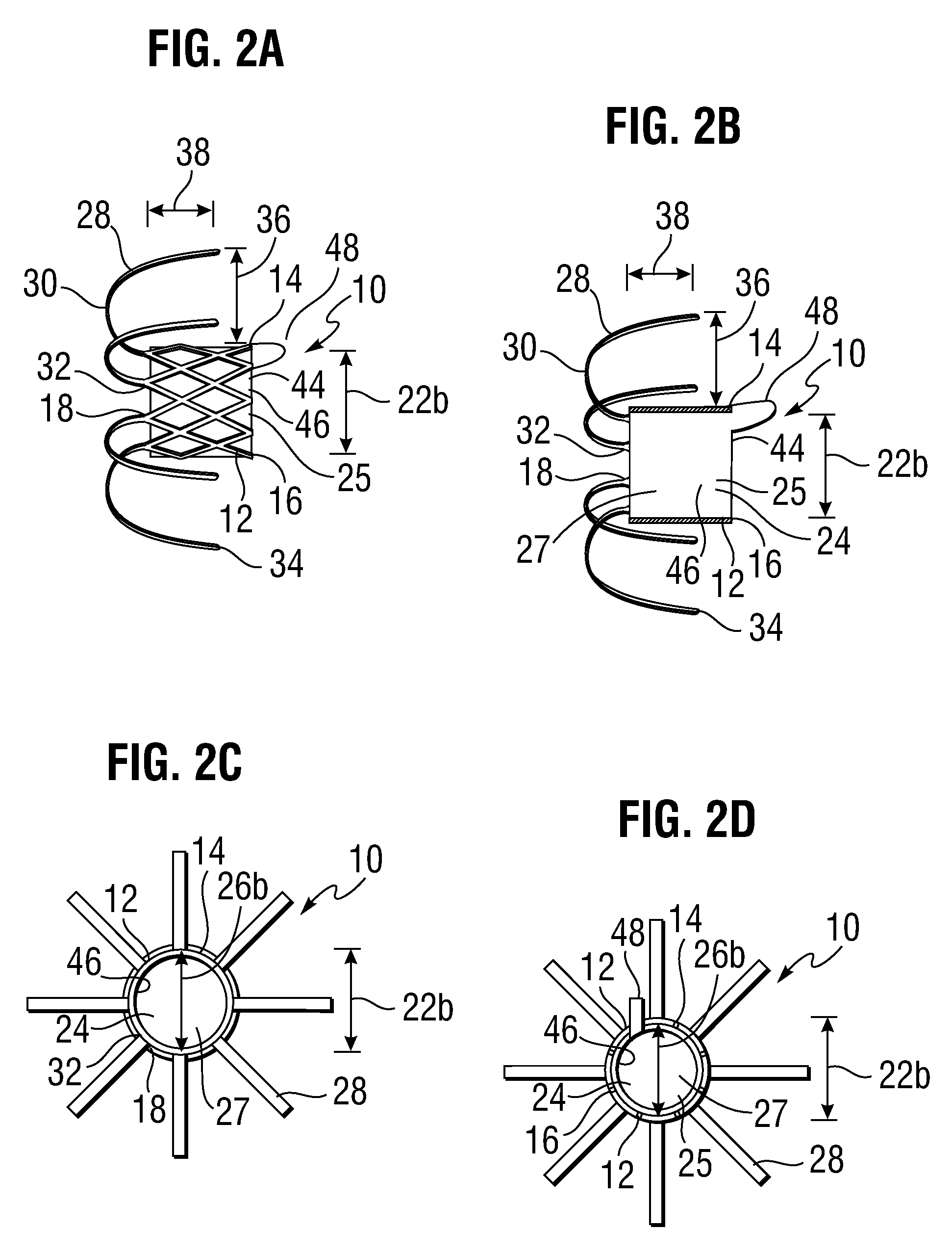
[0048]FIGS. 1A-1D, 2A-2D, 3A-3D, and 4 depict a hemostatic closure / access device 10 according to an embodiment of the invention. In FIGS. 1A-1D, the device 10 is in a delivery configuration for delivery to a treatment site in a patient. In FIGS. 2A-2D, the device 10 is expanded in an access configuration to provide an access passage to the interior of a target body organ, and in FIGS. 3A-3D the device 10 is in a closed configuration to seal the access passage. FIG. 4 depicts an exemplary cutting pattern used to create the stent-like framed structure 12 (aka support stent) of the device 10.

[0049]The device 10 includes a stent-like framed structure 12 having a main body 14 with a proximal end 16 and a distal end 18. The main body 14 in this embodiment is generally cylindrical, having a length 20 and an outside diameter 22a, 22b. An inner lumen 24 extends through the main body 14. The inner lumen 24 has a proximal opening 25, and includes a lumen diameter 26. The main body 14 can be ra...


PUM


Abstract

A device, system, and method for providing access to, and sealing of, a body organ includes an implant device. An implant device has a main body having an internal access lumen, with a plurality of prongs extending from a distal end of the main body. The main body can include two lumens, one slidable within the other, to form a single continuous lumen with an adjustable length. The main body has an expanded configuration with an expanded diameter, and an unexpanded configuration with an unexpanded diameter. The prongs have a generally straight configuration where they extend distally of the distal end of the main body, and a bent configuration where the prongs bend around so that their tips extend proximally of the distal end of the main body. The device may include a hemostatic barrier to prevent fluid leakage therethrough when the main body is in the unexpanded configuration.

Description

RELATED APPLICATION DATA[0001]The present application claims priority under 35 U.S.C. §119(e) to U.S. Provisional Application No. 61 / 470,930, filed Apr. 1, 2011.FIELD OF THE INVENTION[0002]The present invention relates to methods and systems for cinching a tissue puncture around an implement and closing the puncture after removal of the implement, in particular for punctures in heart tissue.BACKGROUND OF THE INVENTION[0003]The heart is a hollow muscular organ of a somewhat conical form; it lies between the lungs in the middle mediastinum and is enclosed in the pericardium. The heart rests obliquely in the chest behind the body of the sternum and adjoining parts of the rib cartilages, and typically projects farther into the left than into the right half of the thoracic cavity so that about one-third is situated on the right and two-thirds on the left of the median plane. The heart is subdivided by septa into right and left halves, and a constriction subdivides each half of the organ ...

Claims


Application Information

Patent Timeline
Patent Type & Authority Patents(United States)
IPC IPC(8): A61B17/02A61B17/00A61B17/34A61F2/958
CPCA61B17/0293A61B17/0057A61B2017/00247A61B2017/00867A61B17/3478A61B2017/3425A61B2017/00592A61B2017/00606A61B2017/00623A61B2017/00628A61B2017/00637A61B2017/00668A61B2017/00601A61B2017/00991A61B17/00234A61B17/02A61B2017/00243A61B2017/00579A61B2017/00597A61B2017/0237
Inventor ROWE, STANTON J.WU, MINGSCHNEIDERCORSO, JR., PHILIP P.
Owner EDWARDS LIFESCIENCES CORP
Learn More