Genetic barcodes

a technology of fluorescence probes and barcodes, applied in the field of fluorescence probes and genetic barcodes, can solve the problems of no single experiment assay, no diagnostic assay that allows an accurate detection of rearrangements, etc., and achieves accurate and inexpensive fluorescence in situ hybridization, facilitate clinical application, and enhance the detection of chromosomal change

Active Publication Date: 2015-08-04
RGT UNIV OF CALIFORNIA
View PDF4 Cites 4 Cited by
  • Summary
  • Abstract
  • Description
  • Claims
  • Application Information

AI Technical Summary

Benefits of technology

[0016]In various embodiments, an accurate and inexpensive fluorescence in situ hybridization (FISH)-based assay is provided for the detection of chromosomal changes in a gene of interest including oncogenes and the unbiased quantitation of the number of gene copies in small samples of tissue. The FISH-based technique and assay can be tailored to query alterations of other particular genetic loci of interest in cancer studies. The assay is a relatively efficient; 2 day assay and requires few cells or slide mounted tissue slices. The present FISH assay does not require the generation of metaphase spreads, which facilitates the applicability to clinical settings and in the investigation of archived tissue specimens, although in some examples, the use of metaphase spreads may enhance detection of chromosomal change by barcode detection.
[0017]Thus, the present invention also provides an accurate and inexpensive fluorescence in situ hybridization (FISH)-based assay for the detection of chromosomal changes involving either ErbB1, ErbB2 or any other oncogene and the unbiased quantitation of the number of gene copies in small samples of tissue.

Problems solved by technology

No diagnostic assays that allow an accurate detection of rearrangements while scoring of the number of copies of the EErbB 1 and ErbB2 genes are presently available.
Yet, no assay exists to test cells in a single experiment for alterations of both, ErbB1 and ErbB2, abnormalities.

Method used


Image

  • Genetic barcodes
  • Genetic barcodes
  • Genetic barcodes

Examples


example 1

Using Genetic Barcodes to Detect ER Status in a Panel of Cancer Cell Lines

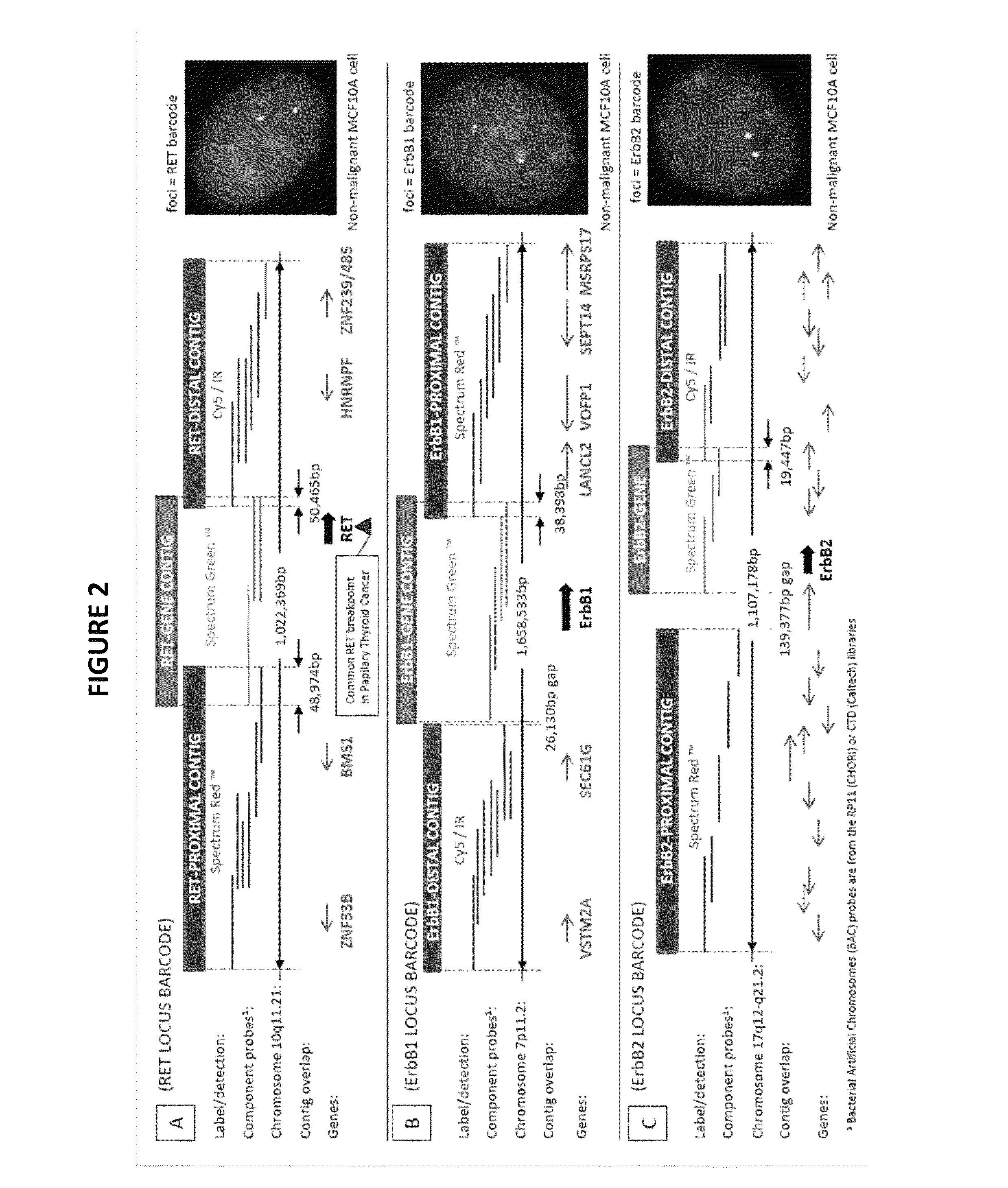
[0066]Probes to ErbB1 gene and the proximal and distal regions were made from BAC probes from the RP11(CHORI) or CTD (Caltech) libraries. Methods to make such probes are described above and in International application WO 2006 / 091979, which is US Patent Publication No. US-2009-0098534-A1, hereby incorporated by reference.

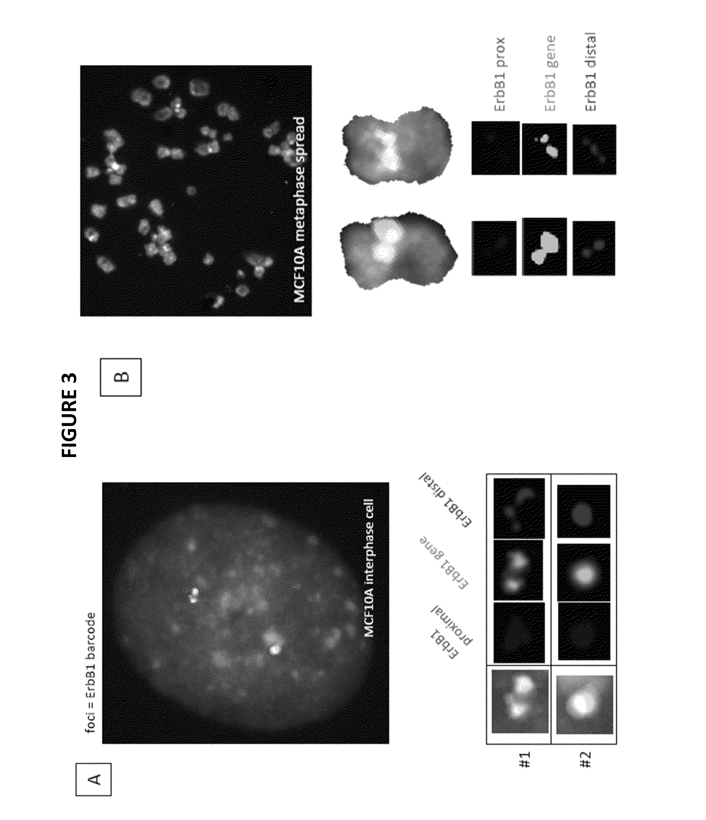
[0067]Detection of specific abnormalities and DNA probes preparation was a carried out in cancer cell lines. In FIG. 3, a FISH barcode staining pattern in MCF10A non-malignant cells. MCF10A cells were shown to have 2 foci of the ErbB1 barcode with each foci containing each component contig, with each of the foci clearly on a separate chromosome and the predominant (86% of cells) pattern in the culture is 2 foci.

[0068]Referring now to FIG. 4 genetic amplifications / translocations were identified by a genetic barcode in cancer cells. MDA-231 cells have 5 foci identified by the ErbB1 barcode, with...

example 2

Using Genetic Barcodes to Detect ER Status in a Patient Biopsy

[0071]Present work focuses on the development of clinical assays for the detection and characterization of genetic alterations in tumor cells, and investigation of a potential correlation between tumor cell genotype and disease progression and outcome.

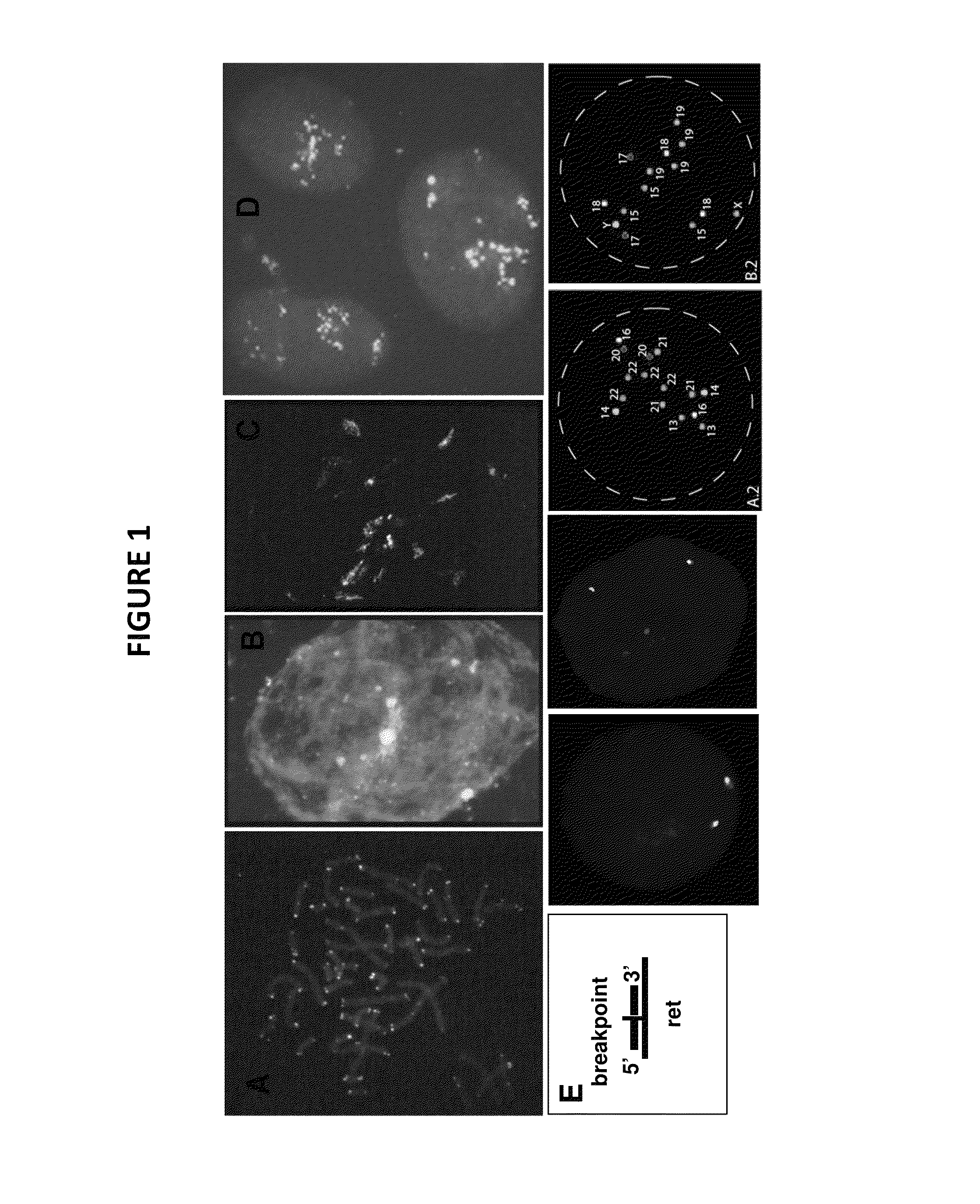
[0072]Detection of specific abnormalities and DNA probes preparation in cancer cell lines is described in Example 1 and FIG. 1. Clinical application of the assay is expected. In collaboration with scientists at the Hormone Receptor Laboratory, University of Louisville (U Louisville), KY, (Dr. James Wittliff, P.I.), we will select tissue specimens obtained from breast cancer patients for which extensive follow-up data is available, and investigate whether ErbB gene alterations besides estrogen and progesterone receptor status (‘ER and PR status’) are important prognostic parameters.

example 3

Using Genetic Barcodes to Detect Gene Status in Cell Lines

[0073]We also began investigating chromosomal rearrangements in breast cancer cell lines. We now have FISH probe sets (2-3 colour) for several additional cancer / disease associated genetic loci (in addition to the previous ones for the ErbB 1, ErbB2, AURKA and RET loci).

[0074]FIGS. 9A, 9B and 9C show examples of analyzing the A) HIPK2, B) PTEN, and C) XPG / ERCC5 gene loci. FIGS. 9A and 9B show the Homo Sapiens homeodomain interacting protein kinase 2 (HIPK2) gene locus in interphase cell nuclei, a protein kinase implicated in cancer chemoresistance, through either regulation of p53 mediated apoptosis or HIF1A mediated angiogenesis and cellular proliferation. This serine / threonine kinase involved in a preapoptotic pathway is a possible cancer therapy target, since it was found rearranged in 40-60% of various human cancers (Yu, J.; Deshmukh, H.; Gutmann, R. J.; Emnett, R. J.; Rodriguez, F. J.; Watson, M. A.; Nagarajan, R.; Gutman...


PUM

PropertyMeasurementUnit
diameteraaaaaaaaaa
temperatureaaaaaaaaaa
sizeaaaaaaaaaa

Abstract

Herein are described multicolor FISH probe sets termed “genetic barcodes” targeting several cancer or disease-related loci to assess gene rearrangements and copy number changes in tumor cells. Two, three or more different fluorophores are used to detect the genetic barcode sections thus permitting unique labeling and multilocus analysis in individual cell nuclei. Gene specific barcodes can be generated and combined to provide both numerical and structural genetic information for these and other pertinent disease associated genes.

Description

CROSS-REFERENCE TO RELATED APPLICATIONS[0001]This application is a non-provisional of and claims priority to U.S. Provisional Patent Application No. 61 / 614,456, filed on Mar. 22, 2012, hereby incorporated by reference in its entirety.STATEMENT OF GOVERNMENTAL SUPPORT[0002]This invention was made with government support under Contract No. DE-AC02-05CH11231 awarded by the U.S. Department of Energy and under Grant Numbers CA123370A and CA132815A awarded by the National Cancer Institute and the National Institutes of Health. The government has certain rights in the invention.REFERENCE TO TABLE APPENDIX[0003]Table 1 is attached and hereby incorporated by reference.BACKGROUND OF THE INVENTION[0004]1. Field of the Invention[0005]The present invention relates to fluorescent probes and genetic barcodes and methods for detection and analysis of gene characterization, rearrangement, translocation and expression in cancer.[0006]2. Related Art[0007]Our ageing population and the increasing cost o...

Claims


Application Information

Patent Timeline
Patent Type & Authority Patents(United States)
IPC IPC(8): C12Q1/68
CPCC12Q1/6883C12Q1/6841C12Q2600/156C12Q2537/157C12Q2537/16C12Q2600/158
Inventor WEIER, HEINZ-ULRICH G.
Owner RGT UNIV OF CALIFORNIA
Why Eureka
Try Eureka