
Method for diagnosing a protein misfolding disease using nerve cells derived from iPS cells

a technology of nerve cells and ips cells, which is applied in the field of protein misfolding disease diagnosis using nerve cells derived from ips cells, can solve the problems of difficult to distinguish between progression, difficult to preliminarily prepare data sets showing the amount of causative protein or the like at the respective ages over a wide range of ages in a control subject, and difficulty in detecting the onset of a protein misfolding diseas

Active Publication Date: 2015-08-04
KYOTO UNIV
View PDF4 Cites 0 Cited by
  • Summary
  • Abstract
  • Description
  • Claims
  • Application Information

AI Technical Summary

Benefits of technology

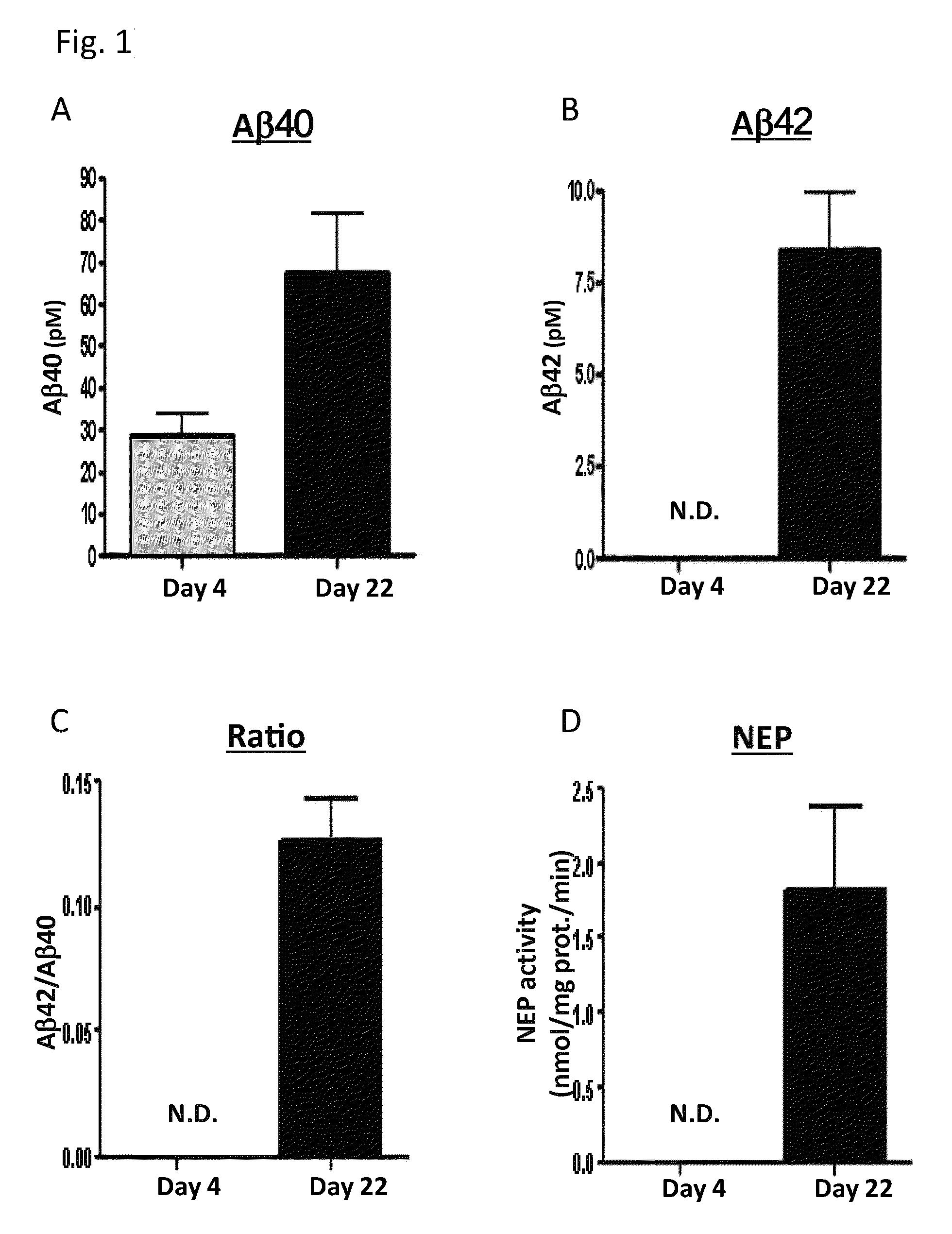
[0023]From the above results, the inventors of the present invention discovered that the onset of, or the risk of development of, Alzheimer's disease can be diagnosed by using nerve cells produced by differentiation induction of iPS cells derived from a test subject to measure the amount of Aβ or the activity or the expression level of neprilysin, followed by comparing the obtained value with a measured value in such nerve cells from a control subject wherein the age of the onset of Alzheimer's disease is known. As another embodiment, the inventors of the present invention discovered that it is useful for prediction of the age of onset of Alzheimer's disease in a test subject to compare the amount of Aβ or an activity index or the expression level of neprilysin in cells derived from a control subject wherein the age onset of Alzheimer's disease is known with the amount of Aβ or the activity index or the expression level of neprilysin in nerve cells produced by differentiation induction of iPS cells derived from the test subject, thereby completed the present invention.

Problems solved by technology

Since, in such a case, it is sometimes difficult to distinguish between progression of the disease condition and aging, it is necessary, for example, to compare a control subject whose onset of the disease is known with a test subject at the same age.
However, since the amount of the protein at the desired age cannot be measured after confirming the onset of the disease, it is very difficult to preliminarily prepare data set showing the amounts of the causative protein or the like at the respective ages over a wide range of ages in a control subject whose onset of the disease is known.
Further, in order to predict the age of the onset of the disease, the amount of the causative protein or the like needs to be preliminarily measured at the same age as the test subject in control subjects with a wide range of ages of onset, and, needless to say, it is difficult.
Because biopsy of nerve cells imposes a heavy burden to a living body, preliminary preparation of control subjects at the respective ages for diagnosis of a protein misfolding disease is problematic from an ethical point of view.

Method used


Image

  • Method for diagnosing a protein misfolding disease using nerve cells derived from iPS cells
  • Method for diagnosing a protein misfolding disease using nerve cells derived from iPS cells
  • Method for diagnosing a protein misfolding disease using nerve cells derived from iPS cells

Examples


example 1

iPS Cells

[0139]253G4 described in Nakagawa M, et al., Nat Biotechnol. 2008 January; 26(1):101-6. was used as the iPS cell. Briefly, the iPS cells were established by introducing Oct3 / 4, Sox2 and Klf4 to fibroblasts derived from a Caucasian female of 36 years old.

Formation of Neurosphere

[0140]The formation of a neurosphere was carried out by the method described in Wada T, et al, PLoS ONE 4(8), e6722, 2009 with a slight modification. More particularly, the iPS cells were divided into small clusters and cultured using N2B27 medium (Gibco) prepared by mixing DMEM / F12 and Neurobasal medium A together at a volume ratio of 1:1 and adding 1% N2, 2% B27 and 200 μM glutamine thereto, in a dish coated with poly-L-lysine / laminin (PLL / LM) (Sigma-Aldrich). Further, to this medium, Noggin (R&D systems) was added at 100 ng / ml. The medium was replaced with a medium containing 100 ng / ml Noggin every 3 days, and, after 10 days of culture, the cells were subcultured to a PLL / LM-coated dish. Thereafter...

example 2

Method of Differentiation into Nerve Cells

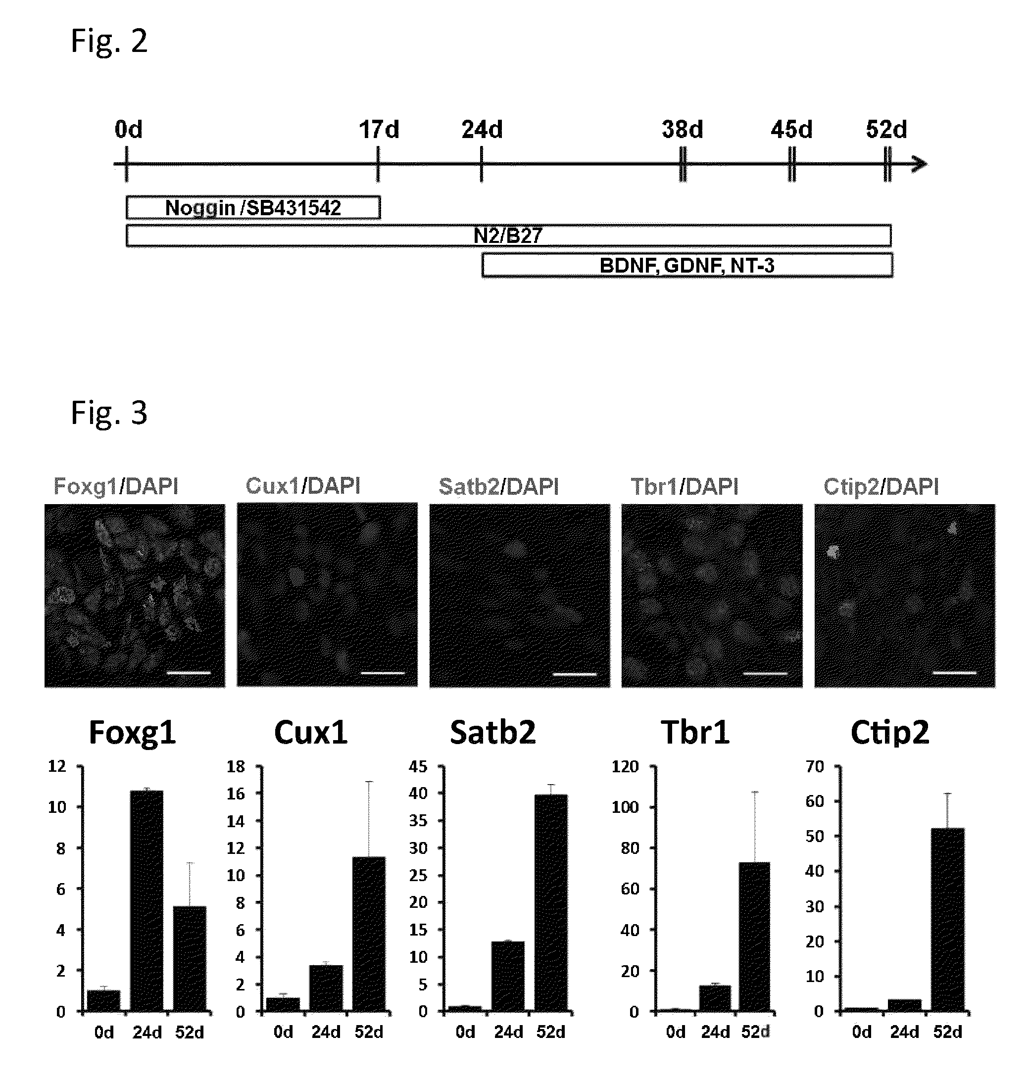
[0152]Human iPS cell line 253G4 were cultured on mitomycin C-treated mouse embryonic fibroblasts in primate ES medium (ReproCELL) supplemented with bFGF (Wako). To obtain neurons derived from the iPS cell line, the reported method was partially modified (Wada, T. et al 2009, Chambers, S. M. et al 2009). Briefly, small clumps of iPS cell colonies (40-100 μm in diameter) were selected with Cell Strainer (BD Falcon) and plated on poly-L-lysine (Sigma) / Laminin (BD Falcon) (PLL / LM) coated dishes in N2B27 medium [DMEM / F12, Neurobasal, N2 supplement, B27 supplement, L-Gln], supplemented 100 ng / ml human recombinant Noggin (R&D systems) and 1 μM of SB431542 (Sigma) for 10 days. Then the colonies were dissociated into small clumps by 200 U / ml Collagenase with CaCl2 and plated into PLL / ECL (Millipore) coated dishes. After 7 days culturing, the cells were dissociated by Accutase (Innovative Cell Technologies) and cultured on PLL / ECL coated dishes for 7 ...

example 3

iPS Cells

[0158]Alzheimer's disease (AD)-iPS cell line was established by using the method described in Takahashi K, et al, Cell. 131:861-72, 2007. Briefly, the iPS cells were established by introducing Oct3 / 4, Sox2, Klf4 and c-Myc to fibroblasts derived from Sporadic AD patient who has the ApoE 3 / 4 isoform and has an onset of the disease in 50's.

Method of Differentiation into Neurons

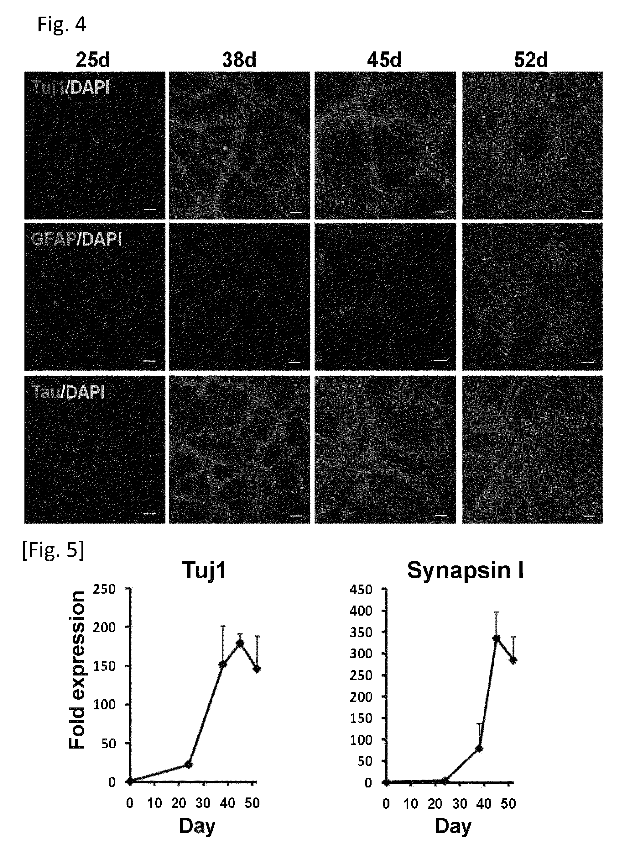
[0159]To obtain neurons derived from AD-iPS cell line, the method described in Example 2 was used. The neurons were evaluated with expression of Tuj1, Tau, SATB2, CUX1, CTIP2 and TBR1 (FIGS. 13 and 14). It was observed that expression of the neuron related genes in AD-iPS cell line was indistinct from that of normal iPS cell line described in Example 2.

Analysis of Aβ Concentration in Nerve Cells

[0160]The contents of Aβ40 and Aβ42 in the nerve cells derived from AD-iPS cells were measured with the ELISA method described in Example 1. The concentration of Aβ40 and Aβ42, ratio (Aβ42 / Aβ40) and relative amoun...


PUM

PropertyMeasurementUnit
concentrationaaaaaaaaaa

Abstract

The present invention provides a method for detecting onset of, or the risk of development of, a protein misfolding disease, and a method for predicting the age of onset of a protein misfolding disease using nerve cells derived from iPS cells. The present invention also provides a kit to be used in these methods.

Description

REFERENCE TO SEQUENCE LISTING[0001]This application incorporates by reference the sequence listing submitted as ASCII text filed via EFS-Web on Aug. 31, 2012. The Sequence Listing is provided as a file entitled “TOYA204—001APC_SL.TXT”, created on Aug. 31, 2012 and which is approximately 5 kilobytes in size.TECHNICAL FIELD[0002]The present invention relates to a method for diagnosing the onset of a protein misfolding disease and a risk of development thereof, which method uses nerve cells derived from induced pluripotent stem (iPS) cells, and to a kit to be used in this method. The present invention further relates to a method for prediction of the age of the onset of a protein misfolding disease and a kit to be used in this method.BACKGROUND ART[0003]Protein misfolding diseases are known as neurodegenerative diseases caused by cytotoxicity due to abnormally aggregated proteins (misfolded proteins) (1). Alzheimer's disease is a representative protein misfolding disease, which causes ...

Claims


Application Information

Patent Timeline
Patent Type & Authority Patents(United States)
IPC IPC(8): G01N33/48C12N5/0793G01N33/68G01N33/573G01N33/50
CPCC12N5/0619G01N33/6896C12N2501/11C12N2501/115C12N2501/13C12N2501/155C12N2501/385C12N2501/41C12N2501/91C12N2503/02C12N2506/45C12N2533/32G01N2333/4709G01N2333/96486G01N2800/2821C12N2506/00
Inventor INOUE, HARUHISAKITAOKA, SHIHOYAHATA, NAOKIIWATA, NOBUHISASAIDO, TAKAOMI
Owner KYOTO UNIV
Learn More