
Chest radiography image contrast and exposure dose optimization

a radiographic image and contrast technology, applied in the field of radiographic imaging, can solve the problems of less than optimal, complicated chest radiographic imaging task, and inability to achieve optimal contrast, so as to improve the contrast of lung tissue, improve imaging parameters and processing, and reduce patient exposure

Active Publication Date: 2015-08-11
CARESTREAM HEALTH INC
View PDF10 Cites 1 Cited by
  • Summary
  • Abstract
  • Description
  • Claims
  • Application Information

AI Technical Summary

Benefits of technology

The present invention provides methods and apparatus for chest x-ray imaging using techniques that reduce patient exposure to radiation and improve the contrast of lung tissue. This means that the invention provides better quality images with less risk to patients and improved accuracy in diagnosing lung disease.

Problems solved by technology

This can complicate the task of radiographic imaging for the chest region, since the different types of bone and tissue materials have different densities.
However, the contrast of the lung parenchyma may be viewed as some as being less than optimal.
This can complicate diagnosis and features may be misinterpreted.
Scatter reduces image detail contrast and increases noise levels, both of which hinder diagnostic accuracy.
X-ray anti-scatter grids are frequently used to reduce scattering, but have negative effects.
Grids of higher ratios are required at higher energy levels, increasing the amount of incident exposure that is required to compensate the exposure loss, but at the expense of increased patient-absorbed dose.
A further problem relates to the need for imaging both bone and soft tissue in some patients.
In reality, the bone suppression algorithm may not work perfectly; part of the rib bones may still not be sufficiently suppressed.
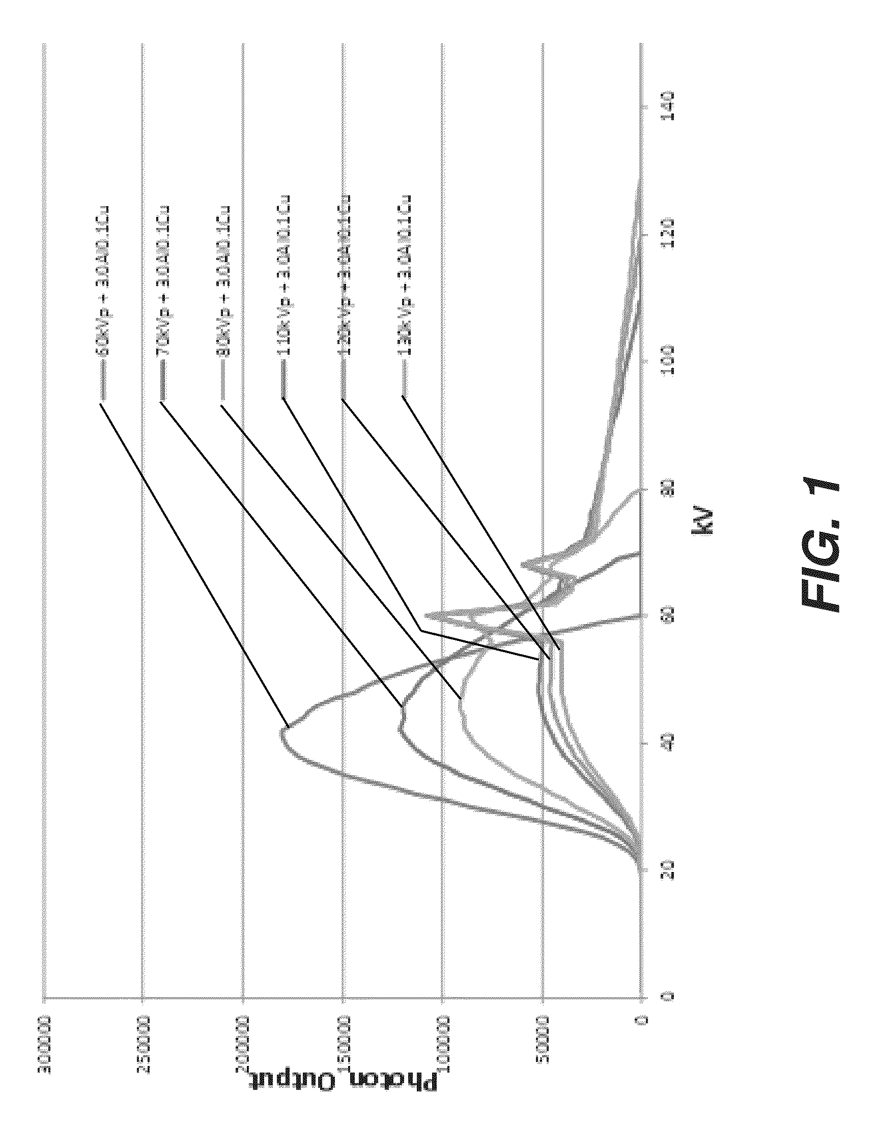
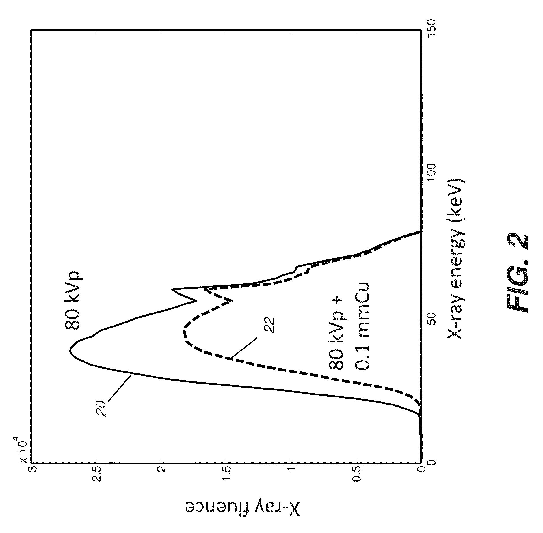
The use of lower energy x-ray photons helps to maximize soft-tissue and bone contrast in chest radiographs, but there can be negative effects if not applied appropriately.
Beam hardening essentially modifies the x-ray spectrum and reduces the effectiveness of radiation that is otherwise optimized for chest imaging.
This effect becomes worse as patient size increases.

Method used


Image

  • Chest radiography image contrast and exposure dose optimization
  • Chest radiography image contrast and exposure dose optimization
  • Chest radiography image contrast and exposure dose optimization

Examples


Embodiment Construction

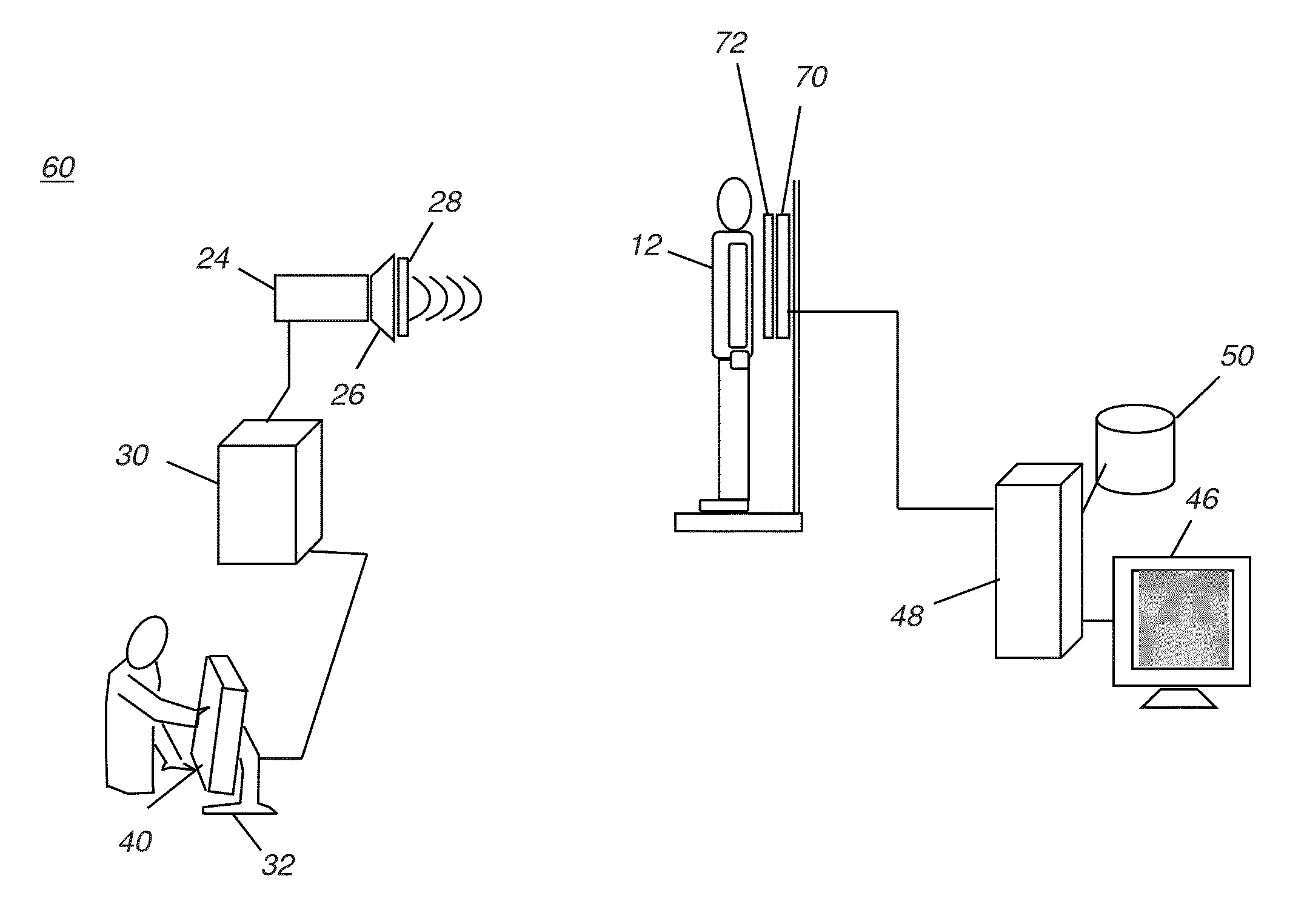
[0027]This application claims priority to U.S. Provisional Ser. No. 61 / 616,455, filed Mar. 28, 2012 in the names of Xiaohui Wang et al. entitled “CHEST RADIOGRAPHY IMAGE CONTRAST AND EXPOSURE DOSE OPTIMIZATION”, incorporated herein by reference in its entirety.

[0028]Reference is made to U.S. application Ser. No. 13 / 527,629 entitled “Rib Suppression in Radiographic Images” to Huo, incorporated herein by reference in its entirety.

[0029]The following is a detailed description of exemplary embodiments of the invention, reference being made to the drawings in which the same reference numerals identify the same elements of structure in each of the several figures.

[0030]In the context of the present disclosure, a digital chest x-ray can be obtained from a digital receiver (DR) or computed radiography (CR) receiver.

[0031]In the context of the present disclosure, the terms “viewer”, “operator”, and “user” are considered to be equivalent and refer to the viewing practitioner, technician, or o...


PUM


Abstract

A method for obtaining a digital chest x-ray image of a patient. The method includes providing a default set of technique settings for the chest x-ray, wherein the default set is selectable by an operator command and includes using a peak kilovoltage exposure setting that is below 90 kVp with beam filtration of the x-ray, and applying a rib contrast suppression algorithm to the digital chest x-ray image data acquired from the exposure.

Description

CROSS-REFERENCE TO RELATED APPLICATIONS[0001]Priority is claimed from U.S. Provisional Ser. No. 61 / 616,455, filed Mar. 28, 2012 in the names of Xiaohui Wang et al. entitled “CHEST RADIOGRAPHY IMAGE CONTRAST AND EXPOSURE DOSE OPTIMIZATION”, incorporated herein by reference in its entirety.FIELD OF THE INVENTION[0002]The invention relates generally to the field of radiographic imaging and more particularly to methods for improving image quality and exposure settings for radiographic images.BACKGROUND OF THE INVENTION[0003]The chest x-ray is used for detecting a number of patient conditions and for imaging a range of skeletal and organ structures. Radiographic images of the chest can be useful for detection of lung nodules and other features that indicate lung cancer and other pathologic structures. In clinical applications such as in the Intensive Care Unit (ICU), chest x-rays can have particular value for indicating pneumothorax as well as for tube / line positioning.[0004]The chest re...

Claims


Application Information

Patent Timeline
Patent Type & Authority Patents(United States)
IPC IPC(8): G06K9/00A61B6/00
CPCA61B6/50A61B6/5252A61B6/542A61B6/545A61B6/4035A61B6/4233
Inventor WANG, XIAOHUIHUO, ZHIMINRICHARD, SAMUEL
Owner CARESTREAM HEALTH INC
Learn More