
Hybrid dual energy imaging and bone suppression processing

a dual-energy imaging and processing technology, applied in the field of chest x-ray imaging, can solve the problems of difficult to interpret chest x-ray images in many cases, complicated chest x-ray imaging task, and rarely used in practice, so as to improve the contrast of lung tissue, improve imaging parameters and processing, and reduce patient exposure

Active Publication Date: 2014-05-22
CARESTREAM HEALTH INC
View PDF4 Cites 13 Cited by
  • Summary
  • Abstract
  • Description
  • Claims
  • Application Information

AI Technical Summary

Benefits of technology

This patent is about improving the way chest x-rays are taken and processed. The invention provides a way to get better quality images with lower patient exposure to radiation. This means that doctors can get better information from the images they take, which can help them diagnose and treat lung disease better.

Problems solved by technology

This greatly complicates the task of radiographic imaging for the chest region, since the different types of bone and tissue materials differ widely in density.
In spite of these image processing techniques, the broad range of densities and structures leads to what has been termed “anatomical clutter”, making it particularly challenging to interpret chest x-ray images in many cases.
However, this is rarely used in practice.
However, as a result of this compromise, the contrast of the lung parenchyma is less than optimal.
This complicates the job of diagnosis and makes it more likely that image features be misinterpreted.
Scatter reduces image detail contrast and increases noise levels, both of which hinder diagnostic accuracy.
X-ray anti-scatter grids are frequently used to reduce scattering, but have negative effects as well.
Grids of higher ratios are employed at higher energy levels, increasing the amount of incident exposure that is needed to compensate the exposure loss, but at the expense of increased patient absorbed dose.
A further issue relates to the need for imaging both bone and soft tissue in some patients.
The use of lower energy x-ray photons helps to maximize soft-tissue and bone contrast in chest radiographs, but there can be negative effects if not applied appropriately.
Beam hardening essentially modifies the x-ray spectrum at different positions in the image and reduces the effectiveness of radiation that is otherwise optimized for chest imaging.
This effect becomes worse as patient size increases.
Motion artifacts often result, leading to misregistration due to breathing, heartbeat, and other movement of the patient.
Even where motion artifacts are minimal, however, overall image quality is compromised in DE image processing.
Because the image analysis is performed on the standard chest image, and soft-tissue and bone structures overlay each other, it can be very difficult to separate the bones from the underlying soft tissue structure.
In some situations, one or more rib bones, or sections of rib bones, are missing from the segmentation analysis and are, therefore, not accurately suppressed.
Thus it can be seen that even though various approaches have been used to mitigate anatomical clutter in chest x-ray images, each of these approaches have their limitations.

Method used


Image

  • Hybrid dual energy imaging and bone suppression processing
  • Hybrid dual energy imaging and bone suppression processing
  • Hybrid dual energy imaging and bone suppression processing

Examples


Embodiment Construction

[0031]The following is a detailed description of the preferred embodiments of the invention, reference being made to the drawings in which the same reference numerals identify the same elements of structure in each of the several figures.

[0032]Where they are used, the terms “first”, “second”, “third”, and so on, do not necessarily denote any ordinal or priority relation, but may be used for more clearly distinguishing one element or time interval from another.

[0033]In the context of the present disclosure, the term “effective dose” relates to a tissue-weighted sum of equivalent dose, a measure of absorbed dose, in specified tissues and organs of the body. Effective dose is typically quantified in sieverts (Sv). The term “standard dose” relates to standard values of effective dose that apply for a particular type of exam and is determined, in part, by practices followed at a particular radiographic imaging site.

[0034]In the context of the present disclosure, a digital chest x-ray can...


PUM


Abstract

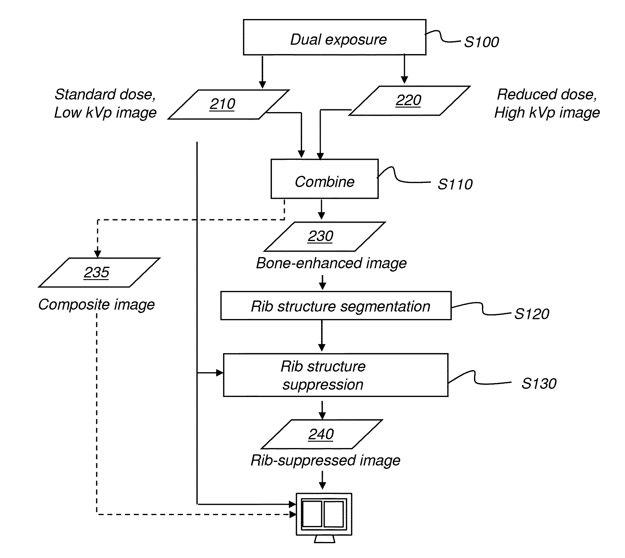
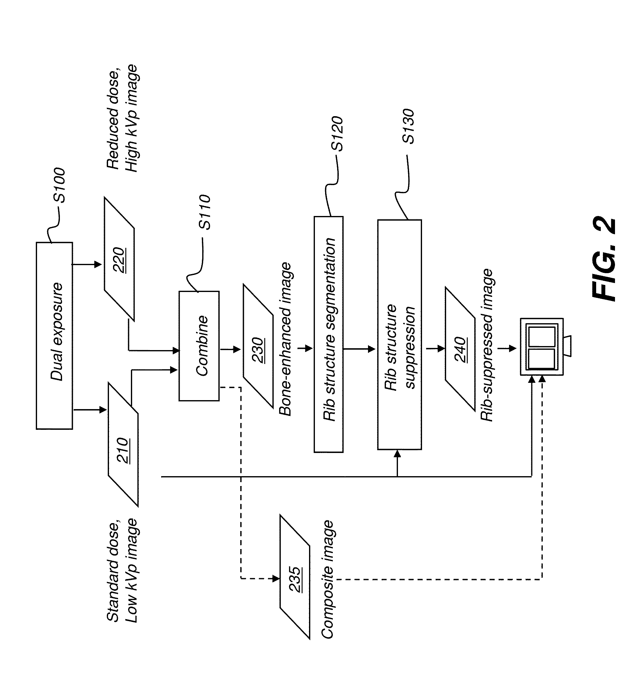
A method for obtaining a digital chest x-ray image of a patient, executed at least in part by a computer system, acquires a standard dose radiographic image and a reduced dose radiographic image, wherein the standard dose radiographic image and reduced dose radiographic images are obtained at peak kilovoltage values that differ by at least 10 kVp. A bone-enhanced image is formed by combining data from the standard dose radiographic image and reduced dose radiographic images and bone structure is segmented from the bone-enhanced image. A processed image is formed by suppressing bone contrast from the standard dose radiographic image according to the segmented bone structure. The processed image is displayed, stored, or transmitted.

Description

CROSS-REFERENCE TO RELATED APPLICATIONS[0001]Priority is claimed from U.S. Provisional Patent Application Ser. No. 61 / 728,821, filed Nov. 21, 2012 entitled “HYBRID DUAL ENERGY IMAGING AND BONE SUPPRESSION PROCESSING” in the names of Wang et al. and incorporated herein in its entirety.FIELD OF THE INVENTION[0002]The invention relates generally to the field of radiographic imaging and more particularly to methods for improving image quality and exposure settings for chest x-ray imaging.BACKGROUND OF THE INVENTION[0003]The chest x-ray is used for detecting a number of patient conditions and for imaging a range of skeletal and organ structures. Radiographic images of the chest are useful for detection of lung nodules and other features that indicate lung cancer and other pathologic structures and other life-threatening conditions. In clinical applications such as in the Intensive Care Unit (ICU), chest x-rays can have particular value for indicating pneumothorax as well as for showing t...

Claims


Application Information

Patent Timeline
IPC IPC(8): A61B6/00
CPCA61B6/482A61B6/505A61B6/5205
Inventor WANG, XIAOHUIHUO, ZHIMINFOOS, DAVID H.
Owner CARESTREAM HEALTH INC
Learn More