Apparatus, method, and system for image-based human embryo cell classification

a human embryo cell and image-based technology, applied in the field of cell classification and/or outcome determination, can solve the problems of low birth rate, miscarriage, and well-documented adverse outcomes for both mother and fetus

Active Publication Date: 2015-11-03
ARES TRADING SA
View PDF3 Cites 12 Cited by
  • Summary
  • Abstract
  • Description
  • Claims
  • Application Information

AI Technical Summary

Problems solved by technology

Infertility is a common health problem that affects 10-15% of couples of reproductive-age.
Multiple gestations have well-documented adverse outcomes for both the mother and fetuses, such as miscarriage, pre-term birth, and low birth rate.
Potential causes for failure of IVF are diverse; however, since the introduction of IVF in 1978, one of the major challenges has been to identify the embryos that are most suitable for transfer and most likely to result in term pregnancy.
However, potential risks of these methods also exist in that they prolong the culture period and disrupt embryo integrity (Manipalviratn S, et al.
However, these methods do not recognize the importance of the duration of cytokinesis or time intervals between early divisions.
However, these methods disclose only a basic and general method for time-lapse imaging of bovine embryos, which are substantially different from human embryos in terms of developmental potential, morphological behavior, molecular and epigenetic programs, and timing and parameters surrounding transfer.
Moreover, no specific imaging parameters or time intervals are disclosed that might be predictive of human embryo viability.
While time-lapse imaging has shown promise in the context of automated analysis of early human embryo development, significant development and / or performance hurdles remain unaddressed by these preexisting methods.
The nature, timing, and other benchmarks of early human embryo development provide challenges for predicting development behavior.
Specifically, automated tracking of individual cells, which forms the basis for each of these determinations, can be difficult due to the inherently noisy nature of biological images, as may arise due to lack of distinct visual features, missing and / or false cell boundaries, changing topology of the cell mass due to the cell division / and or cell movement, cell shape deformation, and so on.
As another example, when the estimated number of cells and / or division timing information is used to determine likelihood of future embryo viability, this automated determination can also be erroneous, and can lead to erroneous decisions, such as whether to proceed with IVF using particular embryos.
In addition, when embryos are of similar quality, it can be difficult to differentiate the embryos to determine, for example, which embryos to implant and which embryos to freeze.

Method used


Image

  • Apparatus, method, and system for image-based human embryo cell classification
  • Apparatus, method, and system for image-based human embryo cell classification
  • Apparatus, method, and system for image-based human embryo cell classification

Examples


example 1

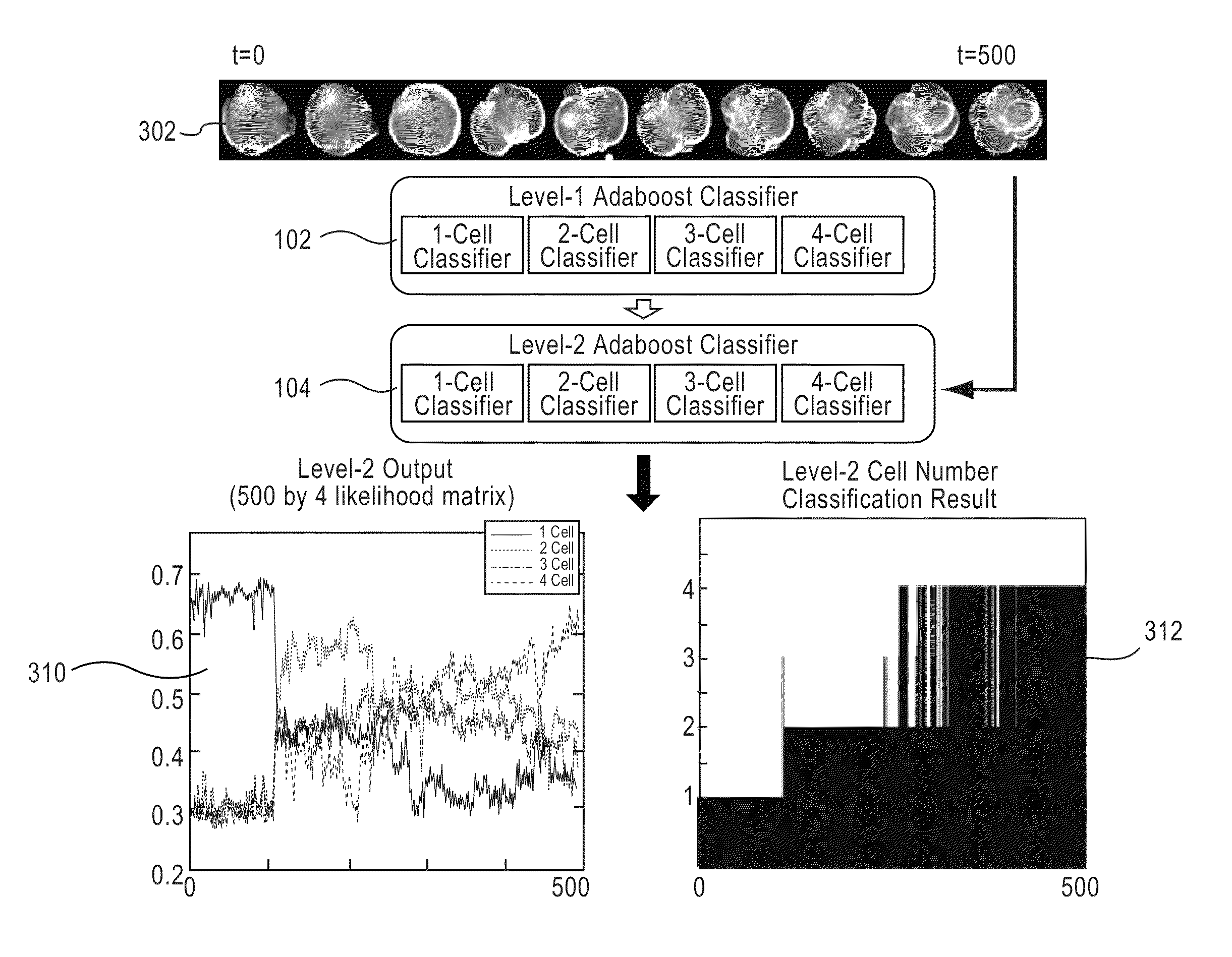
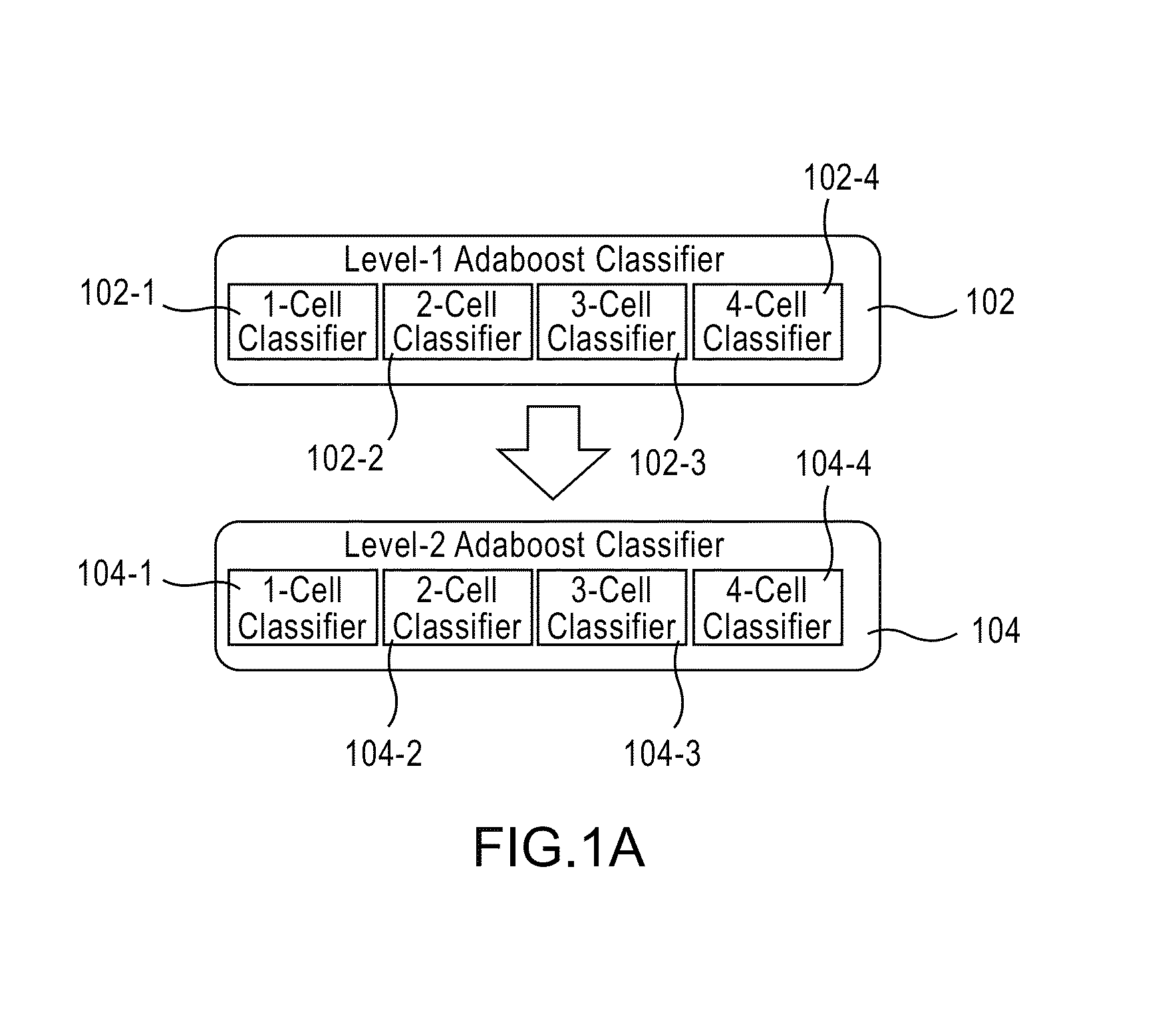
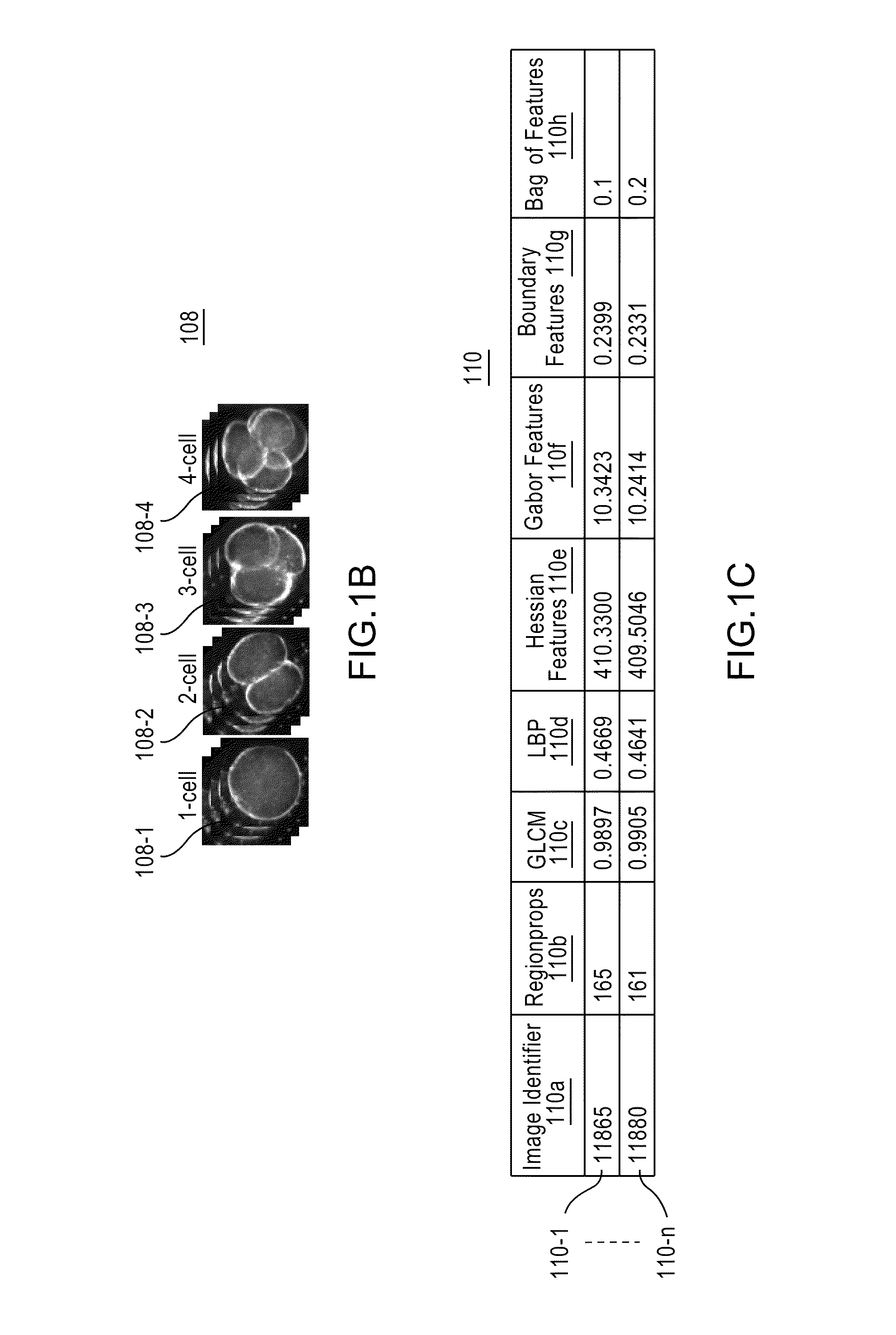
[0212]This example presents a multi-level embryo stage classification method to estimate the number of cells at multiple time points in a time-lapse microscopy video of early human embryo development. A 2-level classification model is proposed to classify embryo stage within a spatial-temporal context. A rich set of discriminative embryo features are employed, hand-crafted, or automatically learned from embryo images. The Viterbi algorithm further refines the embryo stages with the cell count probabilities and a temporal image similarity measure. The proposed method was quantitatively evaluated using a total of 389 human embryo videos, resulting in a 87.92% overall embryo stage classification accuracy.

[0213]Introduction

[0214]Timing / morpho-kinetic parameters measured from time-lapse microscopy video of human embryos, such as the durations of 2-cell stage and 3-cell stage, have been confirmed to be correlated with the quality of human embryos and therefore can be used to select embryo...

example 2

[0251]As noted earlier, some aspects of the invention are also operable for tracking of cell activity. Accordingly, some aspects of the invention are operable for automated, non-invasive cell activity tracking. In some embodiments, automated, non-invasive cell activity tracking is for determining a characteristic of one or more cells without invasive methods, such as injection of dyes. The cell activity tracking can be applied to one or more images of one or more cells. The images can be a time-sequential series of images, such as a time-lapse series of images. The cell(s) shown in the plurality of images can be any cell(s) of interest. For example, the cells can be a human embryo that may have one or more cells. Other examples of such cells of interest include, but are not limited to, oocytes and pluripotent cells.

[0252]In some embodiments, a number of the cells in each image is of interest, and can be determined by an embodiment of the invention. For example, the number of cells c...

example 3

[0376]Human embryo tracking can face challenges including a high dimensional search space, weak features, outliers, occlusions, missing data, multiple interacting deformable targets, changing topology, and a weak motion model. This example address these by using a rich set of discriminative image and geometric features with their spatial and temporal context. In one embodiment, the problem is posed as augmented simultaneous segmentation and classification in a conditional random field (CRF) framework that combines tracking based and tracking free approaches. A multi pass data driven approximate inference on the CRF is performed. Division events were measured during the first 48 hours of development to within 30 minutes in 65% of 389 clinical image sequences, winch represents a 19% improvement over a purely tracking based or tracking free approach.

[0377]Augmented Simultaneous Segmentation and Classification

[0378]Augmented simultaneous segmentation and classification leverages trackin...


PUM


Abstract

Apparatuses, methods, and systems for automated cell classification, embryo ranking, and / or embryo categorization are provided. An apparatus includes a classification module configured to apply classifiers to images of one or more cells to determine, for each image, a classification probability associated with each classifier. Each classifier is associated with a distinct first number of cells, and is configured to determine the classification probability for each image based on cell features including one or more machine learned cell features. The classification probability indicates an estimated likelihood that the distinct first number of cells is shown in each image. The classification module is further configured to classify each image as showing a second number of cells based on the distinct first number of cells and the classification probabilities associated therewith. The classification module is implemented in at least one of a memory or a processing device.

Description

CROSS-REFERENCE TO RELATED APPLICATIONS[0001]This application claims priority to: U.S. Provisional Application No. 61 / 785,170, “APPARATUS, METHOD, AND SYSTEM FOR IMAGE-BASED HUMAN EMBRYO CELL CLASSIFICATION”, filed on Mar. 14, 2013; U.S. Provisional Application No. 61 / 785,216, “APPARATUS, METHOD, AND SYSTEM FOR IMAGE-BASED HUMAN EMBRYO OUTCOME DETERMINATION”, filed on Mar. 14, 2013; U.S. Provisional Application No. 61 / 771,000, “AUTOMATED EMBRYO STAGE CLASSIFICATION IN TIME-LAPSE MICROSCOPY VIDEO OF EARLY HUMAN EMBRYO DEVELOPMENT”, filed on Feb. 28, 2013; U.S. Provisional Application No. 61 / 785,179, “APPARATUS, METHOD, AND SYSTEM FOR AUTOMATED, NON-INVASIVE CELL ACTIVITY TRACKING”, filed on Mar. 14, 2013; U.S. Provisional Application No. 61 / 785,199, “APPARATUS, METHOD, AND SYSTEM FOR HUMAN EMBRYO VIABILITY SCREENING BASED ON AUTOMATED CONFIDENCE ESTIMATION OF ASSESSMENT OF CELL ACTIVITY”, filed on Mar. 14, 2013; and U.S. Provisional Application No. 61 / 770,998, “UNIFIED FRAMEWORK FOR ...

Claims


Application Information

Patent Timeline
Patent Type & Authority Patents(United States)
IPC IPC(8): G06K9/00G06K9/62G06V10/774
CPCG06K9/00147G06K9/6256G06V20/695G06V20/698G06V10/774G01N33/50G06F18/285G06F18/2415G06F18/214
Inventor WANG, YUMOUSSAVI, FARSHIDLORENZEN, PETERGOULD, STEPHEN
Owner ARES TRADING SA
Why Eureka
Try Eureka