Medical imaging

a medical imaging and image processing technology, applied in the field of medical imaging, can solve the problems of pulsed dopplers suffering from aliasing, affecting the accuracy and reliability of the procedure being performed, and not being able to accept real-time applications, and achieve the effect of high degree of accuracy

Active Publication Date: 2015-11-24
KONINKLIJKE PHILIPS ELECTRONICS NV
View PDF33 Cites 15 Cited by
  • Summary
  • Abstract
  • Description
  • Claims
  • Application Information

AI Technical Summary

Benefits of technology

[0030]For therapeutic applications, an incorrect orientation may damage adjacent tissues, so the orientation between the transducer and the reference structure benefits from a high-degree of accuracy.
[0049]Any suitable instrument tracking system known in the art may be employed, allowing the position of the instrument within the patient-specific anatomical model to be accurately and repeatedly determined. It is therefore not required to employ computationally intensive algorithms to determine the instrument position.

Problems solved by technology

While this may be acceptable in off-line applications where the user is processing previously acquired imaging data, it is not acceptable in real-time applications, where the input of the user directly affects the accuracy and reliability of the procedure being performed.
However, pulsed Doppler is known to suffer from aliasing if the velocity of the reference structure and the angle between the measurement beam and the blood flow direction combine to give a Doppler frequency greater than half of the pulse repetition frequency.
This creates ambiguity in the Doppler signal, and may cause misinterpretation of the reference object's movement.
An additional problem of pulsed Doppler is that the depth of measurement is limited by the pulse repetition frequency chosen, because the time interval must be sufficient to allow a pulse to travel from the transducer to the reference structure and back, before the next pulse is emitted.
The measurement of samples and the processing of the results require considerable computational power, making such an instrument expensive.
The position and extent of the measurement volume is conventionally selected by the user, and therefore can result in inaccuracies in the results measured.
Although acceptable for some applications, this system can only provide a limited field of view from the viewpoint of the biopsy needle, making it very easy for the user to navigate the needle incorrectly and to lose orientation.
Such applications typically employ higher energies than for visualization or functional measurement, and therefore inaccuracies in positioning may result in damage to surrounding tissues.

Method used


Image

  • Medical imaging
  • Medical imaging
  • Medical imaging

Examples


Embodiment Construction

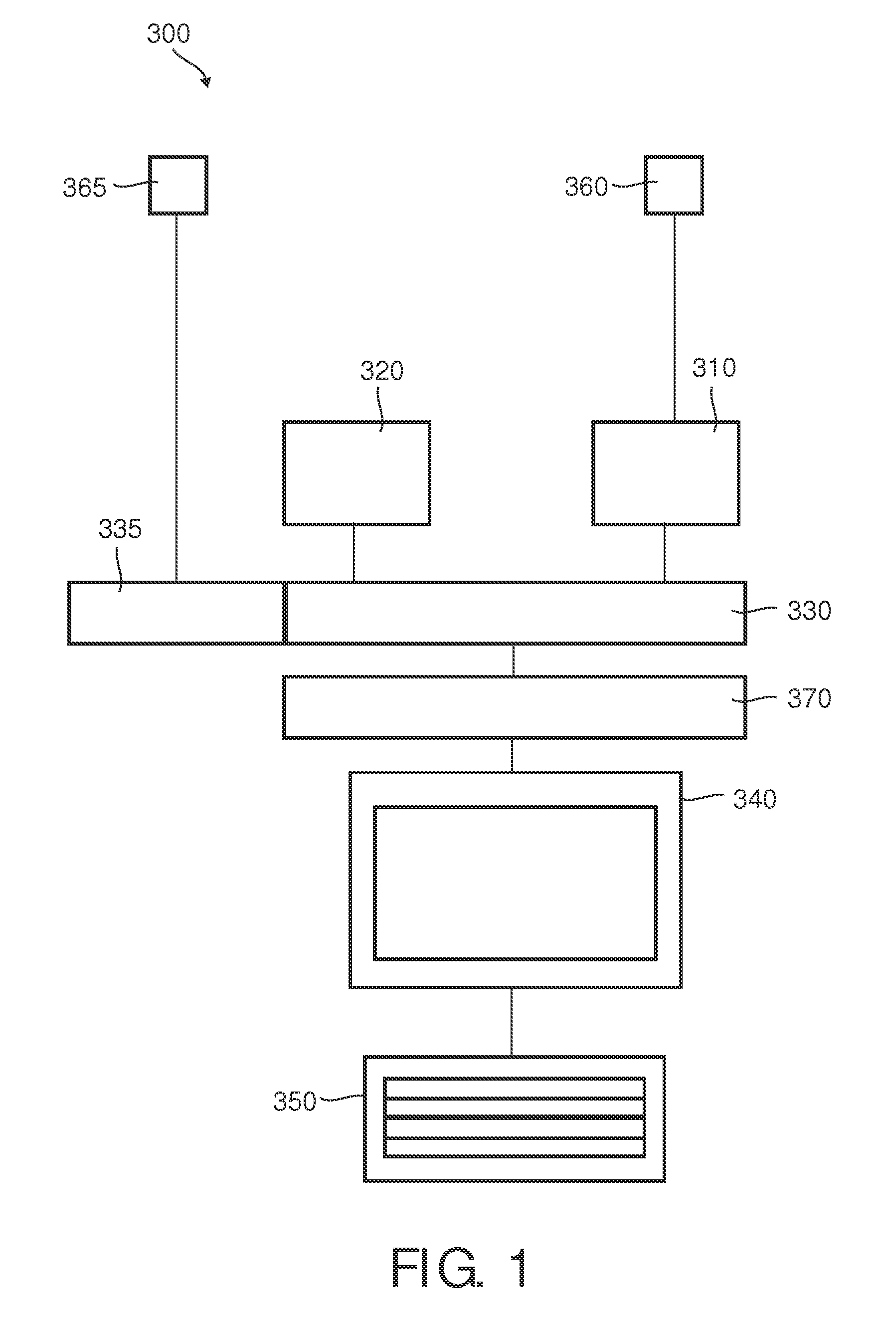
[0069]A system 300 for providing imaging data of a specific anatomical volume is depicted in FIG. 1. The system 300 comprises:[0070]a medical imaging transducer 360 configured to provide imaging data of an anatomical volume 100. For example, this is an ultrasound transducer, suitable for visualization, for functional measurements, or for therapeutic applications. In practice, the anatomical volume may be considered to be the anatomical region of a patient within the range of the imaging transducer 360;[0071]a user input 350 configured to specify a reference structure 120 in the anatomical volume. Typically, the user input 350 provides for interaction with the system in any form known in the art, for example, as icons, thumbnails, menus, and pull-down menus. The user input 350 may also comprise a keyboard, mouse, trackball, pointer, drawing tablet or the like;[0072]an imager 310 configured to receive the imaging data. The imager 310 cooperates with the transducer 360 to provide the i...


PUM


Abstract

Ultrasound imaging, or ultrasonography, is a convenient imaging modality for diagnostic purposes. During procedures requiring visualization, the user coordinates the movement of the transducer through the manual selection of an appropriate representation on the screen. Ultrasound may also be employed for functional measurements. Typically, the measurement requires the manual selection of target planes, lines, or volumes where the measurement is to be performed. Therapeutic applications of ultrasound are also known in the art. Such applications typically employ higher energies, and therefore inaccuracies in positioning may result in damage to surrounding tissues. The invention provides a system and method for repeated determination of a first vector between a reference structure comprised in an anatomical volume and a medical imaging transducer configured to provide imaging data of the anatomical volume. The invention provides an automated tracking system of the reference structure, requiring that the user only selects the reference structure.

Description

FIELD OF THE INVENTION[0001]The invention relates to a system and method for providing imaging data of a reference structure in an anatomical volume.BACKGROUND OF THE INVENTION[0002]Ultrasound imaging, or ultrasonography, is a convenient imaging modality for diagnostic purposes. It may be employed before, during or after a therapeutic intervention. Typically, a healthcare professional uses a hand-held probe, or transducer, which is moved as appropriate to visualize reference structures. In many cases, the transducer is placed on the surface of the body. However, in some specialized procedures, such as endovaginal, endorectal and transesophageal procedures, specific transducers are placed inside the patient's body. Small transducers may even be mounted on catheters and inserted into blood vessels to image the vessel walls.[0003]Recent advances in ultrasound require real-time monitoring in an increasing range of diagnostic and interventional procedures. For example, for the diagnosis ...

Claims


Application Information

Patent Timeline
Patent Type & Authority Patents(United States)
IPC IPC(8): G06T7/00A61B8/00G06T3/00A61B8/08G06T7/20G01S7/52
CPCG06T7/0081A61B8/42A61B8/488G06T3/0068G06T7/0089G06T7/20A61B8/4245G01S7/52063G01S7/52073G06T2207/10132G06T2207/20092G06T2207/20112G06T7/11G06T7/149
Inventor MEYER, CARSTEN
Owner KONINKLIJKE PHILIPS ELECTRONICS NV
Why Eureka
Try Eureka