

Methods for detecting and treating cancer
a cancer and cancer technology, applied in the field of cancer detection and treatment, can solve the problems of perinatal death, lack of foot process formation, and persistent tight junction between podocytes
- Summary
- Abstract
- Description
- Claims
- Application Information
AI Technical Summary
Benefits of technology
Problems solved by technology
Method used
Image

Examples
example 1
Materials and Methods
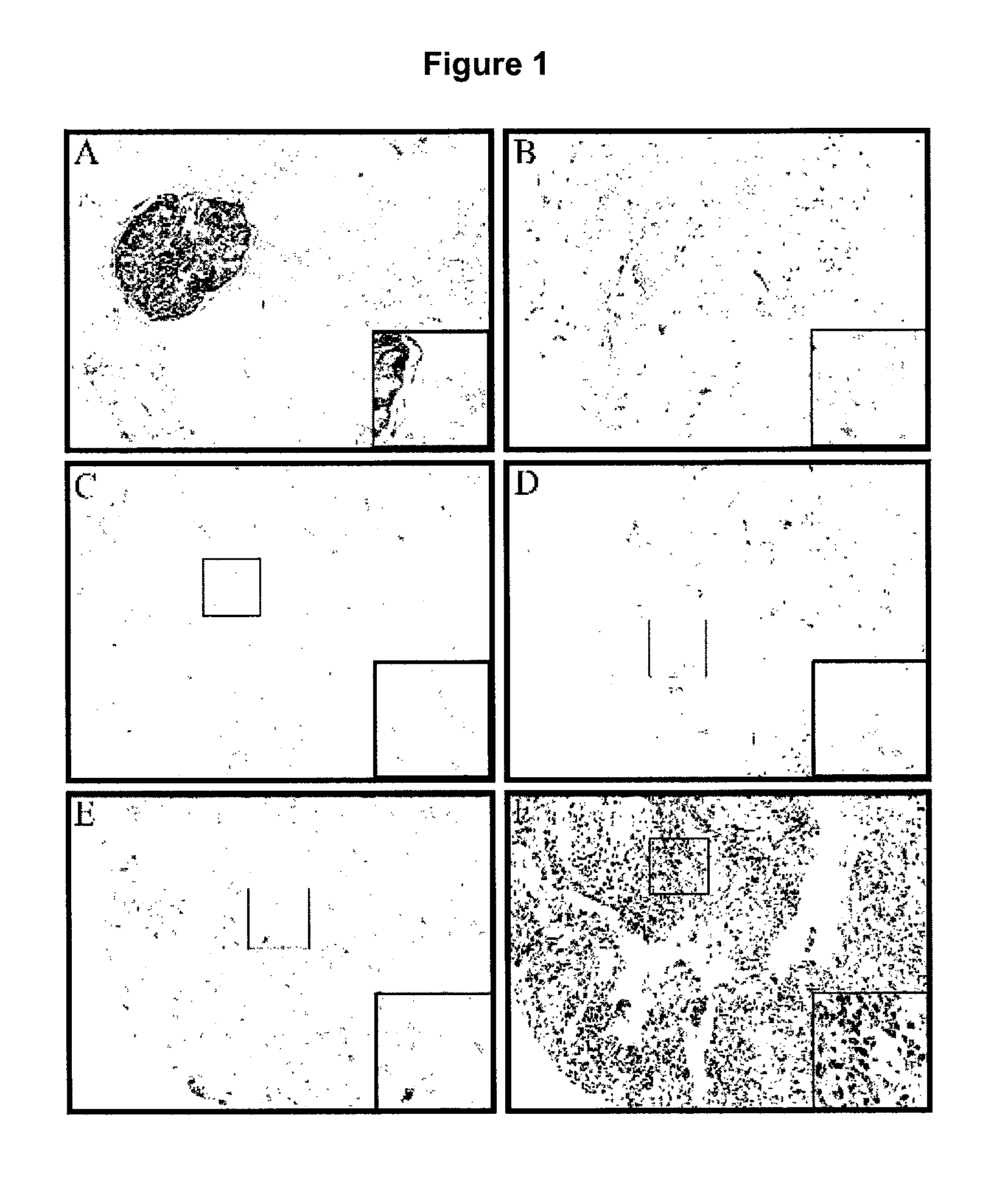
Tissue Microarray Construction
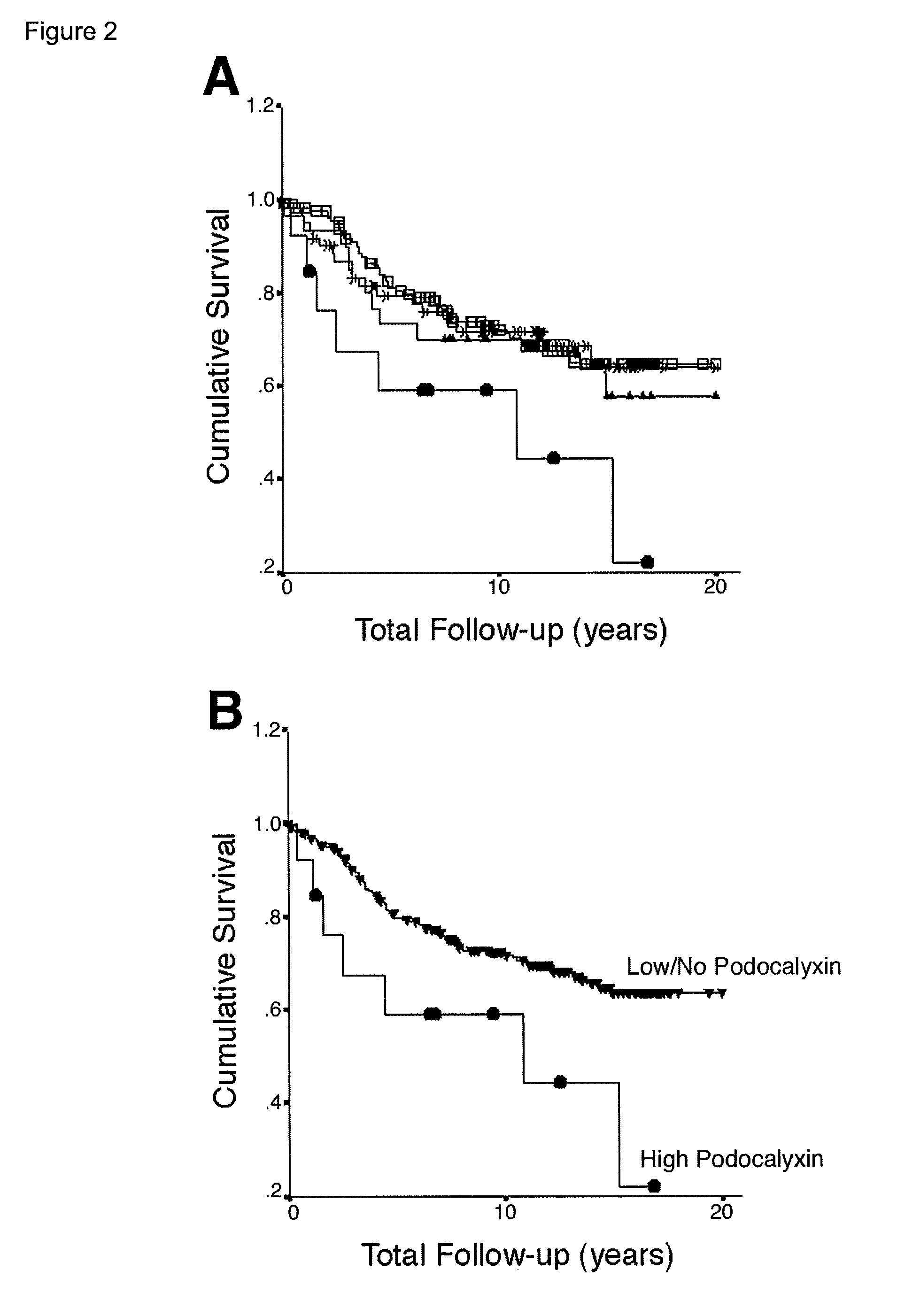
[0257]A total of 270 formalin-fixed, paraffin-embedded primary invasive breast cancer tissue blocks (archival cases from Vancouver General Hospital from the period 1974-1995) that had been graded according to the Nottingham modification of the Scarth, Bloom, Richardson method (Elston and Ellis, 1991) were used to construct a tissue microarray (TMA) as described previously (Parker et al., 2002). Briefly, a tissue-arraying instrument (Beecher Instruments, Silver Springs Md.) was used to create holes in a recipient block with defined array coordinates. Two 0.6 mm diameter tissue cores were taken from each case and transferred to the recipient block using a solid stylet. Three composite high-density tissue microarray blocks were designed and serial 4 μm sections were then cut with a microtome and transferred to adhesive-coated slides. Normal breast and kidney tissues were used as controls.
TMA Immunohistochemistry, Scoring and...
example 2
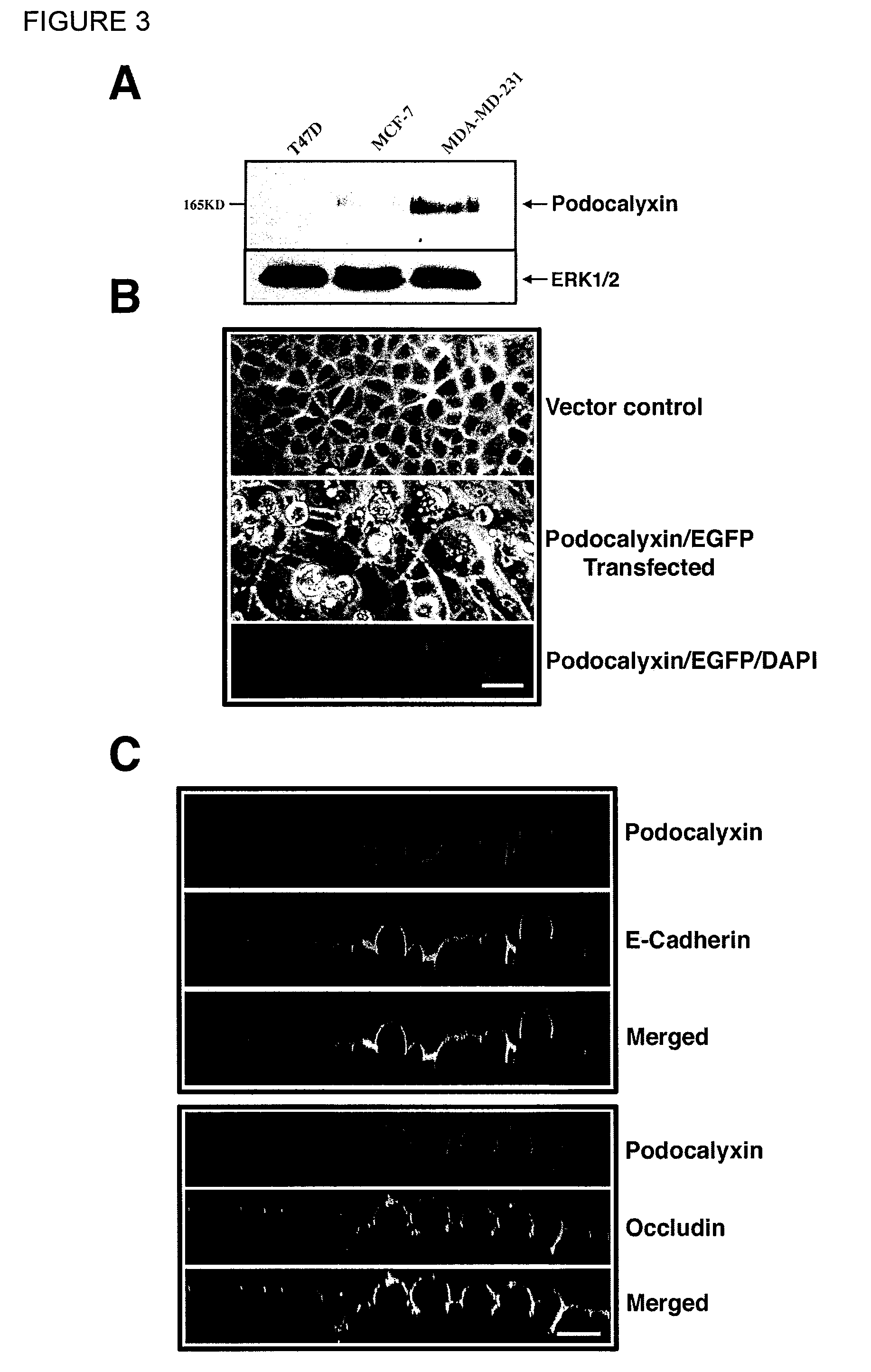
Endoglycan
Results
Tissue Distribution of CD34 Family Members
[0269]Data was compiled from published analyses on human and mouse CD34, Podocalyxin and Endoglycan (Krause 1996, McNagny 1997, Doyonnas 2001, Sassetti 2000) and from our unpublished observations on mouse Endoglycan. Endoglycan and Podocalyxin expression profiles were generated using unpublished data obtained from: 1) Northern blots of hematopoietic lineage cell lines, 2) RT-PCR of sorted hematopoietic subsets from bone marrow, 3) antibody stains and flow cytometry analysis using existing antibodies to CD34 (RAM34) Podocalyxin (PCLP1) and 4) Immunohistochemistry using the same antibodies. Results are shown in Table 4.
Preparation of Monoclonal Antibody with Specific Binding Against Endoglycan
[0270]To make the rat monoclonal antibody, rats were immunized with a peptide corresponding to sequence from the extracellular domain: V A S M E D P G Q A P D L P N L P S I L P K M D L A E P P W H M P L Q G G C (SEQ ID NO:10) linked to KL...
example 3
Material and Methods:
FDC-P1 Cells
[0281]For all experiments and maintenance purposes (unless otherwise indicated), factor dependent cell-Paterson1 cells (FDC-P1) were grown in complete RPMI (Hyclone, Logan, Utah) media (10% FBS, 4 mM L-glutamine, penicillin and streptomycin (all from Gibco-Invitrogen, Burlington, ON)) with the addition (10% v / v) of WEHI-3B conditioned media as a source of mIL-3.
Lentiviral shRNA Infection of FDC-P1
[0282]Silencing-RNA target sequences were designed using PSI Oligomaker v1.5 (http: / / web.mit.edu / jacks-lab / protocols / pSico.html) and the resulting oligos were generated by Invitrogen (Burlington, ON). Lentiviral infection was performed using an adaptation of a protocol previously described (Rubinson et al., 2003). The expression vector, pLL3.7 kb+2.0 kb spacer, was a generous gift from Dr. Fabio Rossi (University of British Columbia, Vancouver, BC). shRNA oligos were annealed at 55° C. for 40 cycles, cloned into pLL3.7 plasmids and propagated in E. coli. Pla...
PUM
| Property | Measurement | Unit |
|---|---|---|
| time | aaaaa | aaaaa |
| diameter | aaaaa | aaaaa |
| pH | aaaaa | aaaaa |
Abstract
Description
Claims
Application Information