

Methods of predicting endometrial receptivity
a technology of endometrial receptivity and embryo implantation, which is applied in the direction of instruments, biochemistry apparatus and processes, material analysis, etc., can solve the problems of limiting art success, implantation failure still remains a limitation obstacle, and the inability to pinpoint the specific involvement of a particular cell type or a specific molecul
- Summary
- Abstract
- Description
- Claims
- Application Information
AI Technical Summary
Benefits of technology
Problems solved by technology
Method used
Image

Examples
example 1
and Methods
Human Endometrial Tissues for Isolation of Primary Endometrial Epithelial Cells
[0303]Ethics approval was obtained from the Human Ethics Committee at Monash Medical Centre (Melbourne, Australia), and all patients provided informed written consent. Endometrial biopsies were obtained from women undergoing hysteroscopy dilatation, curettage or assessment of tubal patency. The menstrual cycle stage was confirmed by routine histologic dating of the tissue.
Isolation of Primary Human Endometrial Epithelial Cells (HEECs)
[0304]Tissues from the proliferative phase (days 6-14) were collected into Dulbecco's modified Eagle's medium / F12 (DMEM / F12, Thermo Fisher Scientific, Mass., USA), and cells were isolated within 24 h of collection. Cells were isolated by enzymatic digestion and filtration as previously described (Marwood et al., 2009). Briefly, endometrial tissue samples were digested with collagenase from Clostridium histolyticum (7.5 U / ml; Sigma) and DNase 1 (2000 U / ml; Roche, Ca...
example 2
Identification of Podocalyxin in Primary Human Endometrial Epithelial Cells
[0341]Primary endometrial epithelial cells (HEECs) from human endometrial tissues were isolated and enriched for plasma membrane proteins as described in Example 1.
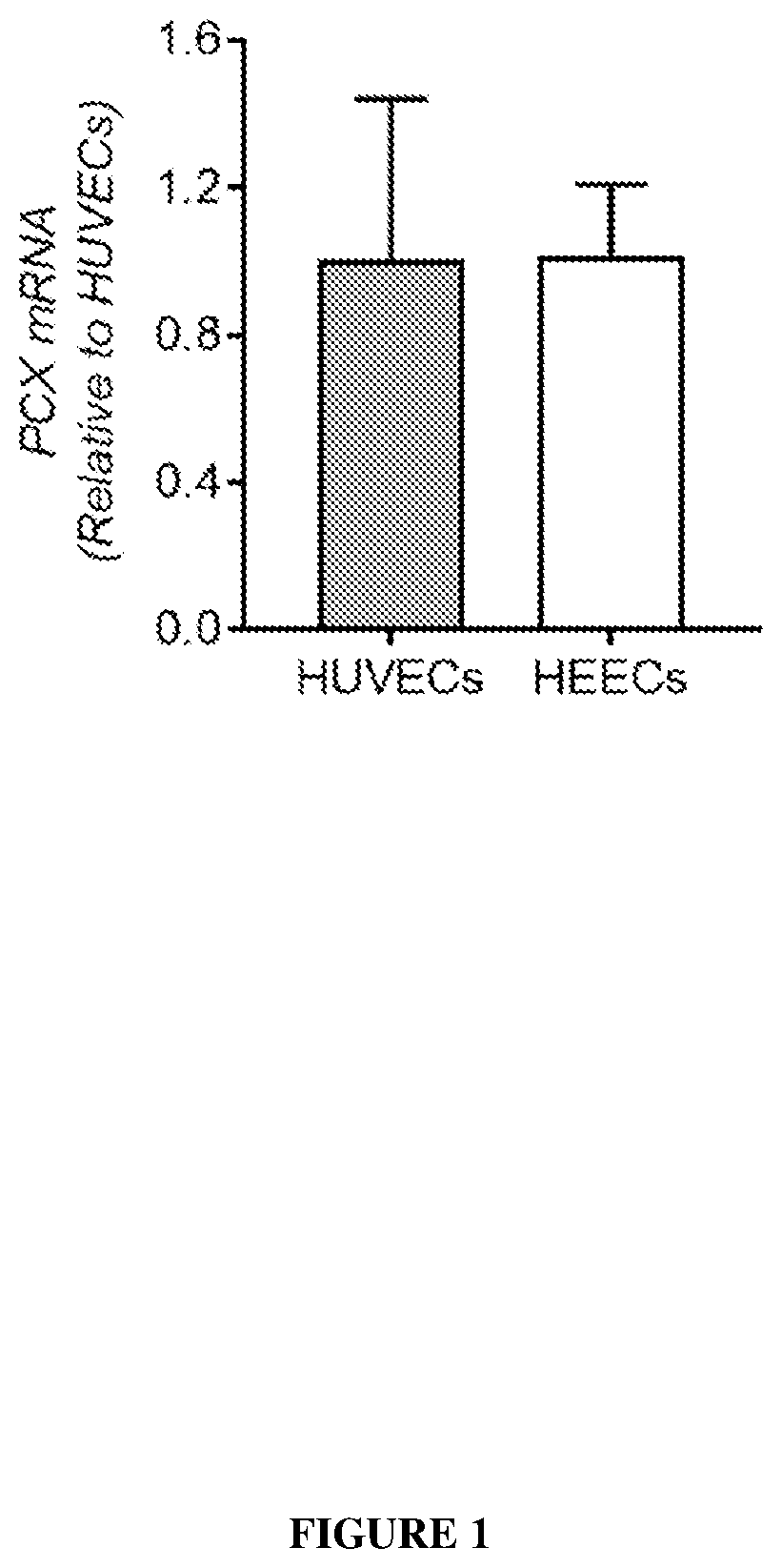
[0342]The resulting proteins were analysed by mass spectrometry and a total of 250 proteins were identified (Table 2). Of these, 47 were deemed to be cell membrane proteins, 10 of which were associated with cell adhesion including podocalyxin (PCX).
[0343]To confirm the proteomic finding, total cell lysates of primary HEECs isolated from the proliferative phase endometrium (as for the proteomic study) were analysed by western blot using 3 antibodies against different regions of human PCX.
[0344]A dominant band of ˜150 kDa was detected by all 3 antibodies with compatible levels in both cell types. Ab1 detected an additional fainter band of ˜80 kDa in both HUVECs and HEECs, whereas Ab2 recognized additional bands of ˜45, 37 and 30 kDa primarily in HUVE...
example 3
calized to the Apical Membrane of Epithelial and Endothelial Cells in the Human Endometrium and is Down-Regulated Specifically in the Luminal Epithelium Coinciding with Receptivity Establishment
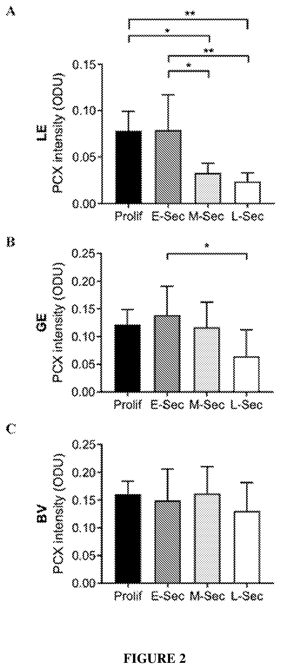
[0346]The cellular localization of PCX in the human endometrium across the menstrual cycle was examined by immunohistochemistry, as described in Example 1.
[0347]All 3 PCX antibodies detected a similar pattern of staining. In the proliferative phase, PCX was localized strongly to the apical surface of both the luminal and glandular epithelial cells (LE and GE respectively), as well as of endothelial cells in blood vessels (BV). The stroma showed no / below detection. This pattern persisted more or less to the early secretory phase, after which drastic differences emerged, especially in LE. In the mid-secretary phase, while PCX staining was still strong in both GE and BV, it was almost non-detectable in LE. In the late-secretory phase, whilst LE continued to be with minimal PCX, GE displayed fain...
PUM
| Property | Measurement | Unit |
|---|---|---|
| Time | aaaaa | aaaaa |
| Level | aaaaa | aaaaa |
Abstract
Description
Claims
Application Information