Fluoroscopy-independent balloon guided occlusion catheter and methods

a technology of fluoroscopy and balloon guide, applied in balloon catheters, medical science, surgery, etc., can solve the problems of neurological death, cardiac arrest, and difficulty in applying vascular clamps, and achieve the effect of reducing the risk of strok

Active Publication Date: 2016-10-25
PRYTIME MEDICAL DEVICES INC
View PDF117 Cites 20 Cited by
  • Summary
  • Abstract
  • Description
  • Claims
  • Application Information

AI Technical Summary

Problems solved by technology

Unlike an extremity injury, wherein a tourniquet could be used for vascular control or direct pressure could be held at select arterial pressure points, vascular injuries to the torso require surgical exposure followed by the often difficult application of vascular clamps for hemorrhage control.
In a patient group presenting in shock, the time it takes to achieve such exposure and control may mean the difference between life and death.
Specifically, the end stages of shock from hemorrhage or cardiac or neurologic causes are accompanied by critically low blood pressure and circulation to the brain and heart, which eventually lead to neurological death, cardiac arrest, or both.
Currently accepted methods of controlling hemorrhage in other areas of the body are not effective in treating torso hemorrhage.
For example, while tourniquets have been developed and used successfully to manage bleeding from injured limbs, they are not successful in controlling torso bleeding.
Manual pressure with and without new topical hemostatic agents and bandages has been taught for extremity and head and neck wounds, but is not successful for torso vascular injury.
However, without similar expeditious maneuvers to address uncontrolled hemorrhage in the torso, this pattern of bleeding remains the leading cause of potentially preventable death on the modem battlefield and occurs frequently in civilian trauma centers.
Moreover, one currently acceptable method of managing non-compressible torso hemorrhage, i.e., open resuscitative thoracotomy with clamping of the thoracic aorta, has major limitations.
As such, resuscitative thoracotomy requires specialized surgical instruments and lighting, and can only be performed by a select group of highly trained medical professionals.
Thoracotomies are considered one of the most difficult surgical incisions to manage post-operatively, as they are extremely painful and frequently lead to lung complications.
Chest wall pain and manipulation of the left lung from the procedure can prevent the patient from breathing effectively, and may lead to pneumonia.
However, the technique of balloon occlusion in the thoracic aorta of young trauma victims was, and continues to be, inadequate because of deficient balloon design and the requirement for fluoroscopy in order to deploy any such devices.
Each of these balloon systems require specialized and often scarce radiographic imaging (i.e. x-ray or fluoroscopy) to place and inflate them in the correct position in the thoracic aorta.
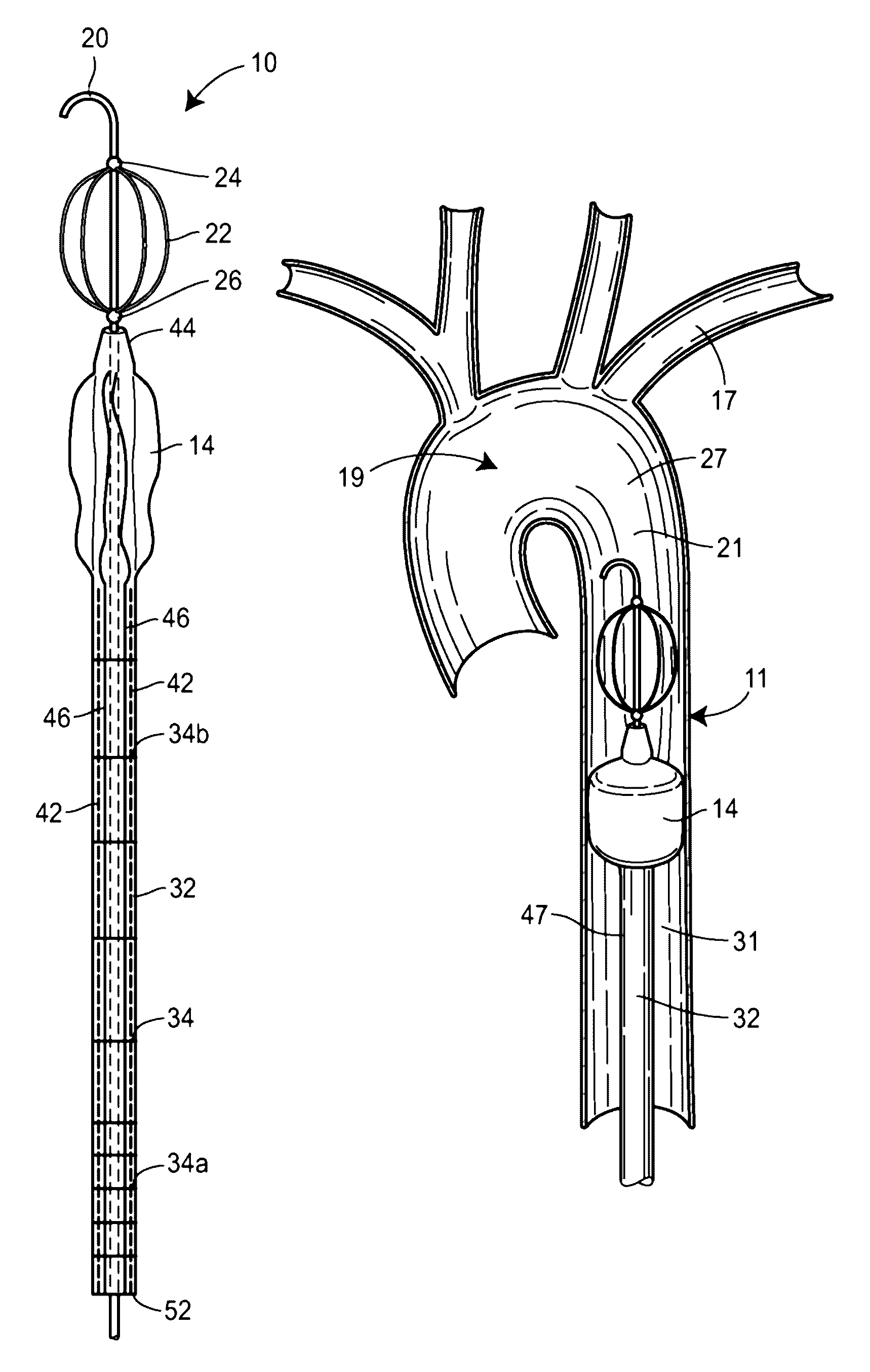
Also, the delivery shafts of currently available balloons are too flexible to remain in position without a supporting sheath.
Further, the balloons mentioned as examples above do not have a mechanism for safeguarding from over-inflation, which is why each must be inflated while being directly visualized using x-ray or fluoroscopy to prevent aortic rupture.
However, there is nothing that prevents over-pressurization of the internal aortic balloon.
The conventional technique of balloon occlusion is also limited by reliance upon x-ray or fluoroscopy to deliver and inflate the balloon within the correct position.
However, fluoroscopy is required to use this balloon catheter for such procedures.
Thus, the requirement of x-ray or fluoroscopy to use currently available balloon occlusion systems restricts performance of this procedure to fixed operating rooms with C-arm capabilities or fixed imaging suites, both of which are typically not available in trauma or emergency settings.
Although fluoroscopy affords visualization of endovascular procedures, the need for this modality carries a significant burden.
Specifically, fluoroscopic imaging is costly and its requirement severely limits where catheter-based endovascular procedures can be performed and who can perform them.
The requirement for fluoroscopy means that valuable and potentially lifesaving interventions can only be performed by a select number of trained providers in adequately equipped facilities often hours from a point of injury.
Even routine or elective endovascular procedures may be delayed as they compete in a resource limited environment among a pool of procedures to be completed using fluoroscopic equipment in the intensive care unit, operating room or endovascular imaging suite.
In addition, in emergency, intensive care or surgical environments, fluoroscopy is often not readily available or the environments in which the patients are positioned, e.g., an intensive care unit (ICU) bed or operating room (OR) table, are not specifically made for imaging, thereby impeding the use of fluoroscopy.
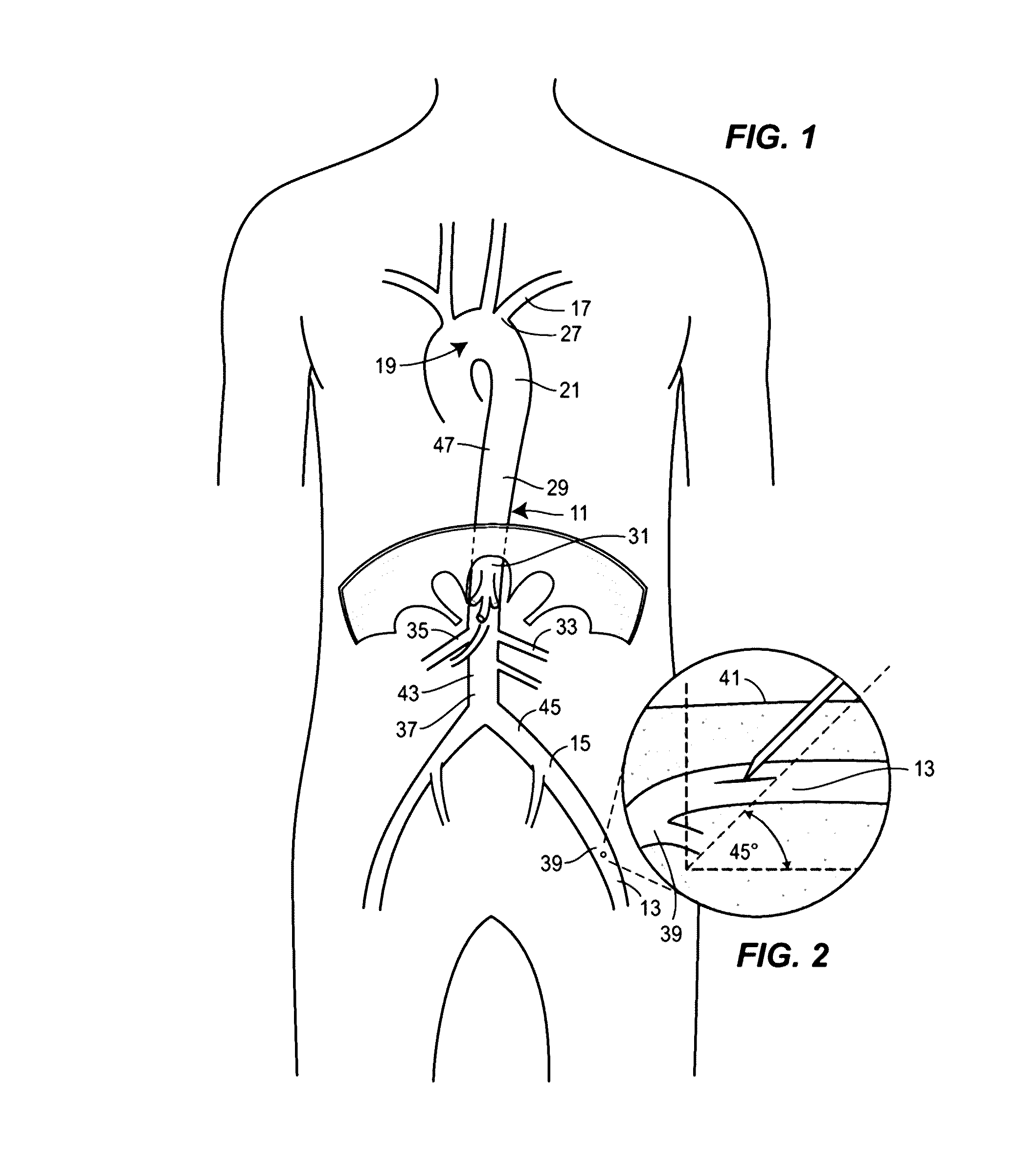
However, there is no similar device for adult torso vascular anatomy, i.e. morphometry, which will facilitate or guide endovascular procedures of the torso.

Method used


Image

  • Fluoroscopy-independent balloon guided occlusion catheter and methods
  • Fluoroscopy-independent balloon guided occlusion catheter and methods
  • Fluoroscopy-independent balloon guided occlusion catheter and methods

Examples


Embodiment Construction

[0035]Using a sufficiently broad pool of human patients from which statistically reliable data may be drawn, it is possible to mathematically derive a correlation (i.e., nomogram) between readily measurable external torso landmarks and the dimensions of the human aorta within the abdomen and thorax. As used herein, the term nomogram includes one or more tables, charts, graphs, or other visual depiction of a correlation of data. More specifically, it is possible to define, using this easily discernable and consistently located external measure of torso extent, the anticipated lengths or distances of arterial anatomy, i.e. arterial morphometry, between functionally important locations within the torso. This mathematical correlation or nomogram will allow determination of the appropriate distance with which to insert an endovascular wire and aortic occlusion balloon into the torso aorta without the need for fluoroscopy (x-ray). In other words, the nomogram will allow a rapid measure of...


PUM


Abstract

A system for deploying and selectively inflating a balloon at a desired location, without the aid of fluoroscopy is described.

Description

STATEMENT REGARDING FEDERALLY SPONSORED RESEARCH OR DEVELOPMENT[0001]This invention was made with government support under Contract No. W81XWH-12-1-0558 awarded by U.S. Army Medical Research Materiel Command. The government has certain rights in the invention.FIELD OF THE DISCLOSURE[0002]This disclosure relates generally to occlusion systems.BACKGROUND OF THE INVENTION[0003]Non-compressible sites of torso vascular injury remain one of the leading causes of potentially preventable death in both active duty troops during wartime conflict and in civilian trauma centers. An example of this type of torso vascular injury is a gunshot wound to the abdomen with a central site of bleeding and a patient in shock. Unlike an extremity injury, wherein a tourniquet could be used for vascular control or direct pressure could be held at select arterial pressure points, vascular injuries to the torso require surgical exposure followed by the often difficult application of vascular clamps for hemorrh...

Claims


Application Information

Patent Timeline
Patent Type & Authority Patents(United States)
IPC IPC(8): A61M29/00A61M25/10A61B17/12A61M25/00A61M25/06A61M25/09A61B17/22
CPCA61M25/1011A61B17/12109A61B17/12136A61M25/0097A61B2017/22048A61B2090/061A61B2090/064A61M25/005A61M25/1018A61M2025/0002A61M2025/0681A61M2025/09175A61M2025/1047A61M2025/1052A61M2025/1095A61M25/10188
Inventor FRANKLIN, CURTIS
Owner PRYTIME MEDICAL DEVICES INC
Why Eureka
Try Eureka