
Transvaginal specimen extraction device

a transvaginal and specimen technology, applied in the field of laparoscopic instruments and use, can solve the problems of increasing costs, unable to remove large specimens, and removing pathologic specimens that are larger than port sites, and achieve the effect of effective specimen removal

Inactive Publication Date: 2017-10-17
UNIV OF SOUTH FLORIDA
View PDF13 Cites 30 Cited by
  • Summary
  • Abstract
  • Description
  • Claims
  • Application Information

AI Technical Summary

Benefits of technology

Enables minimally invasive surgery with enhanced access to the abdominal cavity, allowing for the safe removal of large specimens without morcellation or large incisions, reducing recovery time and minimizing scarring and complications.

Problems solved by technology

One limitation, however, has been the removal of pathologic specimens that are larger than the port sites used to perform these surgeries.
In abdominal laparoscopy, it is impossible to remove large specimens without cutting or morcellating the specimen within the abdominal cavity or making an incision in the abdominal wall that is large enough to accommodate removal of the large specimen.
NOTES has been used for diagnostic and therapeutic procedures including) organ removal, though current articulating instruments for use with NOTES are disposable, increasing costs compared to standard laparoscopic procedures, and removal of large tumors or solid organs cannot be performed using NOTES (Dapri, Single access laparoscopic surgery: Complementary or alternative to NOTES?
Another limitation in traditional laparoscopic surgery is the size of the instruments used.
Larger incisions lead to more scarring and the potential for hernia formation.
However, to date, there are no devices marketed in the U.S. aimed at utilizing the vagina as an access to the peritoneal cavity for the introduction of laparoscopic surgical devices or implants, or the extraction of pathologic specimens.

Method used


Image

  • Transvaginal specimen extraction device
  • Transvaginal specimen extraction device
  • Transvaginal specimen extraction device

Examples


example

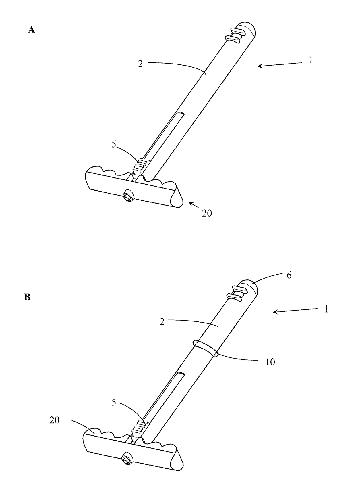
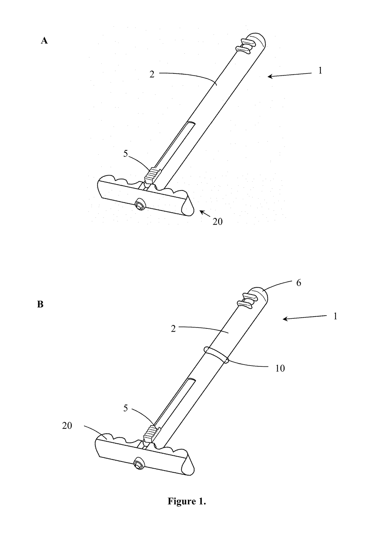
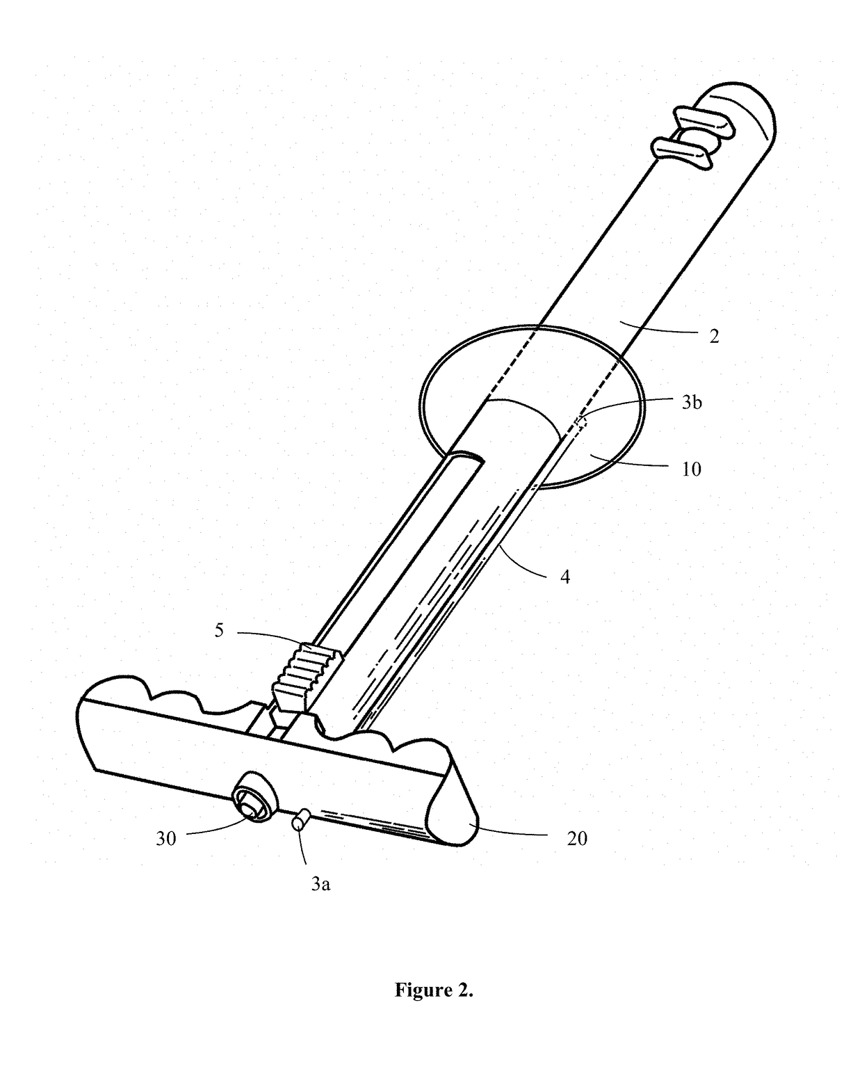
[0067]Laparoscopic instrument 1 optionally comprises a curved sheath 2, comprising interstitial space 60, seen in FIG. 11. Instrument channel 65 is disposed in curved sheath 2, and is adapted to accept laparoscopic surgical tools, such as instruments, implants, sponges, needles or other objects. The instrument channel is fixed within interstitial space 60 on the proximal and distal ends of the instrument channel to the interior walls of curved sheath 2 by means known in the art, such as thermal welding. Alternatively, instrument channel 65 is formed in a solid curved sheath 2, i.e. the instrument channel forms interstitial space 60, as seen in FIG. 12. Instrument seal 50 is disposed on the proximal end of instrument channel 65 and forms a seal against any laparoscopic tools used, thereby maintaining pneumoperiotoneum. Instrument channel 65 may have a curve or be straight, fitting in sheath 2 as seen in FIG. 12, and may end in the distal end of sheath 2 or on the wall of the sheath 2...


PUM


Abstract

In laparoscopic surgery, small (5-12 mm diameter) incisions are made in an abdominal wall through which instruments dissect and remove specimens that may be several centimeters in diameter. Removal of a sample typically requires either enlarging these incisions or morcellating the sample to pass through sub-centimeter ports. A laparoscopic device permits extraction of the sample to be removed in a female using a vagina, which has sufficient elasticity to accommodate removal of large specimens. A posterior portion of the vagina communicates to an abdomen through a few tissue layers, and is distant from vital anatomic structures. Utilizing the vagina is optimal due to its ease of access to the abdomen and repair, minimal scarring and post-operative pain, and faster recovery following surgery. A deployable collection bag is housed in a sheath, which is deployed into the vagina or an abdominal cavity to extract a large (multiple-centimeter) specimen(s) through the vagina. An optional insufflation system and an inflatable balloon to maintain a pneumoperiotoneum may be used to reduce a number of laparoscopic ports required.

Description

CROSS-REFERENCE TO RELATED APPLICATIONS[0001]This application is a continuation of and claims priority to International Patent Application No. PCT / US2012 / 070147, filed on Dec. 17, 2012 which claims priority to U.S. Provisional Patent Application No. 61 / 576,759, entitled, “Transvaginal Specimen Extraction Device,” filed on Dec. 16, 2011, the contents of which are herein incorporated by reference.FIELD OF THE INVENTION[0002]This invention relates to laparoscopic instrumentation and use. Specifically, the invention discloses an endoscopic instrument for transvaginal surgery.BACKGROUND OF THE INVENTION[0003]Laparoscopic surgical techniques have been developed in order to avoid large skin incisions associated with traditional surgery, using small incisions (of 5-12 mm) in which surgical instruments are inserted. These surgical instruments may be used to dissect and remove tissues and organs (specimens) which may be several centimeters in diameter. Such minimally invasive surgical techniq...

Claims


Application Information

Patent Timeline
Patent Type & Authority Patents(United States)
IPC IPC(8): A61M13/00A61B10/04A61B17/42A61B17/00A61B17/34A61M25/00
CPCA61M13/003A61B10/04A61B17/42A61M25/007A61M25/0082A61B2017/00287A61B2017/00424A61B2017/345A61M2025/0096A61M2210/1475
Inventor HART, STUART RICHARDHIPOL, PHILIP JAMESSIMOES, MARIO ALVESZAKARIA, MARK ANTOINE
Owner UNIV OF SOUTH FLORIDA
Learn More