Suppression of malignant mesothelioma by overexpression or stimulation of endothelial protein C receptors (EPCR)

a technology of endothelial protein and c receptor, which is applied in the field of biochemistry, molecular biology and medicine, can solve the problems of failure to induce an aggressive phenotype, achieve the effects of reducing tumor growth, attenuating tumorigenicity, and reducing tumor growth

Active Publication Date: 2017-11-07
BOARD OF RGT THE UNIV OF TEXAS SYST
View PDF5 Cites 0 Cited by
  • Summary
  • Abstract
  • Description
  • Claims
  • Application Information

AI Technical Summary

Benefits of technology

[0011]Malignant pleural mesothelioma (MPM) cells that express TF and PAR1 but not PAR2 generate large tumors in the thoracic cavity. Suppression of either TF or PAR1 reduces tumor growth in this model. However, overexpression of TF in less aggressive MPM cells that lack TF but express PAR1 failed to induce an aggressive phenotype. No EPCR expression was found in aggressive MPM cells whereas abundant EPCR expression was found in non-aggressive MPM cells. Introduction of EPCR expression to aggressive MPM cells by EPCR knock-in completely attenuated their tumorigenicity whereas the knock-down of EPCR expression in non-aggressive MPM cells that expressed TF markedly increased the tumorigenicity. This is the first discovery that EPCR acts as a tumor suppressor in MPM.
[0014]Conversely, silencing of EPCR in non-aggressive MPM cells, which constitutively express EPCR, transformed them to become highly tumorigenic by suppressing tumor cell apoptosis and promoting cell proliferation, generating aggressively growing tumors.
[0018](4) TF-driven tumor growth of MPM depends upon PAR1-mediated signaling, shown by the knock-down of PAR1 in REN cells markedly reducing their tumorigenicity.
[0019](5) Conferring EPCR expression on REN cells markedly reduced the number of tumors they formed, and the few tumors that were formed remained very small and nonadherent.
[0020](6) Knock-down of EPCR expression in TF-overexpressing MS-1 and M9K cells dramatically increased the tumorigenicity of these non-aggressive MPM cells.

Problems solved by technology

However, overexpression of TF in less aggressive MPM cells that lack TF but express PAR1 failed to induce an aggressive phenotype.

Method used


Image

  • Suppression of malignant mesothelioma by overexpression or stimulation of endothelial protein C receptors (EPCR)
  • Suppression of malignant mesothelioma by overexpression or stimulation of endothelial protein C receptors (EPCR)
  • Suppression of malignant mesothelioma by overexpression or stimulation of endothelial protein C receptors (EPCR)

Examples


example i

Materials and Methods

Cell Lines and Reagents

[0201]REN cells were from S. Albelda, University of Pennsylvania, MS-1 cells were from S-M. Hsu, University of Texas Health Science Center at Houston, and M9K cells were from Dr. B. Gerwin, NIH. All cells were grown in RPMI medium+10% fetal bovine serum and 1% penicillin / streptomycin. Preparation of monospecific polyclonal anti-human TF IgG was described earlier (32). EPCR antibodies were obtained from C. T. Esmon, Oklahoma Medical Research Foundation. PAR1 and PAR2 antibodies were from Santa Cruz Biotechnology (Santa Cruz, Calif.). PAR1 and PAR2 agonist peptides were from Biosynthesis, Lewisville, Tex. Recombinant human FVIIa was obtained from Novo Nordisk (Maaloev, Denmark). Factor X was obtained from Enzyme Research Laboratories (South Bend, Ind.).

Generation of Stable Transfectants of MPM Cells Expressing / Lacking TF, EPCR or PAR1

[0202]TF or PAR1 expression in REN MPM cells was selectively knocked-down by shRNA. shRNA clones were generat...

example ii

Status of TF, EPCR, PAR1 and PAR2 Expression Levels in MPM Cells

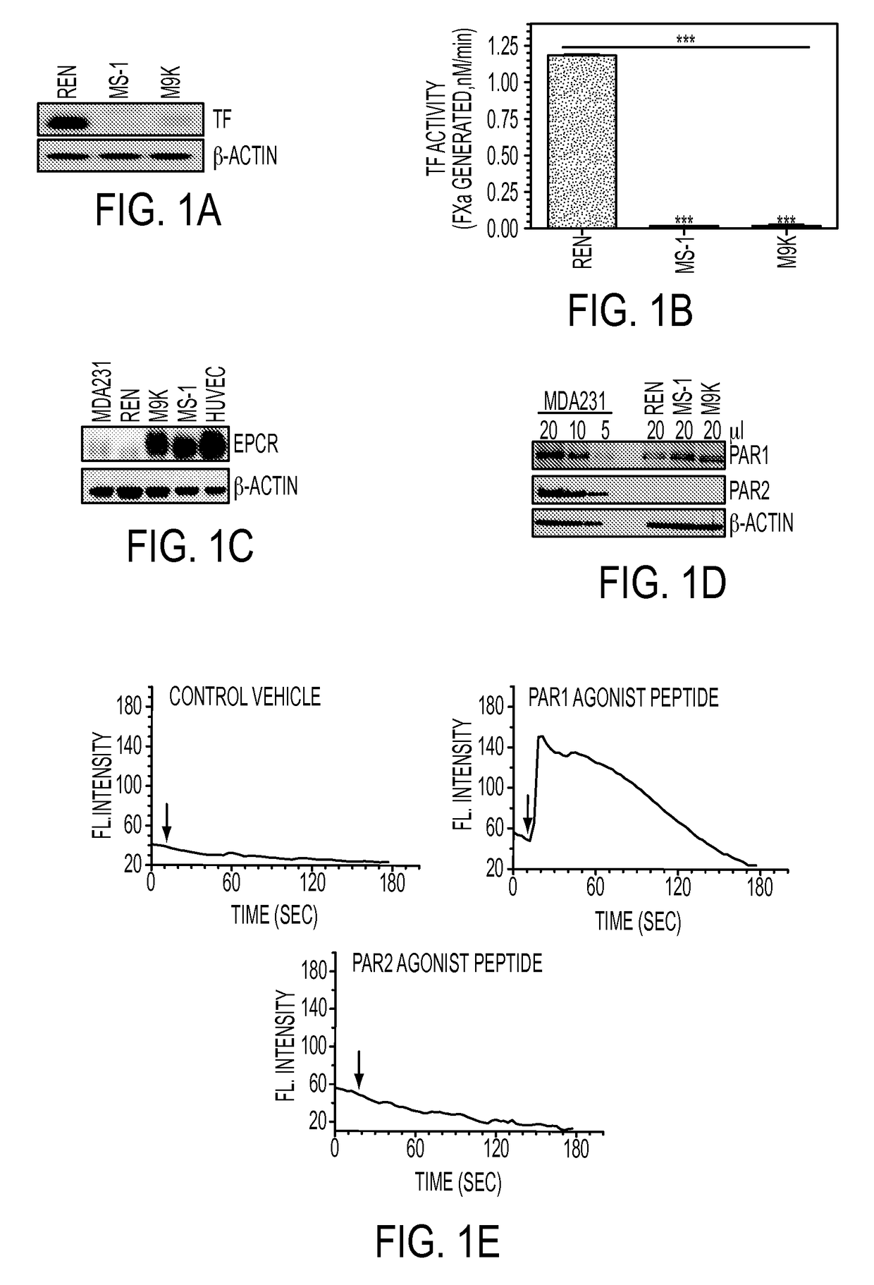
[0211]The expression levels of TF, EPCR, PAR1 and PAR2 in REN, MS-1 and M9K MPM cells were analyzed by Western blot and functional analyses. TF expression was markedly higher in REN MPM cells compared to MS-1 and M9K MPM cells (FIGS. 1A and B). TF expression was barely detectable in MS-1 and M9K cells. In contrast to TF expression, REN MPM cells express very little EPCR whereas both MS-1 and M9K cells abundantly express EPCR, at levels found in endothelial cells (FIG. 1C). Western blot analysis revealed that all three MPM cell types express PAR1 whereas PAR2 expression was undetectable (FIG. 1D). Consistent with the antigen results, a PAR1 but not PAR2 agonist peptide induced intracellular Ca2+ release in REN MPM cells (FIG. 1E). A similar pattern of Ca2+ release was observed in MS-1 and M9K MPM cells in response to PAR1 or PAR2 agonist peptides (results not shown).

example iii

REN MPM Cells Generate Large Invasive Tumors and Knock-Down of TF Reduces Tumorigenicity of REN Cells

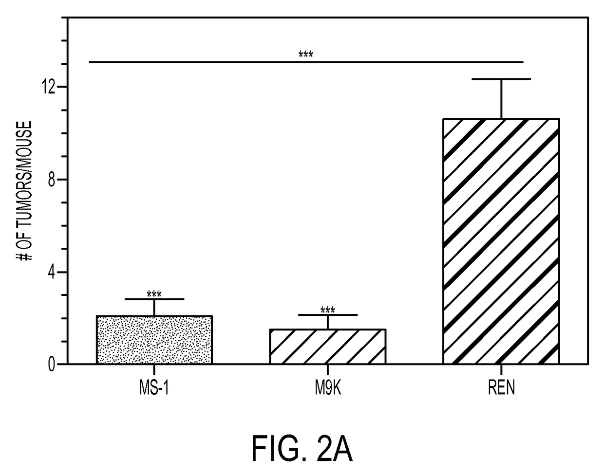
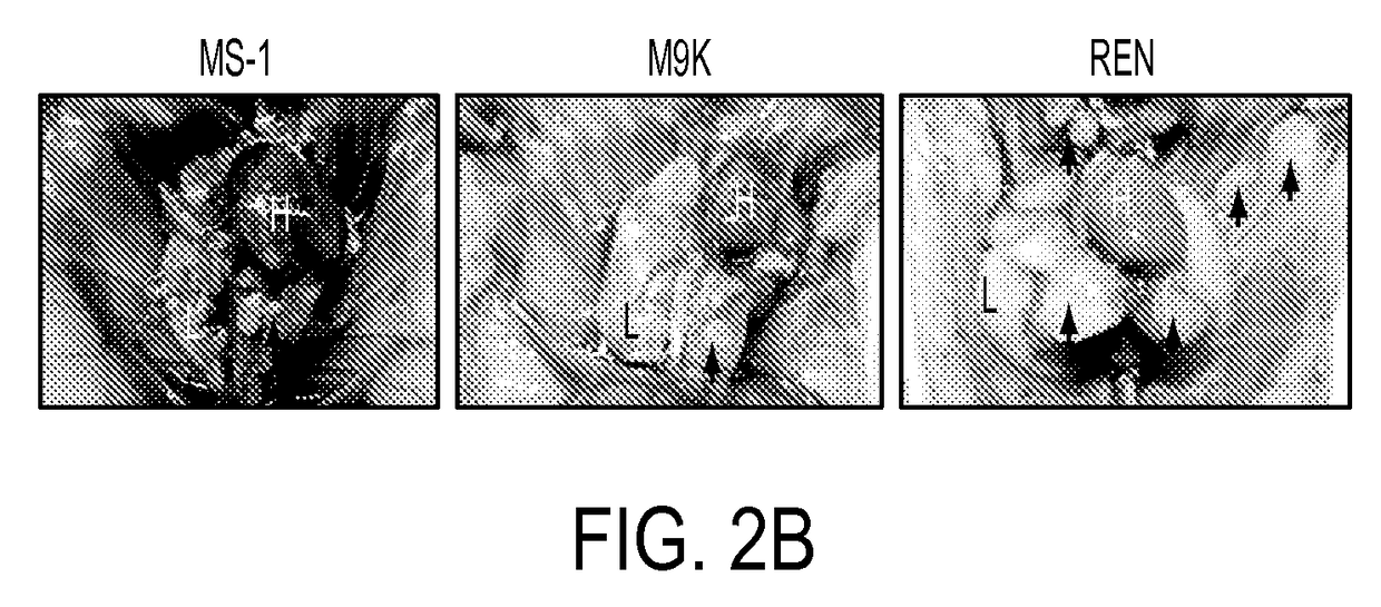
[0212]As reported previously, implantation of REN MPM cells into the thoracic cavity of nude mice resulted in multiple large tumors (>2 mm) within the thoracic cavity (FIG. 2A). The number of tumors in each mouse varied from 6 to 18. Some of the tumors approximated the size of the heart (FIG. 2B). All tumors were limited to the thoracic cavity. These tumors were highly invasive and often penetrated deep into intercostal tissues on which they were attached (FIGS. 2C and 2D). There was no evidence for metastasis as we found no tumors in distant organs such as liver. A variable number of very small tumors (<2 mm) found in the thoracic cavity may reflect dispersed initial seeding of tumor cells than pleural metastases. In contrast to REN, MS-1 and M9K cells in nude mice produced relatively few tumors and some of the mice developed no tumors at all. Most of the tumors that were generated ...


PUM


Abstract

The influence of TF, endothelial cell protein C receptor (EPCR) and protease activated receptor-1 (PAR1) on tumor growth of malignant pleural mesothelioma (MPM) is disclosed. MPM cells that lack or express TF, EPCR or PAR1 and a murine orthotopic model of MPM led to the discovery that intrapleural administration into nude mice of REN MPM cells expressing TF and PAR1 but lacking EPCR and PAR2 generated large pleural cavity tumors. Suppression of TF or PAR1 expression markedly reduced tumor growth. Overexpression of TF in non-aggressive MPM cells expressing EPCR and PAR1 but exhibiting minimal levels of TF failed to alter their tumorigenicity. Introduction of EPCR expression in aggressive MPM cells attenuated tumor growth whereas EPCR silencing in non-aggressive MPM cells overexpressing TF increased tumorigenicity of non-aggressive cells. Expression of EPCR by MPM cells suppresses tumor growth and treats MPM.

Description

BACKGROUND OF THE INVENTION[0001]Field of the Invention[0002]The invention in the field of biochemistry, molecular biology and medicine relates to the suppression of growth and metastasis of malignant pulmonary mesothelioma (MPM) cells and the treatment of MPM using nucleic acid vectors, or endothelial protein C receptor (EPCR) agonists in the form of polypeptides, peptides or small organic molecules.[0003]Description of the Background Art[0004]It has been well recognized for many decades that tumors dramatically increase the risk for hemostatic abnormalities such as disseminated intravascular coagulation, pulmonary and venous thromboembolism (1-3). Prior studies have also established that hemostatic factors play a major role in cancer biology, particularly in tumor dissemination and metastasis (4-6). Tumor cell-associated tissue factor (TF) is known to contribute to tumor growth and progression either directly by TF-FVIIa or TF cytoplasmic tail-mediated cell signaling (7-9) or indi...

Claims


Application Information

Patent Timeline
Patent Type & Authority Patents(United States)
IPC IPC(8): A61K38/17A61K31/5377A61K48/00C12N15/113A61K9/00A61K38/21A61K38/19A61K31/4439
CPCA61K38/177A61K9/007A61K9/0019A61K31/4439A61K31/5377A61K38/191A61K48/005A61K48/0058C12N15/113A61K38/217C12N2710/10371C12N2310/14C12N2310/531C12N2710/10343A61P35/00
Inventor PENDURTHI, USHA R.LELLA, VIJAYA M. R.GADDAM, SHIVAKESHAVAIDELL, STEVEN
Owner BOARD OF RGT THE UNIV OF TEXAS SYST
Why Eureka
Try Eureka