
Method and system for automatic estimation of shear modulus and viscosity from shear wave imaging

a technology of shear wave imaging and automatic estimation, which is applied in the field of medical image-based estimation of mechanical tissue properties, can solve the problems of important challenges in accurate quantitative estimation of tissue elasticity and viscosity from swi

Active Publication Date: 2017-11-14
SIEMENS MEDICAL SOLUTIONS USA INC
View PDF10 Cites 2 Cited by
  • Summary
  • Abstract
  • Description
  • Claims
  • Application Information

AI Technical Summary

Problems solved by technology

However, accurate quantitative estimation of tissue elasticity and viscosity from SWI remains an important challenge.

Method used


Image

  • Method and system for automatic estimation of shear modulus and viscosity from shear wave imaging
  • Method and system for automatic estimation of shear modulus and viscosity from shear wave imaging
  • Method and system for automatic estimation of shear modulus and viscosity from shear wave imaging

Examples


first embodiment

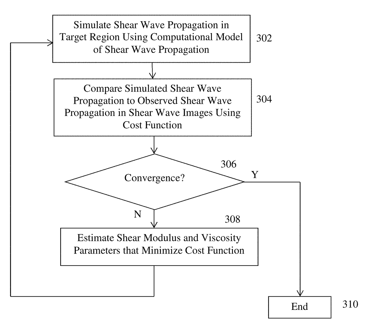
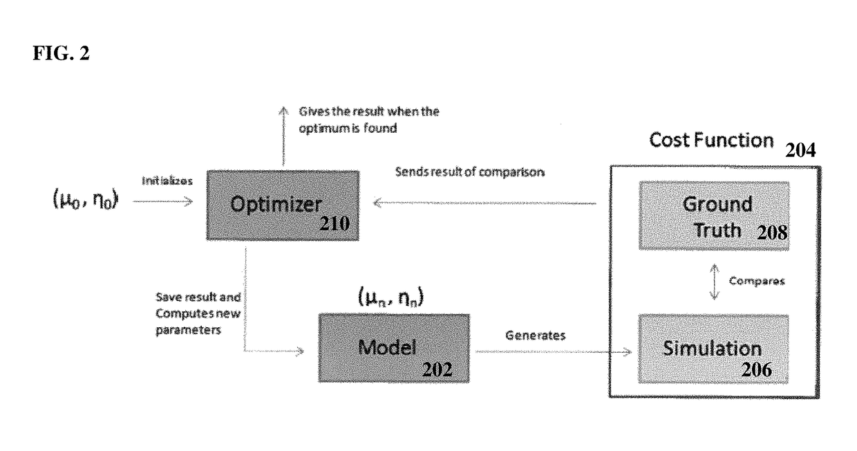
[0020]At step 304, the simulated shear-wave propagation is compared to observed shear-wave propagation in the shear-wave images using a cost function. The shear modulus and viscosity are estimated by minimizing a cost function that calculates a similarity (or difference) between the computed (simulated) shear-wave propagation and the measured (observed) shear-wave propagation in the shear-wave images. In a first embodiment, the computed shear-wave propagation is directly compared with the observed shear-wave displacement in the shear wave images. According to an advantageous implementation, normalized cross correlation (NCC) can be used as a cost function that measures a similarity between the computed and observed shear-wave propagation. However, the present invention is not limited to NCC and any other cost function, such as sum of squared distance, can be similarly employed to measure the similarity between the computed and observed shear-wave propagation. The NCC cost function c...

second embodiment

[0023]In a second embodiment, as an alternative to the cost function directly comparing the simulated and observed shear-wave propagation, indirect estimation can be performed working directly in the radiofrequency (RF) space. Intuitively, shear-wave displacement is captured by a shift in the radiofrequency signal. By tracking this shift over time, shear-wave displacement can be estimated and then displayed to the user. In this mode, shear modulus and viscosity can be estimated by minimizing the differences between the measured RF shift and a computed RF shift obtained as the difference in the amplitude of the simulated shear displacement at a specific location. One advantage of working directly in the RF space is that the cost function is not affected by any post-processing done on the RF signal when estimating shear-wave displacement. Another advantage of working directly in the RF space is that the measured shear-wave displacement can be automatically smoothed through the fitted ...


PUM


Abstract

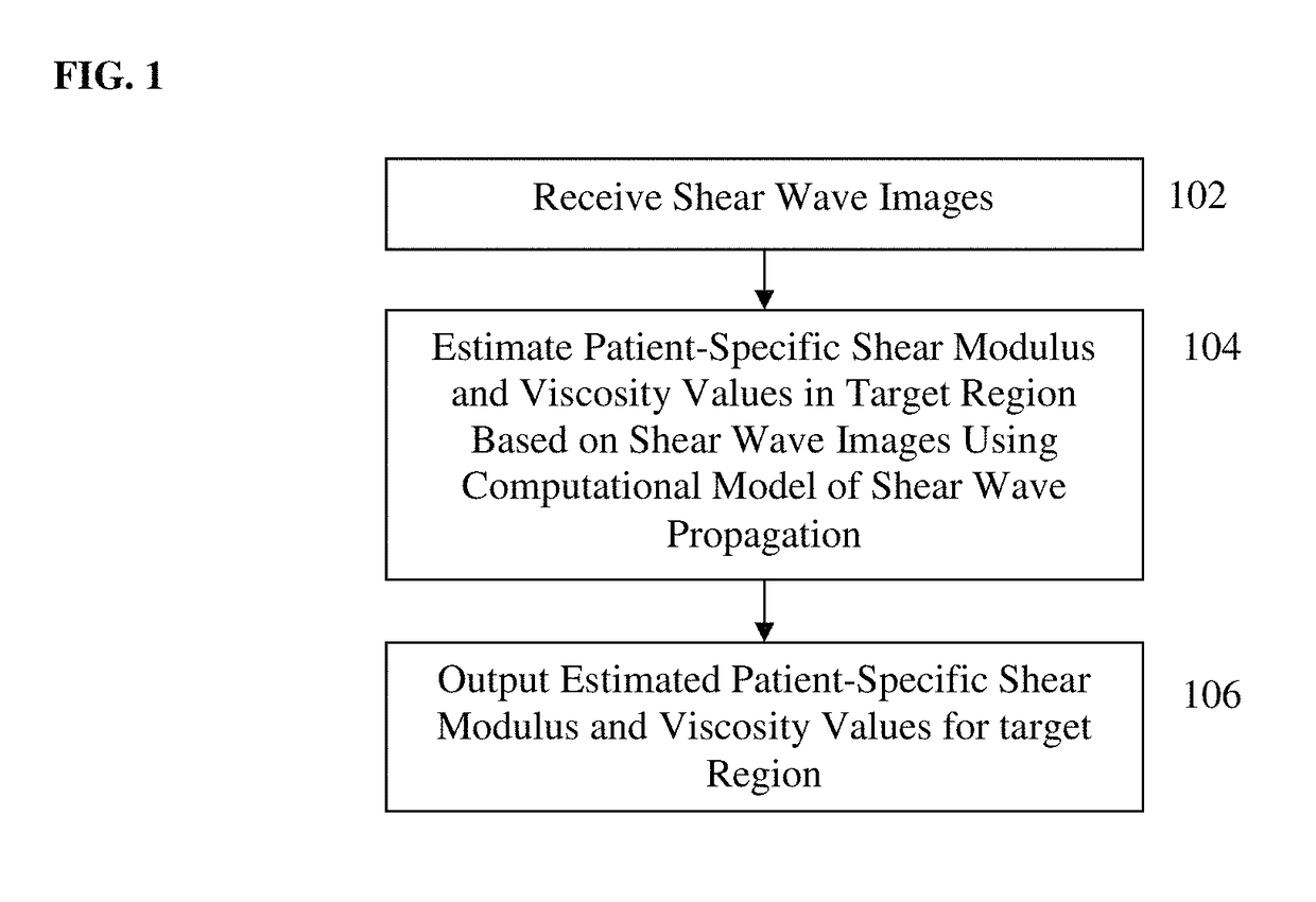
A method and system for automatic non-invasive estimation of shear modulus and viscosity of biological tissue from shear-wave imaging is disclosed. Shear-wave images are acquired to evaluate the mechanical properties of an organ of a patient. Shear-wave propagation in the tissue in the shear-wave images is simulated based on shear modulus and viscosity values for the tissue using a computational model of shear-wave propagation. The simulated shear-wave propagation is compared to observed shear-wave propagation in the shear-wave images of the tissue using a cost function. Patient-specific shear modulus and viscosity values for the tissue are estimated to optimize the cost function comparing the simulated shear-wave propagation to the observed shear-wave propagation.

Description

BACKGROUND OF THE INVENTION[0001]The present invention relates to medical image-based estimation of mechanical tissue properties and more particularly to automatic estimation of shear modulus and viscosity of tissue from shear wave imaging.[0002]Shear-wave imaging (SWI) is an ultrasound-based imaging modality that can provide important information about tissue structure and integrity. In SWI, an ultrasound probe is used to generate and track propagating shear waves in biological tissue. Radiation force from an acoustic radiation force impulse (ARFI) generates the shear waves in the tissue. The velocity of the shear wave propagation in the tissue is measured and a time sequence of images showing the displacement of the shear wave is captured by the ultrasound probe.[0003]Qualitative metrics have been derived for SWI, for example to highlight stiffer or softer tissues in a region of interest. Clinical applications for SWI are numerous, especially in oncology where SWI can be used to d...

Claims


Application Information

Patent Timeline
Patent Type & Authority Patents(United States)
IPC IPC(8): A61B8/00A61B8/08
CPCA61B8/485A61B8/463A61B8/5223A61B8/5269A61B8/5207A61B8/5215G16H50/30
Inventor MANSI, TOMMASORAPAKA, SAIKIRANKAMEN, ALICOMANICIU, DORINFORLOT, FRANCOISFAN, LIEXIANG
Owner SIEMENS MEDICAL SOLUTIONS USA INC
Learn More