Intracavity ultrasonic hardness colour imaging method and its coelom ultrasonic hardness imaging instrument

An intracavity ultrasound and imager technology, used in catheters, operations, etc., can solve problems such as application, and achieve the effect of preventing attenuation, avoiding damage, and facilitating accurate detection

Inactive Publication Date: 2008-04-30
黄晶
View PDF4 Cites 0 Cited by
  • Summary
  • Abstract
  • Description
  • Claims
  • Application Information

AI Technical Summary

Problems solved by technology

However, in order to realize the elastography method, a certain pressure must be applied to the inspected part. The existing technology only achieves success in applying pressure to the superficial part of the cavity, and it is completed by applying pressure on the deeper part. The elastography method has not been reported in the relevant literature

Method used


Image

  • Intracavity ultrasonic hardness colour imaging method and its coelom ultrasonic hardness imaging instrument
  • Intracavity ultrasonic hardness colour imaging method and its coelom ultrasonic hardness imaging instrument

Examples


Embodiment 1

[0031] Example 1 Intracavity Ultrasound Hardness Imaging Method

[0032] In order to achieve the above object, the technical solution one adopted in the present invention is such that the method comprises the following steps:

[0033] (1) Establish an integrated ultrasonic hardness imaging catheter equipped with a pressurized water bladder, design the high-frequency ultrasonic transducer installed in the center of the water bladder to move axially, and install a pressure sensor in the water bladder;

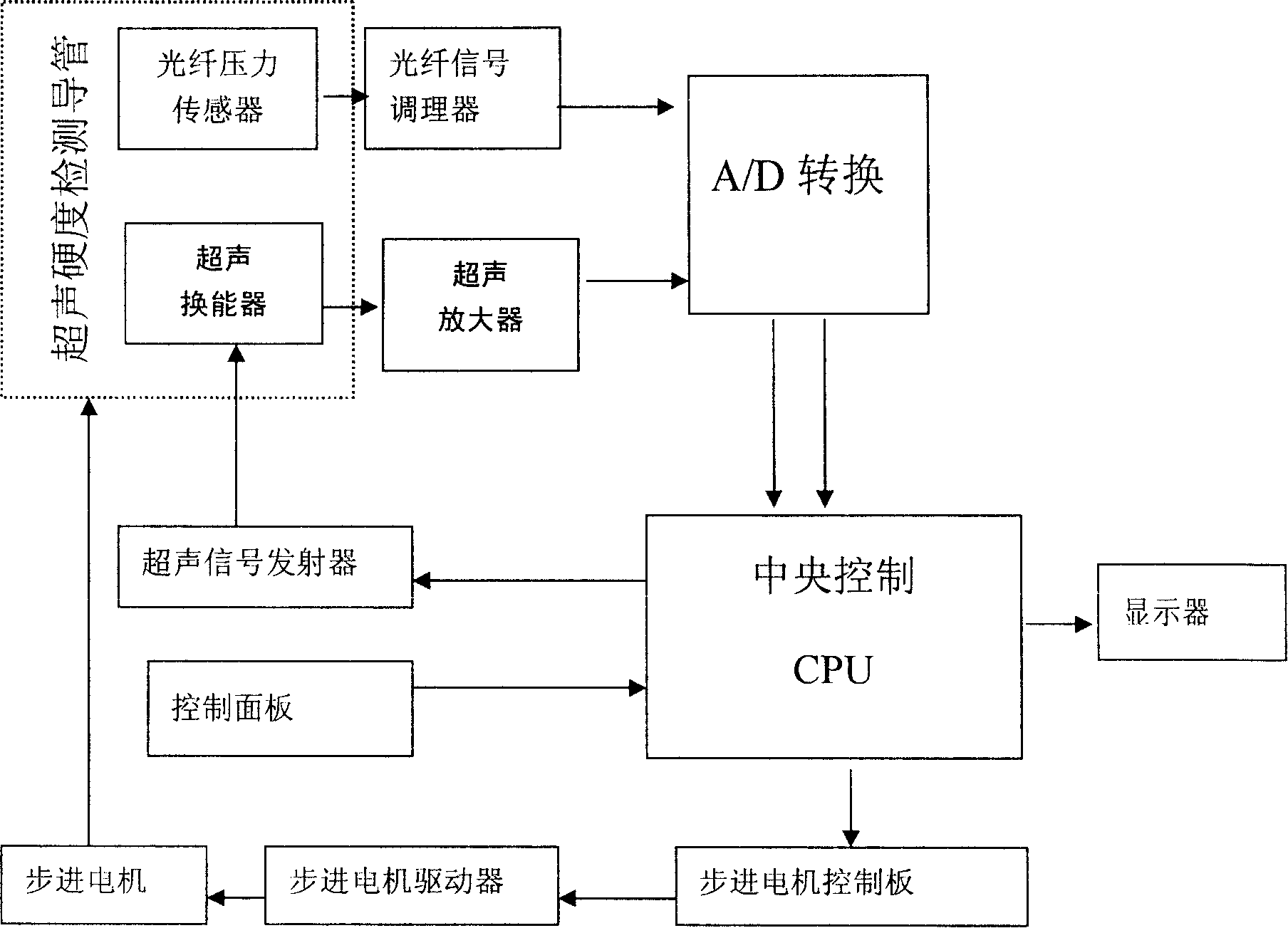
[0034](2) Extend the water bladder and the high-frequency ultrasonic transducer to the detected position in the cavity through the above-mentioned imaging catheter, fill the pressurized water bladder with anaerobic water, and perform initial circumferential pressure on the inner wall of the measured object, by The pressure sensor measures the pressure value in the column capsule; at the same time, the ultrasonic transducer on the catheter emits sound waves at the detection point,...

Embodiment 2

[0041] Example 2 The basic requirements of the intracavity hardness imaging method for the intracavity ultrasound hardness imaging catheter:

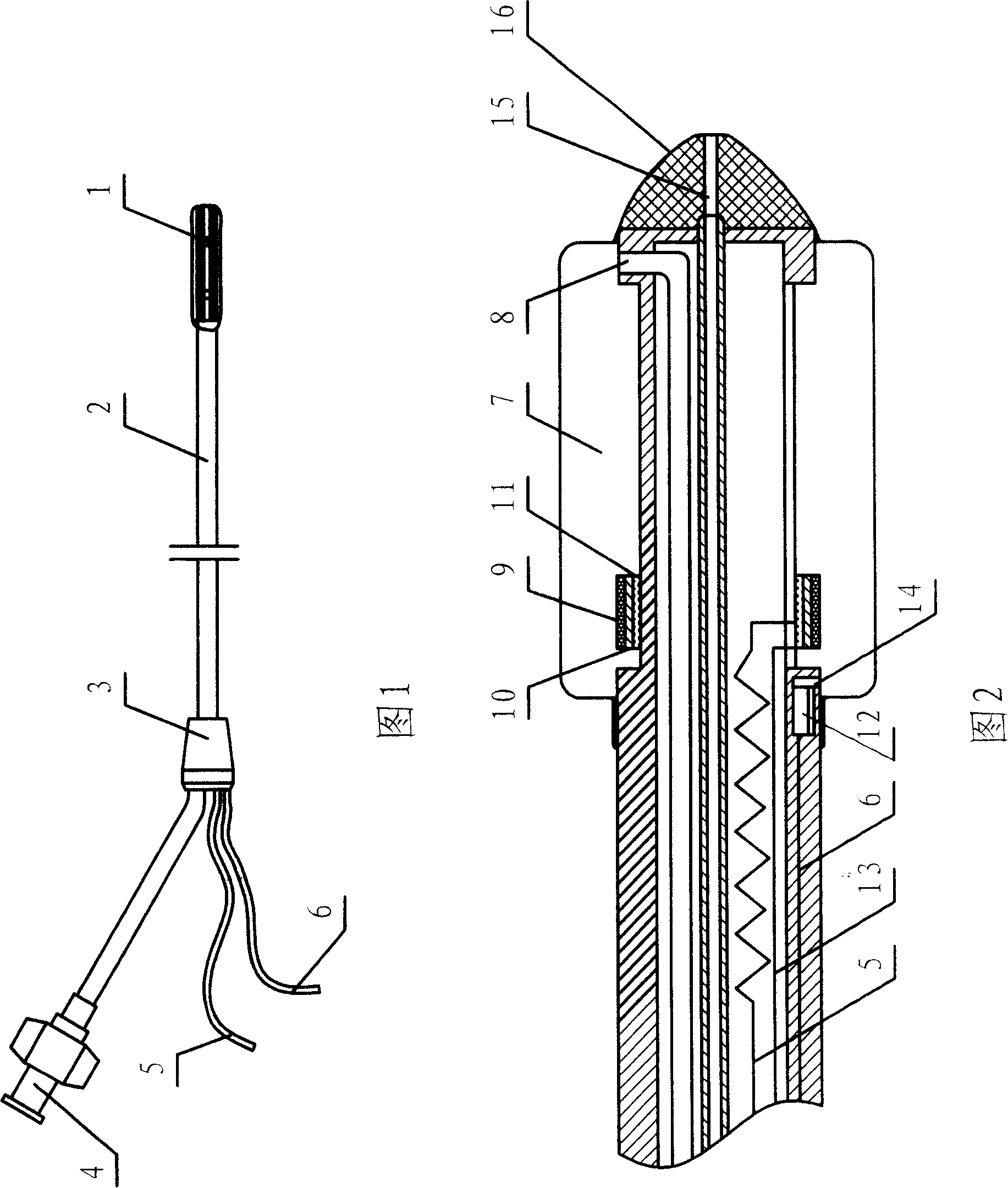
[0042] In an embodiment, the external dimension of the intracavity ultrasonic hardness imaging catheter is similar to that of a microprobe ultrasonic endoscope. The outer diameter of the catheter is 14F or larger, and the intracavity ultrasound hardness imaging catheter is sent into the body cavity. The hardness parameter of the body cavity wall at the corresponding layer is obtained by reaching different depths of the body cavity to be examined.

[0043] The top of the catheter is equipped with a ring-shaped ultrasonic transducer or a single-crystal array rotating ultrasonic transducer made of high-frequency, multi-crystal array piezoelectric ceramics that can work independently. A pressurized water bag is installed at the head end, and a high-frequency ultrasonic transducer installed in the center of the water bag can move axially, a...

Embodiment 3

[0044] Embodiment 3 Body Cavity Ultrasonic Hardness Tester

[0045] (1) The structure of the instrument and the functions of the main components

[0046] See accompanying drawings 1 and 2: the instrument includes an intracavity ultrasonic hardness imaging catheter and a host, the intracavity ultrasonic hardness imaging catheter includes a catheter and a high-frequency ultrasonic transducer installed at the front end of the catheter, the ultrasonic transducer cable, power supply The cable and the signal line are connected to the host by passing through the end of the catheter; the characteristics are: a cylindrical pressure water bladder is installed at the front end of the catheter, and a ring-shaped high-frequency ultrasonic transducer is installed on the catheter in the center of the pressure water bladder; The high-frequency ultrasonic transducer is connected with one end of the mobile control wire, and under its control, it moves axially along the catheter; the cylindrical...


PUM


Abstract

The present invention relates to an intracavitary ultrasonic hardness imaging method. Said method includes the following steps: creating and mounting high-frequency ultrasonic transducer with pressure-applying water bag and its integrated ultrasonic hardness imaging conduit which can be axially moved; utilizing said pressure-applying water bag to apply pressure to inner wall of tested object, according to two different applied pressures on some point calculating pressure variable quantity of said point; and respectively obtaining thickness variable values of said detected point when the applied pressures are different; according to said thickness variable values calculating hardness value and utilizing color hardness graph to make display. Besides, said invention also provides a body cavity ultrasonic hardness imaging apparatus for implementing said method.

Description

technical field [0001] The invention relates to a technology for obtaining information in a body cavity, and relates to the acquisition of hardness parameters of the body cavity wall of a human body and an animal, a color imaging display method and an ultrasonic diagnostic instrument. Specifically, it is a method of using an ultrasonic hardness imaging catheter to enter the body cavity to obtain and analyze the stress-strain relationship of the inner wall tissue of the body cavity, so as to obtain the hardness index of the wall tissue of the body cavity or calculate the Young's modulus of the wall tissue according to the corresponding formula, and then proceed accordingly. The computer color coding method directly displays the overall hardness and hardness distribution information of the digestive tract wall tissue. The obtained information is beneficial to assist medical research and provide reference for clinical diagnosis. The invention also relates to an ultrasonic hardne...

Claims


Application Information

Patent Timeline
Patent Type & Authority Patents(China)
IPC IPC(8): A61B8/12
Inventor 黄晶雷寒白净邓辉胜
Owner 黄晶
Why Eureka
Try Eureka