Tumour angiogenesis external co-culture model

A technology of co-cultivation of tumor blood vessels, which is applied in the field of co-culture models of microencapsulated tumor cells and endothelial cells, can solve the problems of high price and the inability of the bottom membrane to isolate molecules purposefully, and achieve the effect of reducing scientific research manpower

Inactive Publication Date: 2008-04-09
THE AFFILIATED DRUM TOWER HOSPITAL MEDICAL SCHOOL OF NANJING UNIV
View PDF0 Cites 13 Cited by
  • Summary
  • Abstract
  • Description
  • Claims
  • Application Information

AI Technical Summary

Problems solved by technology

Cells inside and outside the Transwell chamber are easy to separate, but Transwell is expensive and has not been ab

Method used


Image

  • Tumour angiogenesis external co-culture model
  • Tumour angiogenesis external co-culture model
  • Tumour angiogenesis external co-culture model

Examples


Embodiment Construction

[0030] 1. Microencapsulation of tumor cells

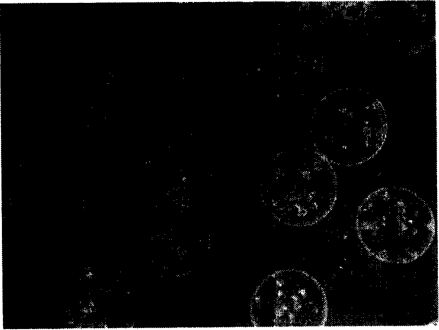
[0031] Resuscitate human liver cancer cell line HepG2 cells and inoculate them in 25ml culture flask; when the cells are 90% confluent, digest and count the cells. to 10 5 Mix a HepG2 cell with 1ml 2.0% sodium alginate solution thoroughly, spray it into 100mmol / L calcium chloride solution through a microcapsule generator to make it gel; absorb the calcium chloride solution after 10 minutes, and wash it with normal saline solution Wash 3 times, then react with 0.1% polylysine solution, wash 3 times with normal saline solution; react with 0.15% sodium alginate solution, wash 3 times with normal saline solution; Cystic core, treated for 6 minutes; washed 3 times with normal saline solution, washed once with medium, and placed at 37°C in 5% CO 2 incubator culture, such as figure 1 shown.

[0032] After 3 days, the HepG2 cells in the microcapsules can be seen to divide, and the cells enter a period of vigorous growth. The microcapsu...


PUM


Abstract

The invention relates to a co-culture model of microencapsulated tumor cells and endothelial cells. The preparation method comprises the steps: first, tumor cells are mixed into 2 percent of sodium alginate solution to lead cells suspension which is injected with 100mmol/L of calcium chloride solution to be colloidized through a microcapsule generator then; normal saline is used for washing the obtained material which then reacts with 0.1 percent of polylysine solution and is washed by normal saline; next, 0.15 percent of sodium alginate solution is added and washed by normal saline; the 55mmol/L sodium citrate solution is employed to liquefy microcapsule core and cultured in a CO2 of culture box; the microencapsulated tumor cells are directly used for being cultured or frozen reserved; the endothelial cells are employed to be adherently cultured or led to three dimensional growth; the microencapsulated tumor cells are added into the same culture solution; co-culture is done; tumor cells and endothelial cells are separated; finally, travelling gene detection, protein detection and culture liquid cytokine detection are done. The invention overcomes the limitation of Matrigel method and the defect of Transwell method, has an imitated micro-environment for microencapsulated tumor cells and endothelial cells to interact, and has long service life for one-time making and decreases research personnel and financial resources.

Description

technical field [0001] The invention relates to tumor angiogenesis, in particular to a co-cultivation model of microencapsulated tumor cells and endothelial cells. Background technique [0002] In the early stage of tumor growth, after the solid tumor grows to 1 to 2 mm, its continued growth will depend on the formation of new blood vessels. Prior to this, tumor cells themselves have released a large number of pro-angiogenic factors to induce tumor angiogenesis. These new blood vessels are the basis for material exchange between the tumor and the outside world, and angiogenesis is closely related to the development, invasion and metastasis of malignant tumors. Therefore, it is very necessary and urgent to study the mechanism of angiogenesis, especially the early tumor angiogenesis. [0003] Establishing tumor angiogenesis model is an important means to study its formation mechanism. Among them, the in vitro co-culture model is an effective method to simulate tumor angioge...

Claims


Application Information

Patent Timeline
IPC IPC(8): C12N5/06C12N5/08C12N11/04C12N5/071C12N5/09
Inventor 丁义涛方胜余德才孙喜太
Owner THE AFFILIATED DRUM TOWER HOSPITAL MEDICAL SCHOOL OF NANJING UNIV
Why Eureka
Try Eureka