Revascularization of ischemic retinal tissue and screening method therefor

A technique for retinal ischemia and retina, which is applied in the field of treatment of retinal neovascular diseases, can solve problems such as laborious, unquantified tissue, and time-consuming

Inactive Publication Date: 2008-05-14
THE SCRIPPS RES INST
View PDF0 Cites 1 Cited by
  • Summary
  • Abstract
  • Description
  • Claims
  • Application Information

AI Technical Summary

Problems solved by technology

This method is laborious, time-consuming, and comes with several difficulties, including the need to distinguish cells of abnormal blood vessels from cells of vitreous vessels in the vitreous
Because, in general, only one in every 30 serial sections is evaluated, most of the tissue is not quantified, potentially leading to large sampling errors

Method used


Image

  • Revascularization of ischemic retinal tissue and screening method therefor
  • Revascularization of ischemic retinal tissue and screening method therefor
  • Revascularization of ischemic retinal tissue and screening method therefor

Examples


Embodiment 1

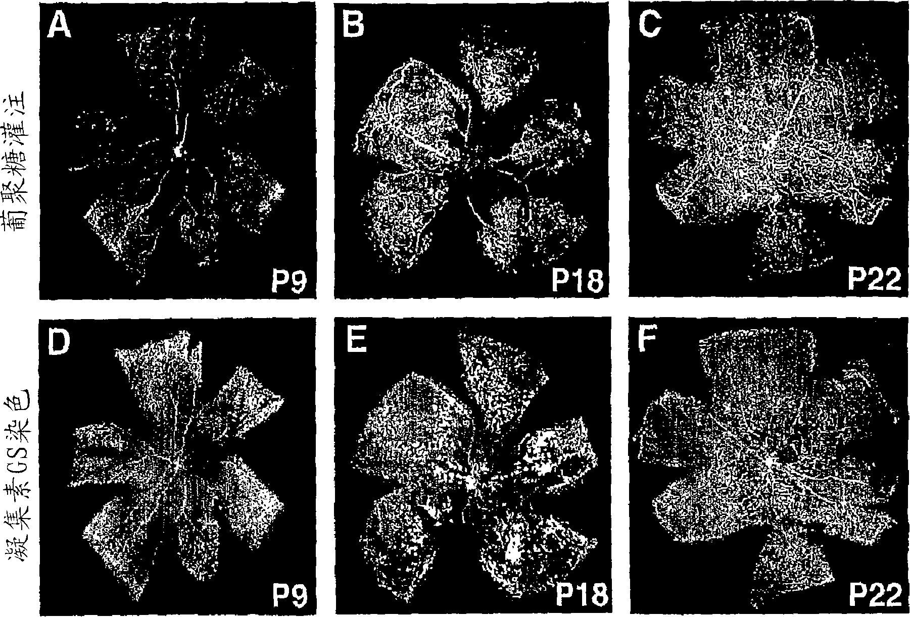
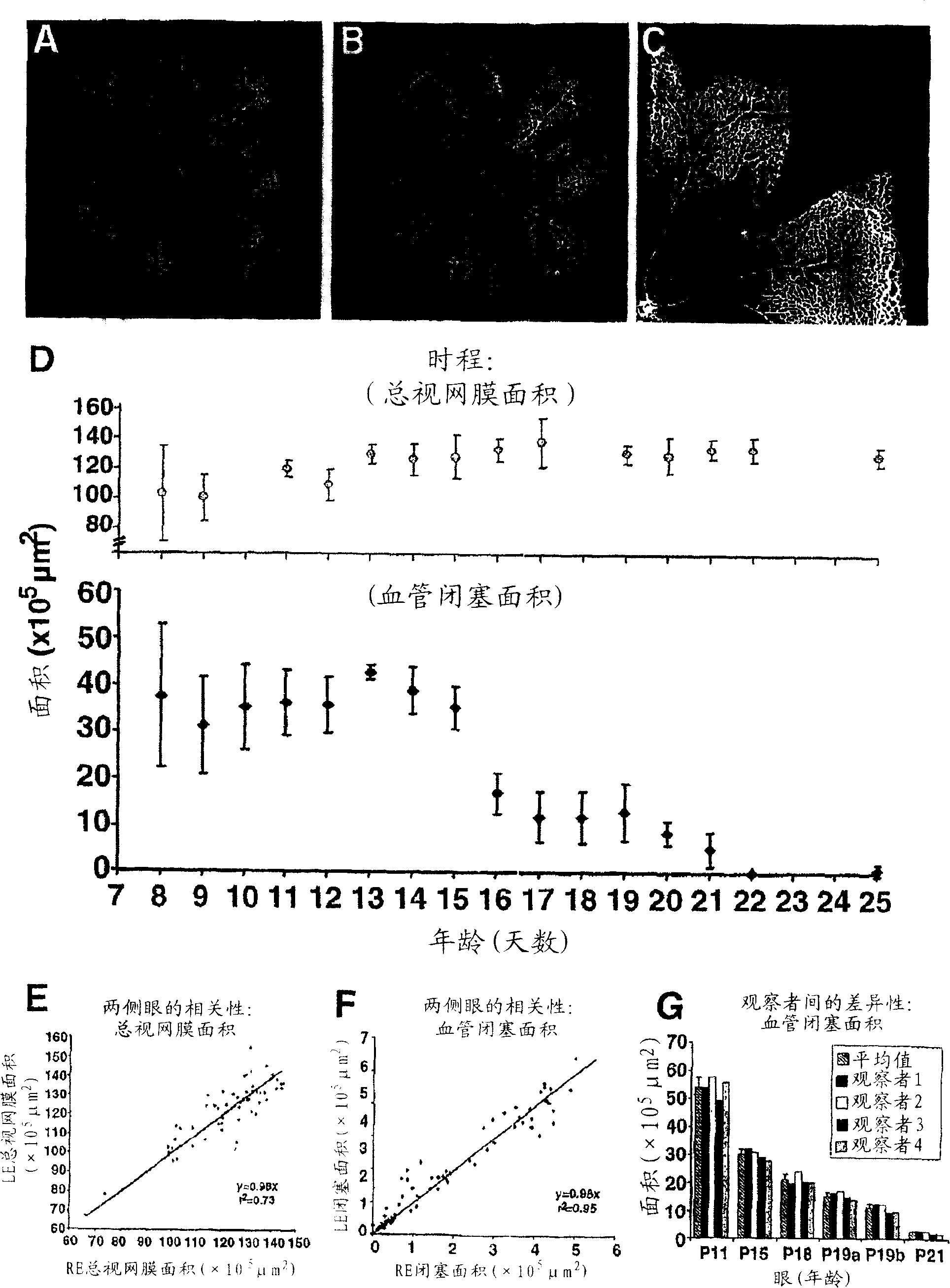
[0067] Example 1 Quantification of the area of ​​vascular occlusion and the rate of retinal vascular regeneration

[0068] To quantify the area of ​​vascular occlusion and the rate of retinal vascular regeneration, dextran-injected or isolectin-stained retinal mounts were photographed at low magnification using a fluorescence microscope. 4 overlapping photos were used to construct the retinal stitched photo ( figure 2 , inset A), occlusion area (yellow) and total retinal (blue line) area ( figure 2 , inset B). Since the border between vascular and avascular retina is usually well defined in these preparations, interobserver variability is very small ( figure 2 , inset G). Be careful not to include areas with cutting or fixation artifacts in the analysis area. The change in total retinal area over time is shown in figure 2 The upper inset D. Retinal growth appeared to continue during hyperoxia, with the total retinal area growing approximately 20% between P8 and P13. ...

Embodiment 2

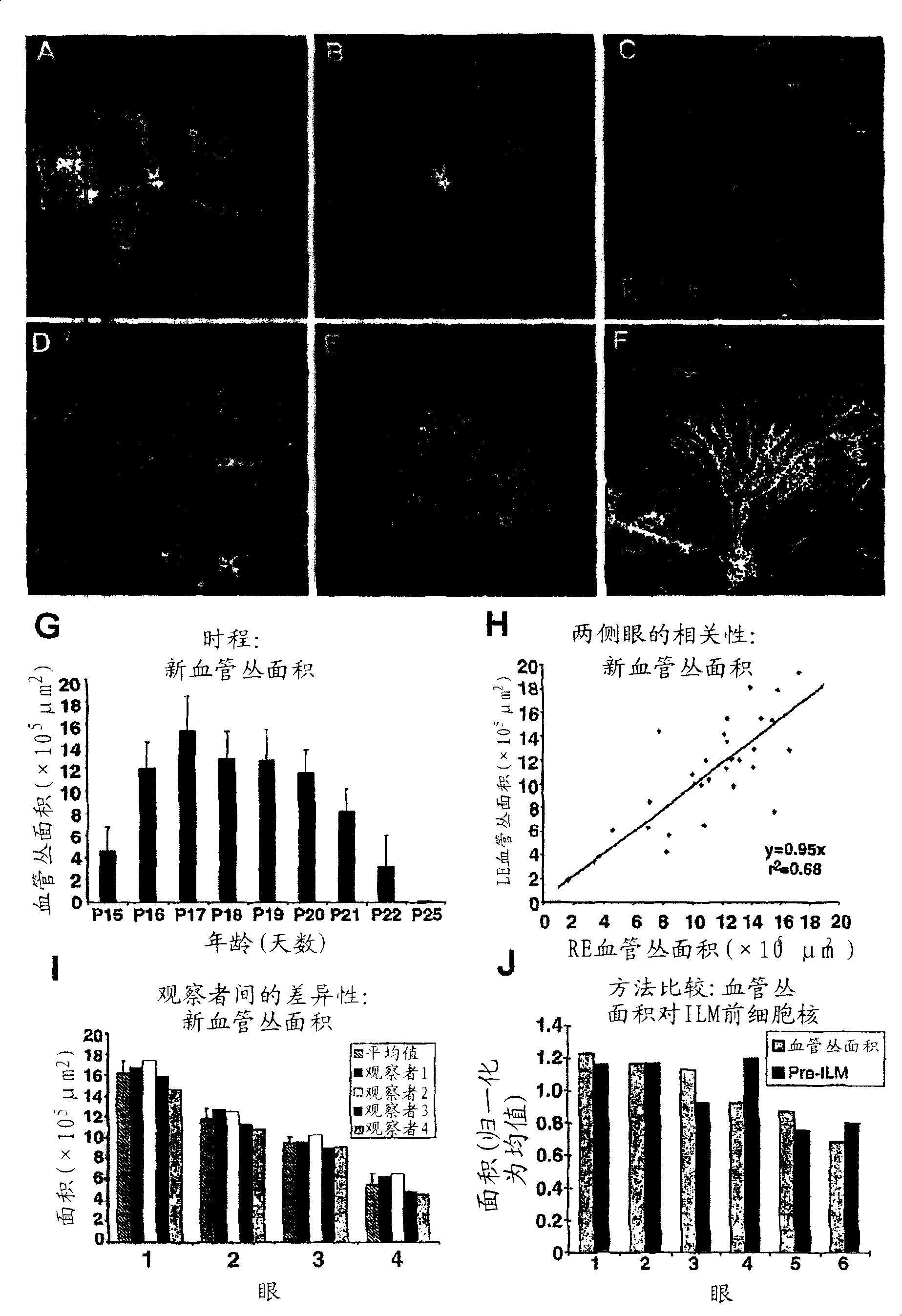
[0069] Example 2. Quantification of Extent of Abnormal Neovascularization

[0070] Between P15 and P22, especially from P17-P19 onwards, abnormal blood vessel growth appeared at the interface of retina and vitreous in mouse OIR model. These preretinal vascular plexuses are particularly prominent at the junction of the vascularized peripheral region and the occluded central region of the retina. These misdirected vascular elements are usually quantified in H&E-stained, paraffin-embedded cross-sections by counting the nuclei of cells projecting toward the vitreous side of the inner limiting membrane (ILM). However, these areas are also clearly visible in isolectin-stained whole-mount mount photographs due to cell populations overlying the superficial vascular network ( figure 1 Inset E, image 3 Illustration A). Using the intensity threshold tool (available in several commercial photo analysis programs), or Adobe PHOTOSHOP The area of ​​preretinal vascular plexus formation ...

Embodiment 3

[0073] Example 3. Angiogenesis Inhibitory Effects of iNOS Inhibitors

[0074] The application of the quantification technique of the present invention was further evaluated by examining the effect of a new angiogenesis inhibitor, the inhibitor iNOS, on retinal vascular regeneration and angiogenesis inhibition, and compared with the prior art pre-ILM nuclei counts. Inhibitors (or control saline) were delivered to mouse eyes between P12-P17 by once daily intraperitoneal injection, eyes were enucleated at P17, and quantified using either of the two techniques described. Figure 4 Inset A shows whole mount and transverse sections of retinas from P17 control mice (upper row) and iNOS inhibitor-treated animals (lower row). The area of ​​vascular occlusion (blue line) is plotted in the left inset, the area of ​​the neovascular plexus (red) is marked on the same whole mount in the middle inset, and an example of the pre-ILM nuclei is shown in the transverse section on the right in (a...


PUM


Abstract

The present invention provides a treatment for promoting beneficial physiological revascularization of ischemic retinal tissue. The treatment comprises administering to a mammal suffering from a retinal vascular ischemia a therapeutically effective amount of an angiostatic fragment of tryptophanyl-tRNA synthetase (TrpRS), thereby simultaneously inhibiting pathological neovascularization while promoting beneficial physiological revascularization of damaged areas of the retina. In a preferred embodiment, the angiostatic fragment of TrpRS is T2-TrpRS or T2-TrpRS-GD. Preferably, the mammal is a human patient suffering from retinal ischemia.

Description

[0001] Cross References to Related Applications [0002] This application claims the benefit of US Provisional Patent Application Serial No. 60 / 655,732, filed February 24, 2005, which is hereby incorporated by reference. [0003] Statement of Government Interest [0004] Portions of the work described herein were supported by National Institutes of Health grants EY11254 and EY14174. The US Government has certain rights in this invention. field of invention [0005] The present invention relates to the treatment of retinal neovascular diseases. More specifically, the invention relates to methods of promoting retinal vascular regeneration by administering to a patient fragments of angiogenic proteins, and to screening methods to identify therapeutic agents for the treatment of retinal vascular diseases. Background of the invention [0006] Retinal vascular diseases, including diabetic retinopathy, exudative age-related macular degeneration (ARMD), retinopathy of prematurity...

Claims


Application Information

Patent Timeline
Patent Type & Authority Applications(China)
IPC IPC(8): A01N37/18A61K38/00G01N33/567
CPCC12Y601/00G01N2800/164A61K49/0008G01N33/5088A61K38/43A61K38/53G01N33/50C12Y601/01002G01N2333/515A61P9/10A61P27/02A61P43/00A61K38/16
Inventor M·弗里兰德E·巴宁E·阿吉拉
Owner THE SCRIPPS RES INST
Why Eureka
Try Eureka