
Three-dimensional modelling approach for children's funnel chest

A technology of 3D modeling and pectus pectus, applied in 3D modeling, image data processing, instruments, etc., can solve problems such as poor quality of spiral CT images, invisible cartilage, invisible cartilage CT images, etc., and achieve a high degree of simulation Effect

Inactive Publication Date: 2009-02-18
NORTHWESTERN POLYTECHNICAL UNIV
View PDF0 Cites 8 Cited by
  • Summary
  • Abstract
  • Description
  • Claims
  • Application Information

AI Technical Summary

Problems solved by technology

[0006] In order to overcome the disadvantages of poor quality of spiral CT images and invisible cartilage due to the small difference between the transparency and density of costal cartilage and thoracic soft tissue in the prior art, the present invention provides a three-dimensional modeling method for pectus pectus excavatum in children, using the region search method Process the cartilage at the joint with the hard bone, analyze the characteristic parameters of the cartilage, and use the region growth method to solve the problem of invisible cartilage CT images, which can realize human cartilage modeling

Method used


Image

  • Three-dimensional modelling approach for children's funnel chest
  • Three-dimensional modelling approach for children's funnel chest
  • Three-dimensional modelling approach for children's funnel chest

Examples


Embodiment Construction

[0023] The establishment method of pectus excavatum thorax three-dimensional model of the present invention, specifically carries out according to the following steps:

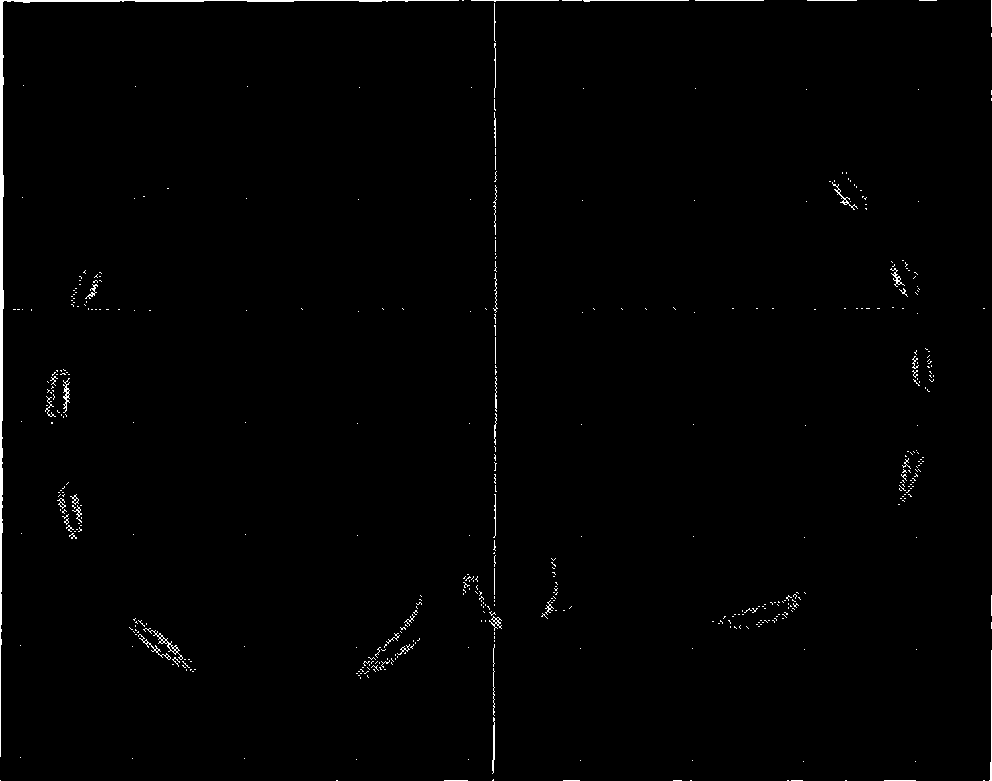
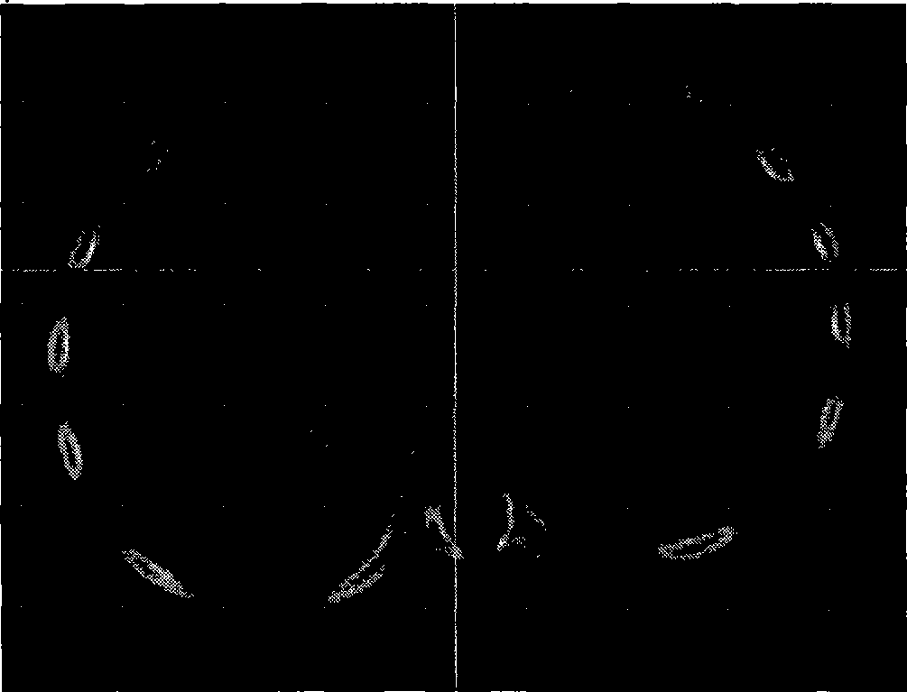
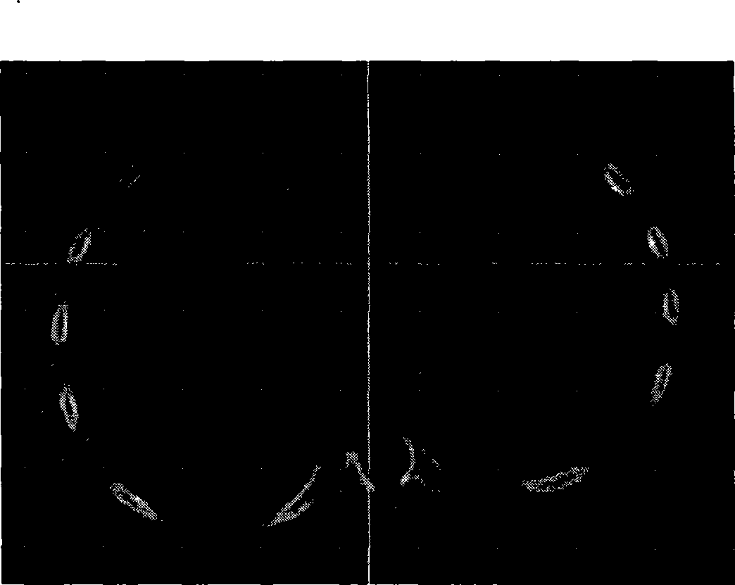
[0024] Will figure 1 The original spiral CT image of pectus pectus excavatum is input into the computer. CT images are sliced ​​images, displayed in grayscale mode, divided into three directions: front view, top view, and left view. The image resolution is 512×512 pixels, the number of slices is 227, and the interval between slices is 0.4mm. From the gray scale of the original CT image, only the difference between rib hard bone and soft tissue can be distinguished, but cartilage and soft tissue cannot be distinguished.

[0025] Firstly, image processing was performed on the original spiral CT images of pectus pectus excavatum in a child. For the case that the original spiral CT image of pectus pectus excavatum is a grayscale image, the costal cartilage grayscale data was extracted first. The gray value ran...


PUM


Abstract

The invention discloses a three-dimensional modeling method for the funnel chest of a child. Firstly, aiming at an original spiral CT picture of the funnel chest of the child, a region search method is adopted to model a thoracic hard bone part; a gray value of a cartilage region of a thoracic three-dimensional model is measured, and masking separation costal cartilage image data are created after the modification; a region growing method is adopted to apply region growing separation costal cartilage image data to a combining end of a hard bone and a cartilage of each rib; the costal cartilage data which is subjected to secondary data separation is three-dimensionally drafted to generate a three-dimensional funnel costal cartilage model; the three-dimensional funnel costal cartilage model is subjected to Gaussian smooth processing, and the model of the thoracic hard bone part of the funnel chest of the child and the three-dimensional costal cartilage model are overlapped to obtain an integral three-dimensional model of the funnel chest of the child. The region search method is adopted to process cartilages at the positions where with hard bones are combined, and the region growing method is adopted to solve the problem that a CT image of the cartilages is invisible, so the created three-dimensional model ensures that costal cartilages are not adhered to each other and each costal cartilage is round and smooth, and has high simulation degree.

Description

technical field [0001] The invention relates to a modeling method of pectus excavatum, in particular to a three-dimensional modeling method of pectus excavatum. Background technique [0002] The incidence rate of pectus excavatum and chicken breast is about 1 to 4‰, of which children with pectus excavatum account for about 90%. The main manifestation of pectus excavatum is the inward depression of the sternal body, which is funnel-shaped at the junction of the sternal body and the xiphoid process. The deformed thorax of pectus pectus excavatum not only causes the depressed sternum to compress the heart, reduces cardiac output, total lung volume and lung capacity, but also easily causes psychological inferiority in children, which seriously affects the mental and physical health of children. Most scholars in the medical field believe that it is caused by abnormal excessive growth of costal cartilage and sternum. [0003] The document "Current Status of Minimally Invasive Su...

Claims


Application Information

Patent Timeline
Patent Type & Authority Applications(China)
IPC IPC(8): A61B6/03G06T7/60G06T17/00G06T7/00
Inventor 史仪凯卫延斌袁小庆唐博张华
Owner NORTHWESTERN POLYTECHNICAL UNIV
Learn More