Hard ultrasonic gallbladder endoscope system and method

A technique of gallbladder and ultrasound, which is applied in the direction of endoscope, laparoscope, internal fixator, etc., to achieve the effect of simple operation and cost saving

Inactive Publication Date: 2009-03-25
乔铁
View PDF0 Cites 5 Cited by
  • Summary
  • Abstract
  • Description
  • Claims
  • Application Information

AI Technical Summary

Problems solved by technology

The micro-ultrasound probe image processing center, micro-ultrasound probe monitor, rigid ultrasound gallbladder endoscope image processing center, rigid ultrasound gallbladder endoscope mo

Method used


Image

  • Hard ultrasonic gallbladder endoscope system and method
  • Hard ultrasonic gallbladder endoscope system and method
  • Hard ultrasonic gallbladder endoscope system and method

Examples


Embodiment Construction

[0019] The present invention will be described in further detail below in conjunction with the drawings:

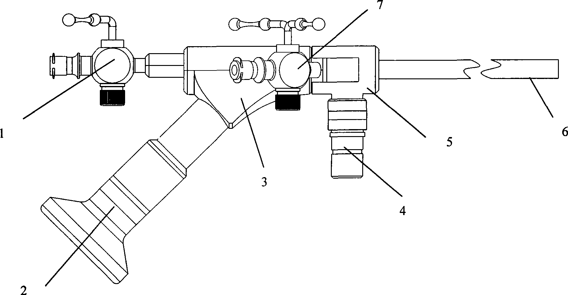
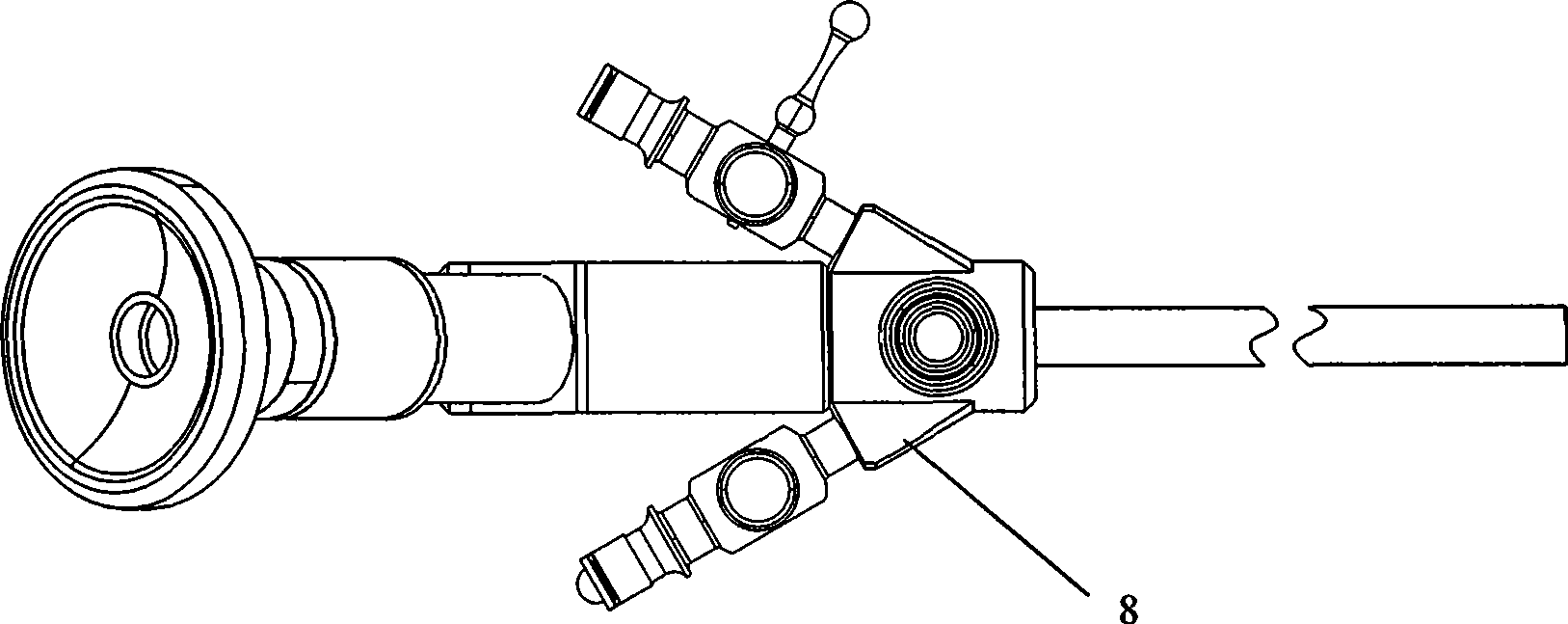
[0020] As shown in Figure 1, combined with Figure 2, the rigid ultrasound gallbladder endoscope of the present invention consists of an instrument channel valve 1, an eyepiece input end 2, an endoscope main body 3, a cold light source input end 4, and a front part of the endoscope main body 5. The endoscope end 6, the liquid channel 7 and the liquid channel 8 are composed.

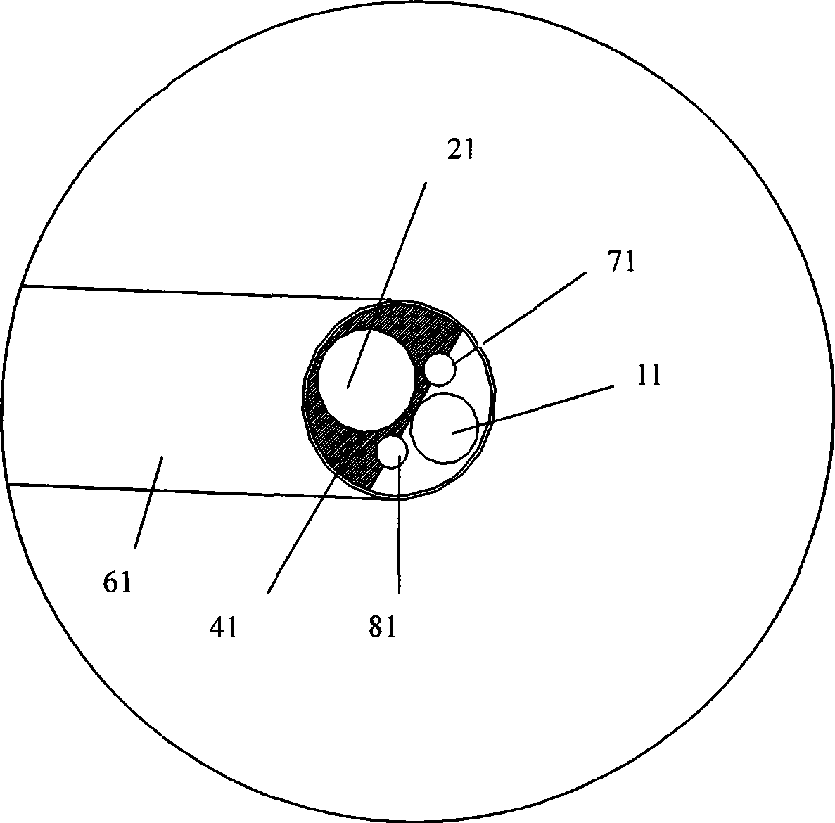
[0021] Fig. 3 shows the structure design of the tip 61 of the rigid ultrasound gallbladder endoscope of the present invention, which includes five parts: optical fiber 41, end exits 71 and 81 of two liquid channels 7 and 8, instrument channel exit 11, and optical lens 21 composition.

[0022] The end 6 and the tip 61 that enter the human body, the length of the end 6 is 250-300mm, the outer diameter of the tip 61 is 5.0-7.0mm, the diameter of the two liquid channel outlets 71 and 81 are both 0.9-1.4mm, an...


PUM

PropertyMeasurementUnit
Diameteraaaaaaaaaa
Lengthaaaaaaaaaa

Abstract

The invention belongs to medical appliance filed and in particular relates to a rigid ultrasonic gallbladder endoscopic system used in minimally invasive endoscopic stone and polypus removing surgeries for protecting the gallbladder. A connection interface of a minisize ultrasonic probe is connected with a minisize ultrasonic probe driver through a data line. The input end of an ocular of an ultrasonic gallbladder endoscope and the minisize ultrasonic probe driver are at the same inputted into an ocular interface and a probe interface at an image processing center of the ultrasonic gallbladder endoscopic system. A high-resolution monitor is connected outside a monitor interface of the image processing center of the ultrasonic gallbladder endoscopic system. A central graphic workstation is connected to a graphic workstation interface, and a keyboard is connected to a keyboard interface. A cold light source host is connected to a cold light source interface of the ultrasonic gallbladder endoscope. Compared with prior art, the rigid ultrasonic gallbladder endoscopic system has the advantage of saving cost and is simple to operate.

Description

Technical field [0001] The invention belongs to the field of medical instruments, and specifically relates to a rigid ultrasound gallbladder endoscopic system for minimally invasive endoscopic bile preservation and stone removal and polyp removal operations. Background technique [0002] At present, the existing biliary endoscopes used for minimally invasive bile preservation and removal of stones and polyp surgery use the connection method of the connection port of the micro ultrasound probe and the micro ultrasound probe driver through a data cable, and the micro ultrasound probe driver is connected to the micro ultrasound probe for image processing Center, the monitoring interface on the back of the micro ultrasound probe image processing center is connected to the micro ultrasound probe monitor, the graphic workstation interface is connected to the micro ultrasound probe graphic workstation, and the keyboard interface is connected to the multifunctional keyboard; at the same ...

Claims


Application Information

Patent Timeline
IPC IPC(8): A61B17/22A61B1/313A61B17/94A61B1/06A61B8/12
Inventor 乔铁
Owner 乔铁
Why Eureka
Try Eureka