

In situ hybridization detection kit for early uterine cancer
A detection kit and in situ hybridization technology, applied in the field of kits, can solve problems such as unreported detection technology, and achieve the effects of high sensitivity, convenient operation and strong specificity
- Summary
- Abstract
- Description
- Claims
- Application Information
AI Technical Summary
Problems solved by technology
Method used
Image

Examples
Embodiment 1
[0042] An in situ hybridization detection kit for early uterine cancer, comprising a hybridization probe, a marker, and a potentiator, wherein the sequence of the hybridization probe is shown in SEQ ID NO.1. Hybridization probes were labeled with digoxigenin. The composition of other liquids and specimens in the kit is as follows:
[0043] Digestive solution 100μl / tube 1 tube / box Colorless transparent liquid
[0044] Protective solution 100μl / tube 1 tube / box Colorless transparent liquid
[0045] Pre-hybridization solution 1300μl / tube 2 tubes / box Colorless transparent liquid
[0046] Sense hybridization solution 10μl / tube 1 tube / box Colorless transparent liquid
[0047] Antisense hybridization solution 10μl / tube 1 tube / box Colorless transparent liquid
[0048] Blocking solution 1000μl / tube 1 tube / box Colorless transparent liquid
[0049] Alkaline phosphatase antibody 1μl / tube 1 tube / box Colorless transparent liquid
[0050] Chromogen A 175μl / tube 1 tube / box Yellow liquid ...
Embodiment 2
[0093] A P2RX7 gene in situ hybridization detection method and kit application
[0094] 1. Specimen processing
[0095] 1. Use a 10ml centrifuge tube to fill 4.5ml of lymphocyte separation solution, then slowly add 3ml of anticoagulated blood into the centrifuge tube containing lymphocyte separation solution (blood: lymphocyte separation solution = 1:1.5), and centrifuge at 2000r / min 10min;
[0096] 2. Take the white blood cells in the middle layer into another centrifuge tube, then add about twice the amount of 1× buffer I to this tube, mix well, and centrifuge at 1500g / min for 10min;
[0097] 3. Discard the supernatant. Add about twice the 1× buffer I to the precipitate, mix well, and centrifuge at 1500g / min for 10min;
[0098] 4. Discard the supernatant, and absorb the excess liquid from the mouth of the test tube with paper towels. Then the precipitate was made into a suspension, dropped on a glass slide, and allowed to dry naturally. (Hospitals with conditions can use...
Embodiment 3
[0133] A comparative experiment between the detection of uterine cancer with the P2RX7 gene kit and the detection of uterine cancer with the HCCR gene kit.
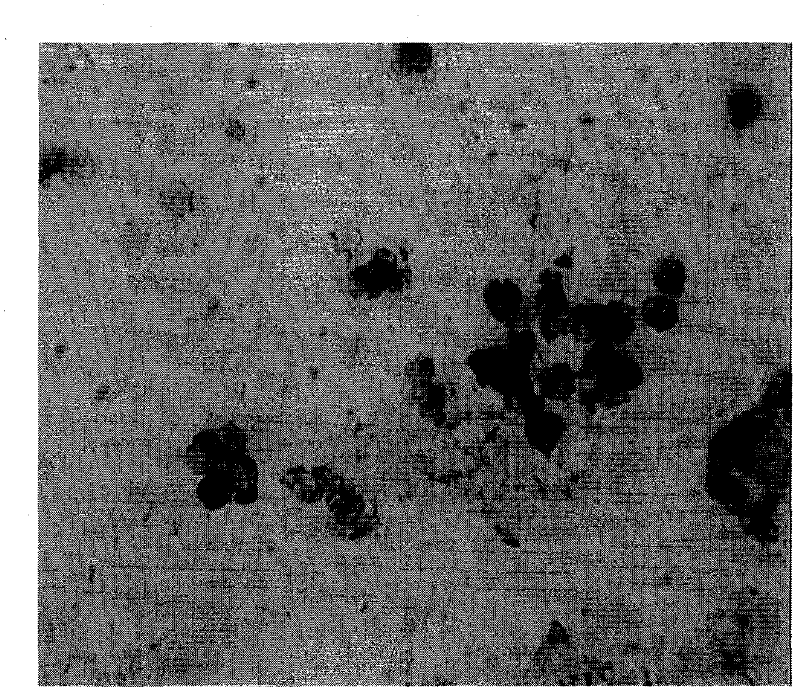
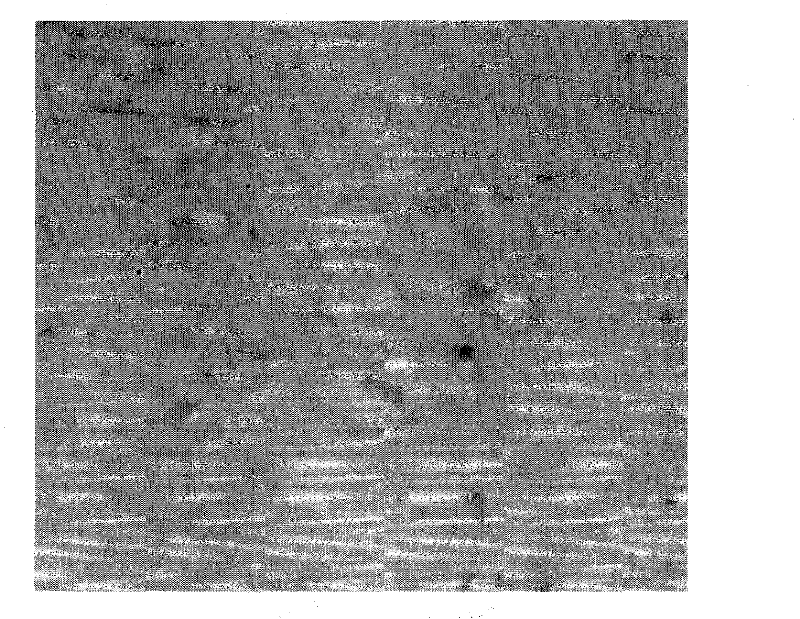
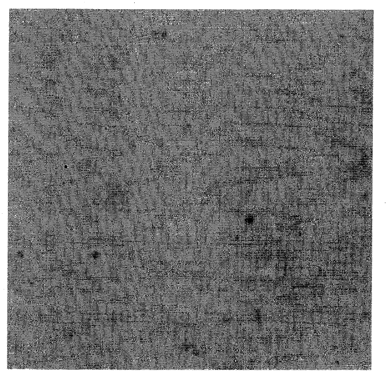
[0134] In order to scientifically evaluate the specificity, sensitivity and accuracy of the above genes in uterine cancer. We use the method of parallel experiment, detect the mRNA of above-mentioned gene at the same time, detection technology adopts nucleic acid in situ hybridization technology, use the peripheral blood of patient with same uterine cancer disease, detect the mRNA of P2RX7 gene and HCCR gene at the same time (carry out nucleic acid in situ hybridization, Immunohistochemical staining, microscopic counting, result reporting, etc. all used the same methods, steps and reagents as the in situ hybridization technique in Example 1 and Example 2). It was found that the expression level of P2RX7 gene in patients with uterine cancer was higher than that of HCCR gene in patients with the same disease. The results s...
PUM
 Abstract
Description
Claims
Application Information