Ultrasonograph

A diagnostic device, ultrasound technology, applied in ultrasound/sonic/infrasonic Permian technology, ultrasound/sonic/infrasonic image/data processing, organ movement/change detection, etc. Outgoing vessel wall thickness and IMT

Inactive Publication Date: 2009-08-12
TOHOKU UNIV +1
View PDF1 Cites 12 Cited by
  • Summary
  • Abstract
  • Description
  • Claims
  • Application Information

AI Technical Summary

Problems solved by technology

Therefore, it is often difficult to distinguish the boundaries of the vessel wall and the membranes that make u...

Method used


Image

  • Ultrasonograph
  • Ultrasonograph
  • Ultrasonograph

Examples


Embodiment Construction

[0024]

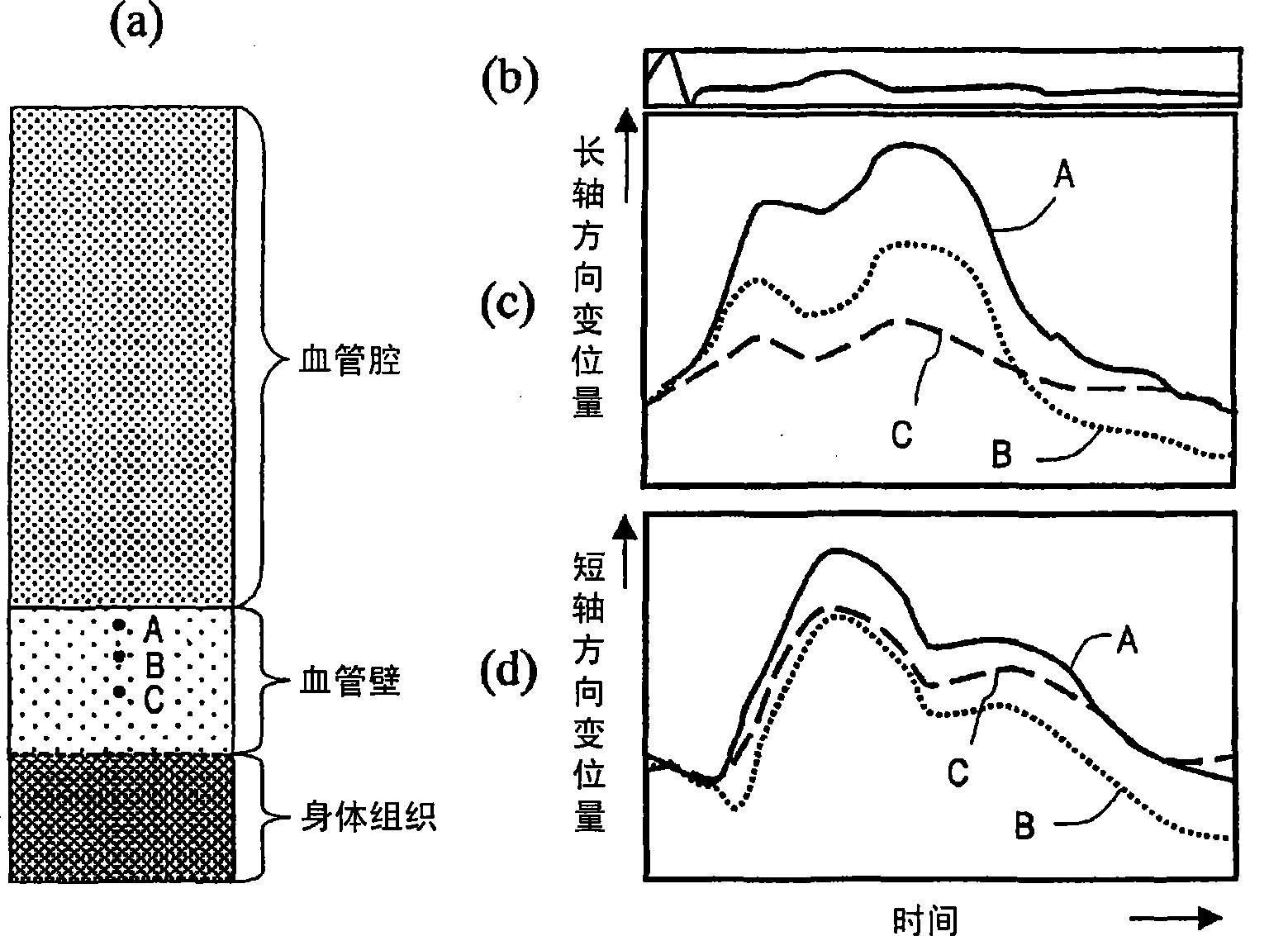
The arterial blood vessel expands and contracts in the radial direction in accordance with changes in the blood flow and blood pressure of the blood moving through the artery. Thus, the arterial wall moves radially in synchronization with the heart rate cycle. The axial movement of the arterial vessel to the extension of the blood vessel is usually not seen, and the axial movement is not considered when analyzing the movement of the arterial wall.

[0025]

However, as described in Nonprofit Documents 2, 3, and 4, it has been confirmed in recent years that the arterial wall of the carotid artery, which is measured as an index of arteriosclerosis, moves slightly axially in synchronization with the heart rate cycle. This movement is considered to be caused by the carotid artery being pulled by the heart along with the contraction and expansion of the heart.

[0026]

Below, refer to figure 1 (a) to (d), describe the movement of the carotid artery wall in the radial ...


PUM


Abstract

An ultrasonograph comprises a transmitting section (102) for driving an ultrasonic probe to transmit ultrasound to a measurement region of a subject including an artery vessel wall, a receiving section (101) for receiving reflected waves produced when the ultrasound is reflected from the subject by means of the ultrasonic probe to generate a reception signal, a movement information calculating section (110) for calculating movement information at least in the axial direction of the artery vessel wall from the reception signal, and a boundary position determining section (109) for determining the position of at least one of the blood flow-inner membrane boundary, the media-outer membrane boundary, and the outer membrane-body connective tissue boundary of the artery vessel wall in the measurement region according to the movement information.

Description

technical field [0001] The present invention relates to an ultrasonic diagnostic apparatus for medical use, and more particularly to an ultrasonic diagnostic apparatus for measuring blood vessel walls. Background technique [0002] An ultrasonic diagnostic apparatus irradiates an object with ultrasonic waves, and analyzes information included in an echo signal (echo signal) to observe the internal state of the object. Ultrasonic diagnostic apparatuses widely used in the prior art convert the intensity of echo signals into the luminance of corresponding pixels to obtain the structure of the subject as a tomographic image. In this way, the internal structure of the subject can be known, and since the condition in the subject can be continuously observed, it has become an indispensable device in clinical practice. [0003] In recent years, the number of patients with arteriosclerosis has been increasing, and in order to diagnose arteriosclerosis, carotid artery echo usin...

Claims


Application Information

Patent Timeline
IPC IPC(8): A61B8/08
CPCA61B8/08
Inventor 金井浩长谷川英之福元刚智
Owner TOHOKU UNIV
Why Eureka
Try Eureka