Analysis of brain patterns using temporal measures

A short-term, brain model technology, applied in the field of neurophysiological analysis, can solve the problems of not being able to analyze the brain state of the brain activity of the subject or diagnose the brain state, without taking the brain model into account, and achieve the effect of low cost and high throughput

Inactive Publication Date: 2009-11-18
RGT UNIV OF MINNESOTA
View PDF1 Cites 13 Cited by
  • Summary
  • Abstract
  • Description
  • Claims
  • Application Information

AI Technical Summary

Problems solved by technology

Although this work forms some interesting insights into the measurement techniques used to take MEG readings (for example, advantageously utilizing 1 kHz sampling, and preprocessing the data to pre-whiten them), the work only utilizes the individual sensor signals interactions without taking into account the entire brain model that must study a large number of sensor groupings
Indeed, for reasons that will be apparent from the disclosure below, the Leuthold et al. disclosure does not enable the analysis of brain activity to characterize or diagnose the brain state of a subject

Method used


Image

  • Analysis of brain patterns using temporal measures
  • Analysis of brain patterns using temporal measures
  • Analysis of brain patterns using temporal measures

Examples


example 1

[0064] Magnetoencephalography (MEG) can be used for elderly subjects as well as subjects with MCI and AD.

[0065] In one example, the gaze task and MEG were used to assess the dynamic state or dynamic function of the brain in three groups: elderly subjects (77.1 ± 1.5 years, mean ± SEM, N = 11), normal subjects (N = 4, 76.5±2.1 years), subjects with MCI (N=4, 75.7±3.7 years) and subjects with AD (N=3, 79.7±0.3 years). While the subject was looking at a certain point for 45 seconds, data were collected from 248 axial gradiometers (Magnets3600WH, 4-D Neuroimaging), and these data were preprocessed to remove the cardiac artifacts or blinking artifacts.

[0066] After pre-whitening the time series by fitting the AutoRegressive Integrative Moving Average (ARIMA) model and taking the residues, calculate the cross-correlation of all pairs of zero lag parts to provide a short resolution of 1 millisecond The estimated value of the strength and sign (positive and negative) of the direct sy...

example 2

[0078] method

[0079] The subject lay on his back on the bed and was asked to look at a point in front of him for 1 minute. Ask the subject to keep their eyes fixed at this point and not blink. Then, the subject closed his eyes for another 3 minutes, thereby ending the test. All analyses were performed using MEG data from the fixation cycle. In one embodiment, the data collected during the closed eye state is suitable for identifying and removing signal artifacts, such as cardiac artifacts.

[0080] MEG instrument

[0081] Use MEG instrument to collect data. The subject lies on a bed in a magnetically shielded room, and takes samples from 248 axial gradiometers (0.1-400 Hz, at a frequency of 1017 Hz, Magnes 3600WH, 4 minutes) for the entire duration of the test (~4 minutes). -D Neuroimaging, San Diego, CA) collect MEG signals.

[0082] data analysis

[0083] data processing. The event-synchronized subtraction method can be used to remove artifacts from the heart. Since the durati...

example 3

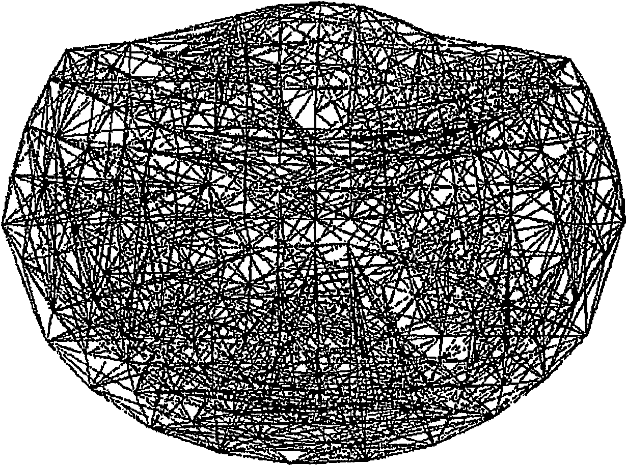
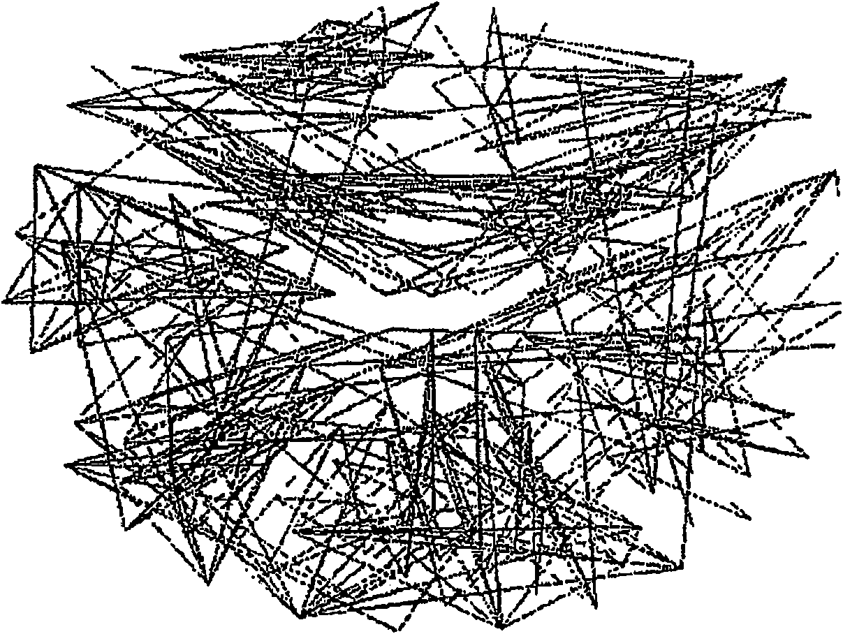
[0099] Use pre-whitened (still) magnetoencephalogram signals to visualize synchronized dynamic brain networks. In one example, data is collected from 248 axial gradiometers. After fitting the autoregressive integral moving average model and taking the residue, calculate the cross-correlation PCC of all pairs of zero lag between the i-th sensor and the j-th sensor IJ O , So as to provide an estimate of the strength and sign (positive and negative) of the direct synchronization coupling between the neural groups with a short resolution of 1 millisecond. In one instance, 51.4% of PCC IJ O Is positive, 48.6% of PCC IJ O Is negative. On average, a positive PCC IJ O Negative PCC IJ O Occurs more frequently with shorter sensor spacing, and is more frequent than negative PCC IJ O Strong 72%. Based on estimated PCC IJ O , Construct a dynamic neural network (one for each subject) to show different characteristics, including multiple local interactions. These characteristics are power...


PUM


Abstract

A set of brain data representing a time series of neurophysiologic activity acquired by spatially distributed sensors arranged to detect neural signaling of a brain (such as by the use of magnetoencephalography) is obtained. The set of brain data is processed to obtain a dynamic brain model based on a set of statistically-independent temporal measures, such as partial cross correlations, among groupings of different time series within the set of brain data. The dynamic brain model represents interactions between neural populations of the brain occurring close in time, such as with zero lag, for example. The dynamic brain model can be analyzed to obtain the neurophysiologic assessment of the brain. Data processing techniques may be used to assess structural or neurochemical brain pathologies.

Description

Technical field [0001] The present invention generally relates to neurophysiologic analysis, and more specifically, relates to, but not limited to, the use of time series representation to analyze brain patterns. Background technique [0002] Like any other organ in the body, the function of the brain needs to be evaluated to evaluate its health and disease status. However, unlike any other organs in the body, there is no better brain function test. Typical behavioral examinations include standard neurological examinations, psychiatric interviews, or neuropsychological tests. Electroencephalogram (EEG) can hardly provide any information unless there is a grand epileptic seizure or a serious disorder of brain function, such as being in a coma. Used to evaluate brain structure (such as magnetic resonance imaging (MRI)), chemical properties (such as magnetic resonance spectroscopy (MRS)), fluoro-deoxyglucose-based positron emission tomography (fluoro-deoxy- Glucose based positron em...

Claims


Application Information

Patent Timeline
Patent Type & Authority Applications(China)
IPC IPC(8): A61B5/04
Inventor A·P·乔治普洛斯
Owner RGT UNIV OF MINNESOTA
Why Eureka
Try Eureka