

Three-dimensional medical image display method based on GPU acceleration
A medical image and display method technology, applied in the field of medical image processing, can solve the problems of difficult interactive display, high cost, and improvement.
Inactive Publication Date: 2009-12-02
UNIV OF ELECTRONICS SCI & TECH OF CHINA
View PDF0 Cites 30 Cited by
- Summary
- Abstract
- Description
- Claims
- Application Information
AI Technical Summary
Problems solved by technology
[0003] The current general-purpose volume rendering technology is generally based on traditional CPU for calculation. For traditional consumer-level mass PCs, it is difficult to perform interactive display in real time.
If you want interactive real-time display calculations, you generally need to run them on a parallel graphics workstation, and the cost is relatively high.
Method used
View moreImage
Smart ImageViewing Examples

Examples
Embodiment Construction
[0039] The present invention adopts the above-mentioned technical scheme to perform display verification for MRI and CT medical DICOM image sequences respectively, and both have better display effects. It should be noted that MRI images need to be de-noised and pre-processed due to their high noise, while CT images are relatively clear and do not need to be de-noised and pre-processed, and can be directly displayed as volume data.
Login to View More PUM
 Abstract
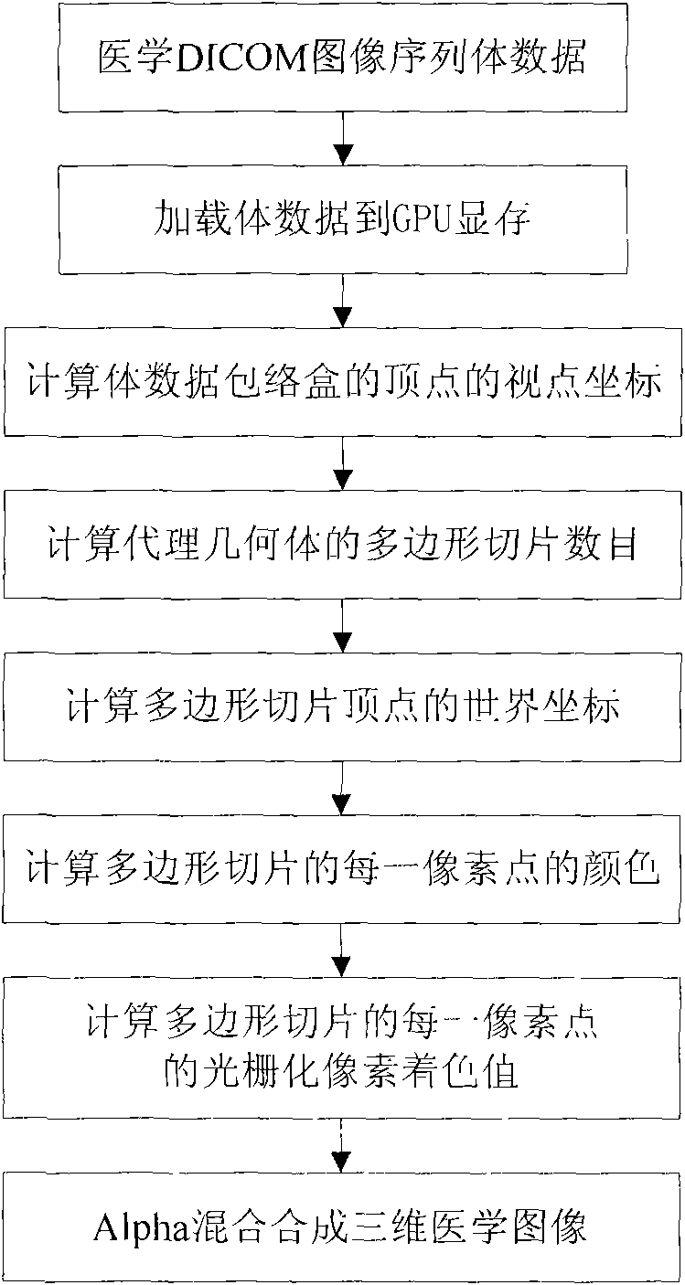
The invention discloses a three-dimensional medical image display method based on GPU acceleration, and belongs to the technical field of medical image processing. The method comprises the following steps: firstly, storing medical DICOM image sequence files in a volume data mode; secondly, expanding an interface function API by three-dimensional graphic base programming of OpenGL or DirectX, and loading the volume data in a GPU memory; thirdly, calculating to generate an agent geometric solid, and performing illumination calculation and color calculation on polygonal slices in the agent geometric solid pixel by pixel; and finally, synthesizing all polygonal slices in the agent geometric solid into a three-dimensional medical image through Alpha blending. Compared with the prior medical image display method based on CPU, the three-dimensional medical image display method based on GPU acceleration has high operation speed, can realize real-time interactive display on PCs of people in common consumption level, and does not need using a graphic workstation to greatly reduce cost.
Description
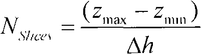
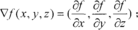
technical field [0001] The invention belongs to the technical field of medical image processing, and mainly utilizes the powerful parallel flow processing capability of GPU (display processing unit) to accelerate volume rendering. Compared with traditional CPU volume rendering technology, this method enables volume rendering to be interactive real-time display. technical background [0002] The auxiliary role of medical images in doctors' diagnosis is becoming more and more obvious, but the human body can only be observed from the two-dimensional cross-sectional direction. In order to improve the accuracy and scientificity of medical diagnosis and treatment planning, it is necessary to transform the sequence of two-dimensional tomographic images into three-dimensional images with intuitive three-dimensional effects. However, the current 3D medical aided diagnosis system needs a workstation-level operating platform to basically meet the real-time requirements, and the price ...
Claims
Login to View More Application Information
Patent Timeline
 IPC IPC(8): G06T1/00G06T1/20G06T15/00
Inventor解梅张帆
OwnerUNIV OF ELECTRONICS SCI & TECH OF CHINA