Three-dimensional acquisition of biopsy tissues and fusion method of multilayer images

An image fusion and three-dimensional image technology, applied in image enhancement, image data processing, measuring devices, etc., can solve the problems of inability to obtain clear tissue images, uneven thickness of sliced ​​tissue, etc.

Active Publication Date: 2009-12-30
苏州优纳医疗器械有限公司
View PDF1 Cites 33 Cited by
  • Summary
  • Abstract
  • Description
  • Claims
  • Application Information

AI Technical Summary

Problems solved by technology

[0005] In order to solve the problem that a clear tissue image cannot be obtained due to the uneven thickness of the sliced ​​tissue
Each focal plane is a layer

Method used


Image

  • Three-dimensional acquisition of biopsy tissues and fusion method of multilayer images
  • Three-dimensional acquisition of biopsy tissues and fusion method of multilayer images
  • Three-dimensional acquisition of biopsy tissues and fusion method of multilayer images

Examples


Embodiment Construction

[0019] specific implementation plan

[0020] The technical solutions of the present invention will be described in further detail below in combination with preferred embodiments according to the accompanying drawings.

[0021] The computer system used in this example is an ordinary PC system, and the operating system is Windows XP HOME version. However, those skilled in the art will understand that the spirit and scope of the present invention are not limited to any computer model and operating system, as well as specific communication protocols.

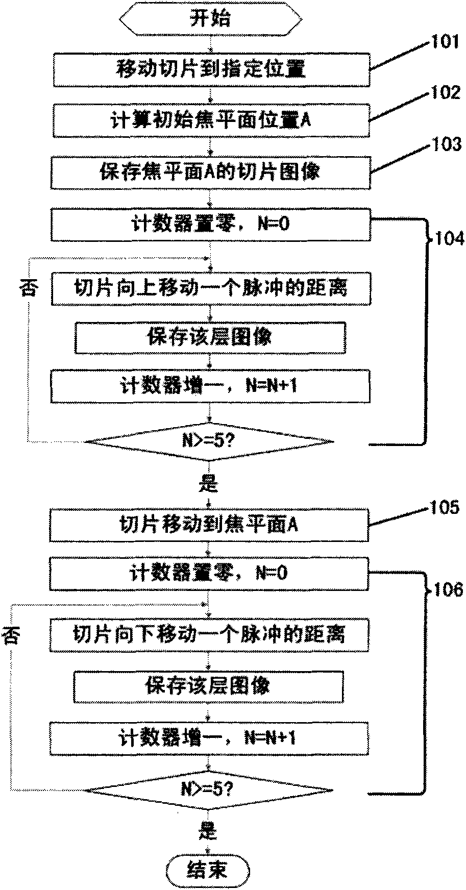
[0022] The slice image acquisition device selected in this example is the slice tissue acquisition system introduced in the utility model patent ZL200620139251.9. The device can transmit tissue images on slices that can be observed under a microscope to a PC through a high-resolution CCD industrial camera, and can be observed on the PC or saved as a file in image format.

[0023] The embodiments and examples given herein illustrat...


PUM


Abstract

The invention discloses three-dimensional acquisition of biopsy tissues and a fusion method of multilayer images after acquisition. During three-dimensional acquisition of the biopsy tissues, the position of the initial focalplane is taken as the center, multilayer image acquisition is carried out on the update positions within the range of biopsy thickness and simultaneously the biopsy image on each layer is stored. The multilayer images of the invention adopt a partitioned area fusion method; based on the definition of the pixels in the images, the areas with relatively high definition in the images to be fused are selected and the template images are updated to finally achieve the purpose of optimizing the definition of the image of the whole biopsy. At the same time, a method of assignment weight is adopted to smoothen the pixel points in the boundary areas, thereby effectively inhibiting false edges. Being adopted, the method of the invention improves the reproduction accuracy of the digital images towards the characteristics of the biopsy tissues. The invention effectively alleviates the workload of the doctors and provides a basis for remote diagnosis.

Description

technical field [0001] The invention belongs to the technical field of multi-layer acquisition and fusion of medical and biological slice images. More specifically, the present invention relates to a multi-layer fusion image processing method for sliced ​​tissue. Background technique [0002] Image fusion technology is the fusion of visual information in multi-sensor information fusion. It uses different imaging methods of various imaging sensors to provide complementary information for different images, increase the amount of image information, reduce the amount of original image data, and improve adaptability to the environment. In order to obtain more reliable and accurate useful information for observation or further processing. It is an emerging technology that integrates sensors, signal processing, image processing and artificial intelligence. In recent years, image fusion has become a very important and useful image analysis and computer vision technology. It has b...

Claims


Application Information

Patent Timeline
Patent Type & Authority Applications(China)
IPC IPC(8): G06T5/00G06T5/50G01N13/10
Inventor 郑众喜刘明星韩隽
Owner 苏州优纳医疗器械有限公司
Why Eureka
Try Eureka