
Urinary cell micro staining analysis method

An analysis method and quantitative analysis technology, applied in the field of medical inspection, can solve the problems of waste of dye solution, inability to achieve individualized dyeing of a single sample, and inability to implement micro-control of dye solution dosage, etc.

Active Publication Date: 2010-01-20
彭艾 +2
View PDF4 Cites 6 Cited by
  • Summary
  • Abstract
  • Description
  • Claims
  • Application Information

AI Technical Summary

Problems solved by technology

However, there are the following defects: the single-cylinder design cannot avoid cross-contamination of urine samples; the dosage of the dye solution cannot be controlled in a small amount; the dyeing is done in batches, and only one kind of dyeing can be supported at the same time, and the personalized dyeing of a single sample cannot be realized; during operation It is necessary to frequently replace the dye solution and clean the dye tank, resulting in a large amount of waste of dye solution; there is no automatic quantitative counting program, no waste water and toxic gas treatment device, so this machine is only suitable for staining small samples of pathological tissue sections

Method used


Image

  • Urinary cell micro staining analysis method
  • Urinary cell micro staining analysis method
  • Urinary cell micro staining analysis method

Examples


Embodiment 1

[0135] Example 1 Urine Cell Analysis

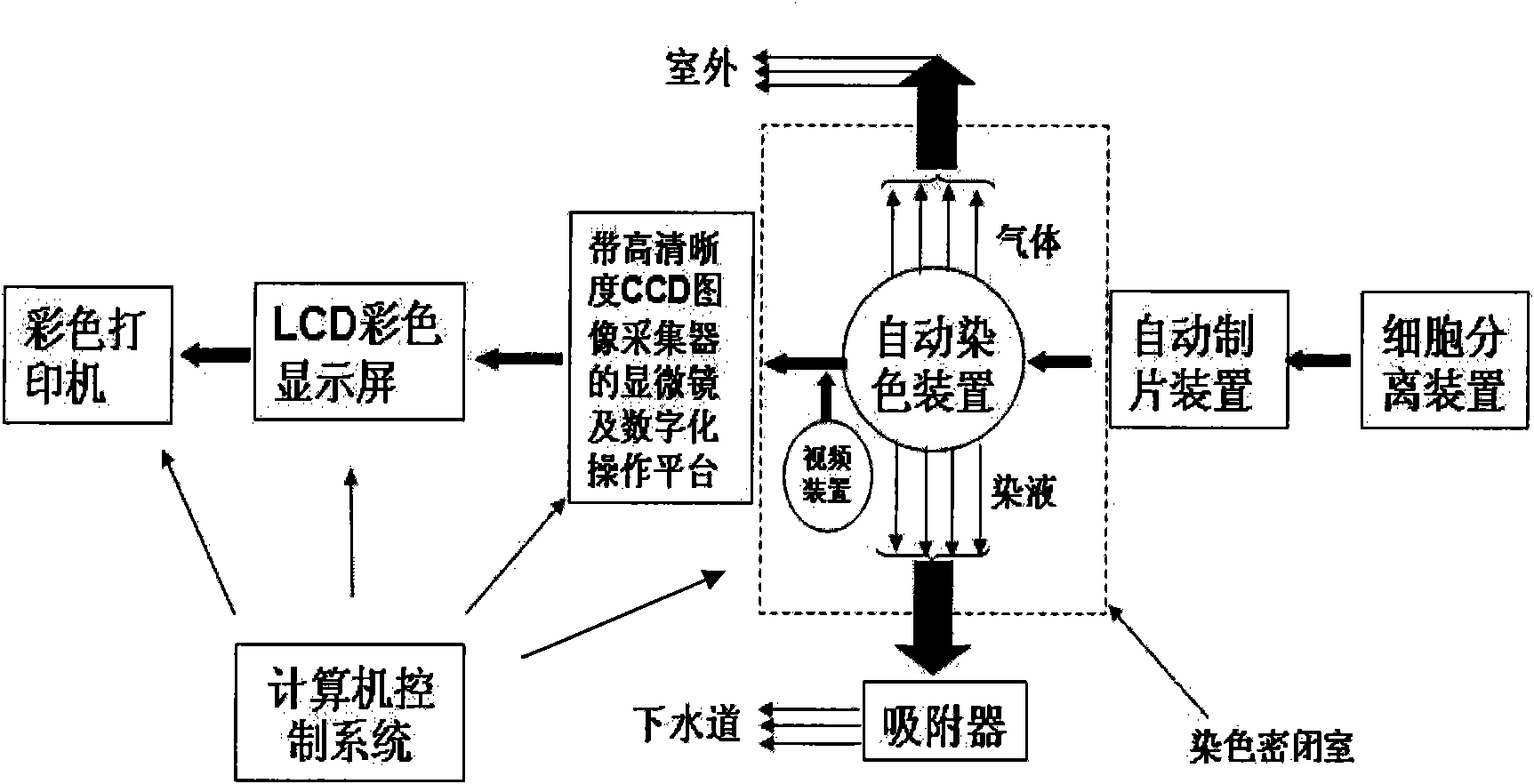
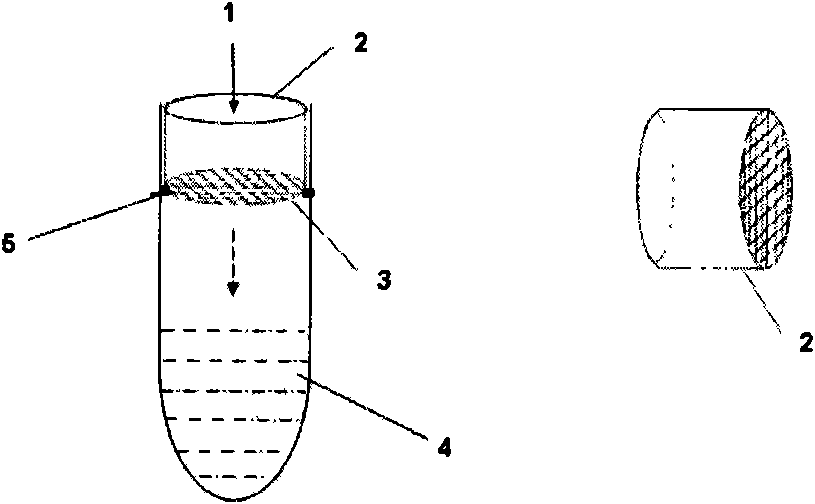
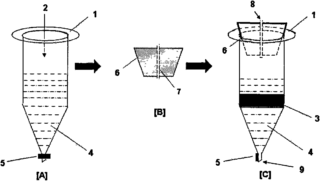
[0136] 10-50ml of fresh urine was centrifuged at 2000rpm for 10min, the sediment was mixed with 1ml of Hanks solution, and passed through a 30-300 mesh filter ( figure 2 , 3), in the centrifuged state, slowly inject 1ml of urine sediment Hanks solution into a continuous density gradient test tube prepared by Ficoll at 1.095-1.055g / ml, suck out the layered cell mass, centrifuge at 2000rpm for 10min, and use 0.5ml Hanks solution to weigh hanging. After diluting the sample with Hanks solution, take 200μl urine cell sample and add 3-8ml Ficoll (1.065g / ml) solution to mix well, then transfer it to a clean cell flake device, and centrifuge at 3000rpm for 5min to automatically make a thin cell sheet ( Figure 4 , 5). Fix with absolute ethanol and dry before use. When staining, place 1 slide in the automatic staining device[ Figure 6 , 7] in the staining tank, input each staining program respectively, such as Papanicolaou staining, then the...

Embodiment 2

[0162] Example 2 Urine Cell Analysis

[0163] 10-50ml of fresh urine was centrifuged at 2000rpm for 10min, the sediment was mixed with 1ml of Hanks solution, and passed through a 30-300 mesh filter ( figure 2 , 3), in the centrifuged state, slowly inject 1ml of urine sediment Hanks solution into a continuous density gradient test tube prepared by Ficoll at 1.095-1.055g / ml, suck out the layered cell mass, centrifuge at 2000rpm for 10min, and use 0.5ml Hanks solution to weigh hanging. After diluting the sample with Hanks solution, take 200μl urine cell sample and add 3-8ml Ficoll (1.065g / ml) solution to mix well, then transfer it to a clean cell flake device, and centrifuge at 3000rpm for 5min to automatically make a thin cell sheet ( Figure 4 , 5). Fix with absolute ethanol and dry before use. When staining, place 24 slides in the automatic staining device[ Figure 6 , 7] in the staining tank, input each staining program respectively, such as Papanicolaou staining, then t...


PUM


Abstract

The invention belongs to the field of medical tests, and relates to a urinary cell micro staining analysis system, in particular to a urinary cell micro staining analysis method and an involved analyzing device. Combining with a computer automatic control and recognition technique, the invention provides a urinary cell micro staining quantitative system which can achieve cell separation, extraction and thin layer flaking of arena, support a single-flow micro staining technique, and aim at clinical large samples and rapid analysis. The system overcomes the defects of the prior art, and the results of urine sample clinical medical inspection practices show that the urinary cells are stained clearly, are distributed evenly and are easy to recognize and quantitate. The method can prevent sample cross contaminations and can perform precise quantitative analysis without staining solution environmental pollution. The method is in accordance with the requirements on arena examination standardization established by the inspection branches of International (NCCL) and Chinese Medical Association.

Description

technical field [0001] The invention belongs to the field of medical examination, and relates to micro-staining analysis of urine cells. It specifically relates to a urine sediment analysis method including supporting micro-staining technology, preventing sample cross-contamination, and accurate quantitative analysis. technical background [0002] The practice of clinical testing shows that the composition of urine sediment is complex, and it is often necessary to use relevant analysis devices for the detection and analysis of urine sediment. So far, there is no urine sediment analyzer in the prior art to carry out separate extraction, treatment and analysis of each component in the urine sediment. Because these existing analyzers cannot avoid the interaction of urine sediment components when staining, do not support micro-staining and thin-layer film preparation, large staining errors, cross-contamination of samples, high false positive rate, and often require manual opera...

Claims


Application Information

Patent Timeline
Patent Type & Authority Applications(China)
IPC IPC(8): G01N33/493G01N1/30
Inventor 彭艾王力生夏红根
Owner 彭艾
Learn More