
Jugular vein microdialysis probe and probe guiding device

A jugular vein and microdialysis technology, applied in catheters, guide wires, medical science, etc., can solve the problems of dialysis membrane performance degradation, experimental failure, concentric probes that are difficult to fix in blood vessels and soft tissues of internal organs, etc. Achieve the effect of improving performance and reducing soaking time

Inactive Publication Date: 2010-05-12
ZHEJIANG UNIV
View PDF0 Cites 7 Cited by
  • Summary
  • Abstract
  • Description
  • Claims
  • Application Information

AI Technical Summary

Problems solved by technology

[0004] Concentric probes are difficult to secure on blood vessels and soft tissues of internal organs, and movement of the animal may cause the probe to dislodge from blood vessels or soft tissues of internal organs
Therefore, the existing commercial concentric circle probes for experiments are produced by foreign companies, such as the CMA20 Elite type of the American CMA Company and the A-I-X-Y series probes of the Japanese EICO Company, which can only be used for intravascular microdialysis under anesthesia.
However, there is no commercial jugular vein microdialysis probe for freely moving animals in China.
At the same time, the current general method of microdialysis experiments is to directly implant the probe into the animal body, and perform the microdialysis experiment after a few days of recovery. Studies have shown that this method of long-term implantation of the microdialysis probe in the body, first, the probe is fragile and causes the experiment to fail. , the second is that the performance of the dialysis membrane decreases

Method used


Image

  • Jugular vein microdialysis probe and probe guiding device
  • Jugular vein microdialysis probe and probe guiding device
  • Jugular vein microdialysis probe and probe guiding device

Examples


Embodiment 1

[0018] Example 1: A probe and probe guiding device for rat jugular vein microdialysis

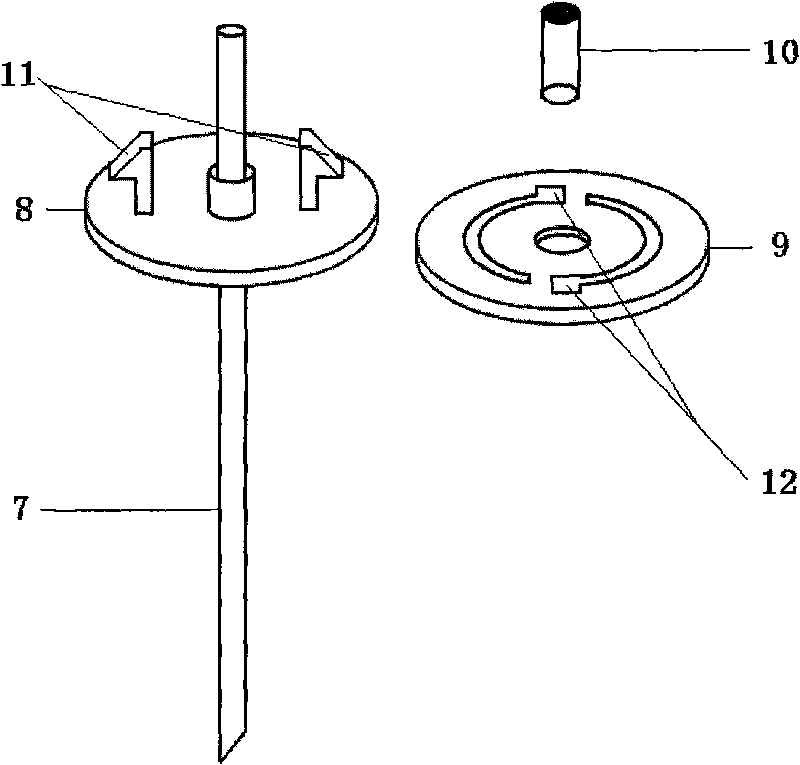
[0019] The total length of the jugular vein microdialysis probe is 125 mm, and the material of the dialysis membrane 1 is polycarbonate, with a length of 10 mm, an outer diameter of 0.15 mm, and a molecular weight cut-off of 13,000 Daltons. Dialysis membrane ends sealed. The liquid inlet pipe 3 has an outer diameter of 0.1mm, an inner diameter of 0.05mm, and a length of 125mm. Outlet pipe 4 has an outer diameter of 0.1mm, an inner diameter of 0.05mm, and a length of 105mm. The connecting pipe 2 has an outer diameter of 0.4mm, an inner diameter of 0.25mm, and a length of 5mm. The inner casing 5 has an outer diameter of 0.6 mm, an inner diameter of 0.2 mm, and a length of 10 mm. Outer casing 6 has an outer diameter of 0.8 mm, an inner diameter of 0.3 mm, a length of 15 mm at the liquid inlet end, and a length of 10 mm at the liquid outlet end. The guide tube 7 has an outer diameter of 1 m...

Embodiment 2

[0020] Embodiment 2: a kind of probe and probe guide device for rabbit jugular vein microdialysis,

[0021] The total length of the jugular vein microdialysis probe is 225mm, and the material of the dialysis membrane 1 is polycarbonate, with a length of 30mm, an outer diameter of 0.15mm, and a molecular weight cut-off of 13,000 Daltons. Dialysis membrane ends sealed. The liquid inlet pipe 3 has an outer diameter of 0.1 mm, an inner diameter of 0.05 mm, and a length of 245 mm. Outlet pipe 4 has an outer diameter of 0.1 mm, an inner diameter of 0.05 mm, and a length of 205 mm. The connecting pipe 2 has an outer diameter of 0.4mm, an inner diameter of 0.25mm, and a length of 5mm. The inner casing 5 has an outer diameter of 0.6 mm, an inner diameter of 0.2 mm, and a length of 10 mm. Outer casing 6 has an outer diameter of 0.8 mm, an inner diameter of 0.3 mm, a length of 15 mm at the liquid inlet end, and a length of 10 mm at the liquid outlet end. The guide tube 7 has an outer...


PUM


Abstract

The invention discloses a jugular vein microdialysis probe and a probe guiding device. The probe part comprises a dialysis membrane, a connecting pipe, a liquid inlet pipe, a liquid outlet pipe, an inner sleeve and an outer sleeve; and the probe guiding device part comprises a guiding pipe, a fixed base, a fixed spiral plate and a guiding pipe cap. The jugular vein microdialysis probe and probe guiding device of the invention can be used to reduce the soaking time of the microdialysis probe and increase the operational performance of the probe.

Description

technical field [0001] The invention relates to a biomedical microdialysis device, in particular to a jugular vein microdialysis probe and a probe guiding device used in an animal's free movement state. Background technique [0002] The microdialysis device is a biological sampling device designed on the basis that small molecular substances can pass through the dialysis membrane and diffuse along the concentration gradient, while macromolecular substances cannot pass through the dialysis membrane. When the microdialysis probe is implanted in the designated area of ​​the animal body, the micropump in vitro makes the perfusate flow through the dialysis membrane, and the substance to be measured in the tissue diffuses into the perfusate through the dialysis membrane and reaches a dynamic equilibrium. The concentration of the analyte is used to study the level and change process of the analyte in the tissue extracellular fluid. [0003] Microdialysis probes are the heart of mi...

Claims


Application Information

Patent Timeline
IPC IPC(8): A61M25/00A61M25/09A61D7/00
Inventor 吕良张恒义王恩禹
Owner ZHEJIANG UNIV
Learn More