Combination of fusion protein for separating fluorescent protein, expression vector and application thereof

A fluorescent protein and fusion protein technology, which is applied in the direction of fluorescence/phosphorescence, the use of vectors to introduce foreign genetic material, hybrid peptides, etc., can solve the complex establishment, the small number of events analyzed at the same time, and the lack of easy screening of viral envelope proteins, etc. question

Inactive Publication Date: 2010-06-23
THE UNIV OF TOKYO +1
View PDF0 Cites 5 Cited by
  • Summary
  • Abstract
  • Description
  • Claims
  • Application Information

AI Technical Summary

Problems solved by technology

This method is both sensitive and quantitative, but has a lag time due to the transcription and translation of the reporter gene
At the same time, those protein fragments with self-complementary ability, especially split enzymes, are often used by researchers due to their versatility; but because most enzyme substrates are not membrane-permeable, such analysis methods are usually Requires disruption of cell membranes (Holland, 2004; Jun, 2007)
This prevents continuous monitoring of membrane fusion events
Some people also use the method of electrophysiological recording (Monck, 1992), but this method is not widely used because the method is very complicated to set up and the number of events that can be analyzed simultaneously is too small
[0006] Therefore, there is no technology in the prior art, that is, a method for quickly and accurately identifying the phenotype of Env-mediated membrane fusion for a large number of samples
At the same time, there is also a lack of a method for easily screening receptors for viral envelope proteins, and a method for easily screening promoters or inhibitors of membrane fusion

Method used


Image

  • Combination of fusion protein for separating fluorescent protein, expression vector and application thereof
  • Combination of fusion protein for separating fluorescent protein, expression vector and application thereof
  • Combination of fusion protein for separating fluorescent protein, expression vector and application thereof

Examples


Embodiment 1

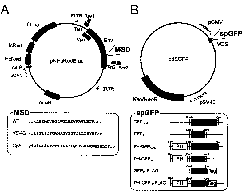
[0059] (1) Split fluorescent protein and PH structural domain and the construction of expression vector:

[0060] In this example, the PH domain of human phospholipase Cδ was assembled and synthesized by 10 oligonucleotides with a length of 79 bases. These oligonucleotide combinations were assembled by PCR (94°C for 30 seconds, 50°C for 30 seconds, 72°C for 40 seconds for 30 cycles). Similarly, the optimized GFP gene is assembled and synthesized by 30 40nt oligonucleotides each with 18 base overlaps, named GFPopt 1-11 . These two amplified sequences were then cloned into CR4Blunt-TOPO and sequenced. GFPopt 1-11 Split into GFP by PCR 1-10 (1-642bp) and GFP 11 (643-696bp), and cloned into pCR4Blunt-TOPO. PH-GFP 1-10 and PH-GFP 11 The gene is obtained by combining the PH domain gene and the split GFP gene, and then cloned into pdEGFP to construct the corresponding expression vector pdPH-GFP 1-10 and pdPH-GFP 11 ( figure 1 B). By introducing the FLAG tag sequence in th...

Embodiment 2

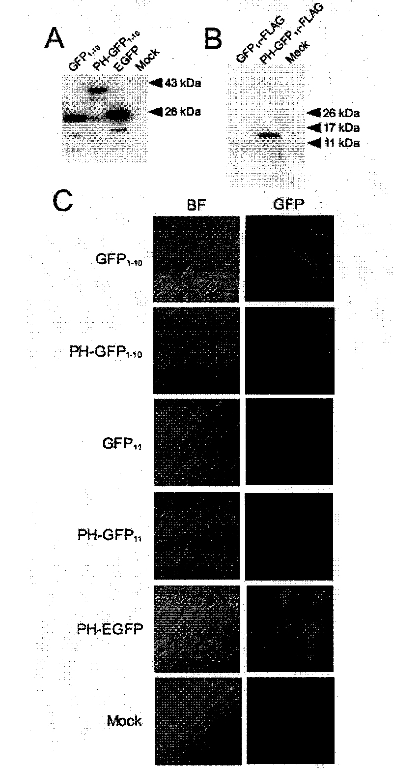
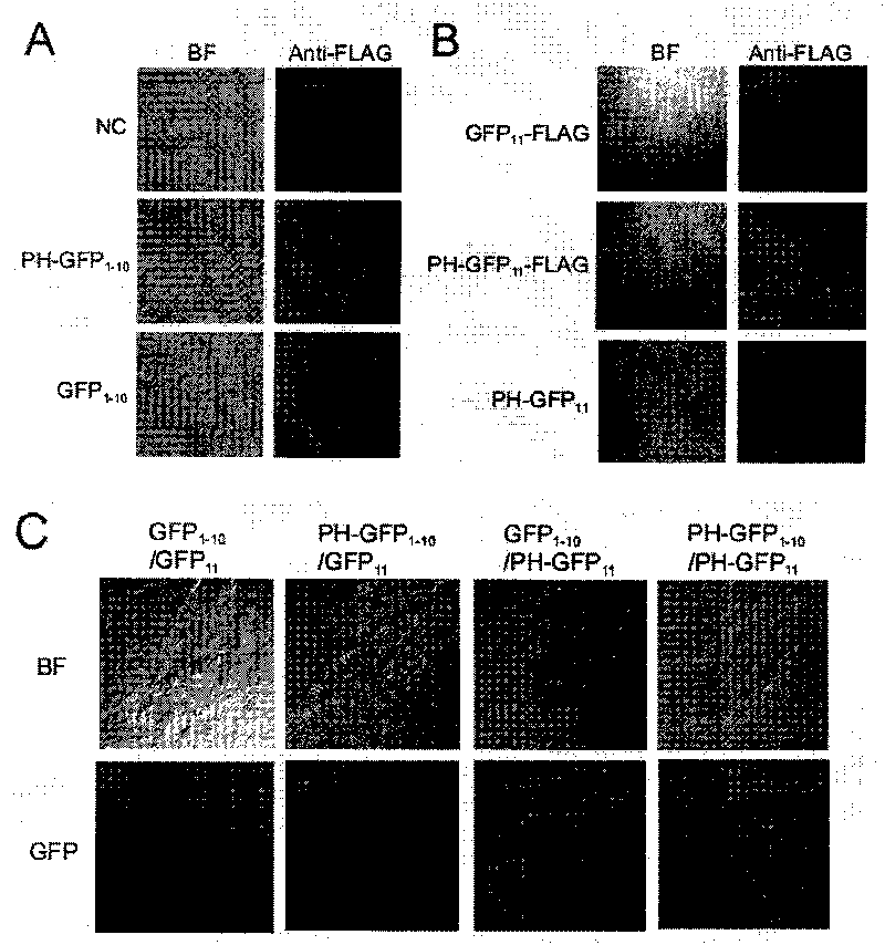
[0076](1) Complementation experiments of split GFP protein and PH-fused split GFP protein in mammalian cells.

[0077] The inventors co-transfected a pair of expression plasmids for the split GFP protein into 293FT cells. When two split GFP protein fragments are co-transfected, a green fluorescent signal can be observed ( image 3 C). and figure 2 and image 3 Consistent with the data shown in the PH-GFP 11 Can produce green fluorescent signal, while transfer into GFP 11 You can't. When PH-GFP 11 with GFP 1-10 When co-transfected, a uniform green fluorescent signal can be observed in the co-transfected cells. This data turns out to suggest that the association of the two fragments occurs before their localization to the plasma membrane. PH-GFP 1-10 and PH-GFP 11 During co-transfection, the main green fluorescent signal was observed at the edge of the co-transfected cells, and some green fluorescent signals were also observed in the cytoplasm. The latter may reflec...


PUM


Abstract

The invention relates to a combination and an expression vector of fusion protein for separating fluorescent protein, a method for screening receptors of viral envelope protein by detecting fluorescence of self re-combined fluorescent protein in the combination of the fusion protein for separating the fluorescent protein in cell fusion, and a method for screening accelerants or inhibitors in membrane fusion.

Description

technical field [0001] The present invention relates to a combination of a fusion protein of split fluorescent proteins and an expression vector thereof, and a method for screening receptors of viral envelope proteins through the combination of fusion proteins of split fluorescent proteins, and a method for screening promoters or promoters of membrane fusion Inhibitor method. In particular, it is possible to screen for receptors of viral envelope proteins by displaying fluorescence on cell membranes, and to screen for promoters or inhibitors of membrane fusion. Background technique [0002] Membrane fusion is a very common phenomenon in biological systems. Membrane fusion is involved in myogenesis, fertilization, and vesicle trafficking. Enveloped viruses that infect host cells also rely on membrane fusion. Membrane fusion is the process by which two separate compartments separated by two membranes merge to form a single compartment. One can monitor this process using a ...

Claims


Application Information

Patent Timeline
Patent Type & Authority Applications(China)
IPC IPC(8): C07K19/00C12N15/63G01N21/64C12Q1/02
Inventor 松田善卫近藤直幸岩本爱吉王健琪
Owner THE UNIV OF TOKYO
Why Eureka
Try Eureka