Cell opto-acoustic microscopic imaging method and device thereof

A technology of photoacoustic microscopy and imaging method, applied in measurement devices, generation of ultrasonic/sonic/infrasonic waves, material analysis using sonic/ultrasonic/infrasonic waves, etc. lower problem

Inactive Publication Date: 2010-07-21
SOUTH CHINA NORMAL UNIVERSITY
View PDF3 Cites 10 Cited by
  • Summary
  • Abstract
  • Description
  • Claims
  • Application Information

AI Technical Summary

Problems solved by technology

However, due to the relatively low resolution of current photoacoustic imaging technology, which can only reach a resolution of 15 microns, it is impossible to observe structural and functional images at the cellular level.

Method used


Image

  • Cell opto-acoustic microscopic imaging method and device thereof
  • Cell opto-acoustic microscopic imaging method and device thereof
  • Cell opto-acoustic microscopic imaging method and device thereof

Examples


Embodiment Construction

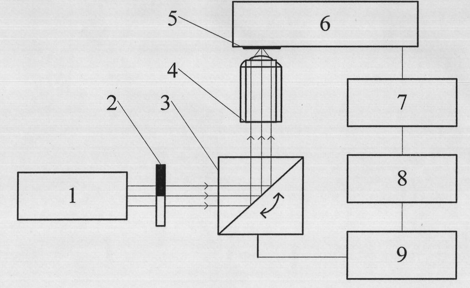
[0021] Such as figure 1 As shown, the device of the present invention includes three parts: a laser scanning imaging mechanism, a photoacoustic sensor and a signal processor. Wherein, the laser scanning imaging mechanism is composed of a laser 1, a scanning vibrating mirror 3, and a microscopic objective lens 4; during operation, the scanning vibrating mirror 3, the microscopic objective lens 4 and the photoacoustic sensor 6 can be installed on the microscope bracket;

[0022] The signal processor is composed of a chopper 2, a lock-in amplifier 7, an acquisition card 8 and a computer 9, which are electrically connected in turn.

[0023] The working process of the present invention is as follows: the laser light emitted by the laser device 1 is modulated by the chopper 2, then enters the laser scanning vibrating mirror 3 for two-dimensional scanning, and then is focused on the surface of the sample 5 by the microscopic objective lens 4. The photoacoustic signal is detected by ...


PUM


Abstract

The invention relates to a cell opto-acoustic microscopic imaging method, which comprises the following steps: placing a cell on the concave surface of microscopic glass, irradiating the cell with laser light permeating the microscopic glass, producing opto-acoustic effect after the cell absorbs the light, transferring acoustic pressure variation produced by the opto-acoustic effect to an opto-acoustic sensor, and detecting the opto-acoustic variation and outputting an opto-acoustic signal by using the opto-acoustic sensor to realize opto-acoustic detection of the single cell; and performing two-dimensional scanning on the cell by using light beam scanning and a micro objective with high resolution to realize the opto-acoustic microscopic imaging of the single cell. The invention also relates to a cell opto-acoustic microscopic imaging device, which comprises a laser light scanning imaging mechanism, the opto-acoustic sensor and a signal processor. In the method and the device, light beam scanning technology without mechanical noise is combined with the micro objective with the high resolution to generate the opto-acoustic signal with high spatial resolution and then the opto-acoustic sensor is adopted for the opto-acoustic detection to perform the opto-acoustic microscopic imaging on the cell, wherein the solution is less than 1 micron.

Description

technical field [0001] The invention relates to microscopic imaging technology, in particular to a cell photoacoustic microscopic imaging method, and also relates to a cell photoacoustic microscopic imaging device. Background technique [0002] Photoacoustic imaging adopts the method of "photoexcitation-induced photoacoustic signal-photoacoustic detection-image reconstruction". Photoacoustic imaging technology has significant advantages over many traditional imaging technologies in terms of image resolution, contrast, and information volume. Currently, photoacoustic imaging methods mainly include the following three imaging methods: acoustic lens imaging, phase-controlled focusing imaging, and inversion Imaging method. The acoustic lens imaging method is limited by the acoustic wave diffraction effect and the acoustic lens aberration, and the resolution can only reach the level of 1 mm; the phased focusing imaging method uses multiple linear array ultrasonic transducers com...

Claims


Application Information

Patent Timeline
Patent Type & Authority Applications(China)
IPC IPC(8): G01N21/63G01N29/34
CPCG01N29/0681G01N29/2418
Inventor 唐志列谭治良吴泳波
Owner SOUTH CHINA NORMAL UNIVERSITY
Why Eureka
Try Eureka