Resonant magnetic induction bioelectrical impedance tomography method and equipment adopted by same

A bioelectrical impedance and tomographic imaging technology, applied in the field of biological tissue electrical impedance tomography and resonant magneto-inductive electrical impedance measurement, can solve the problems that cannot be satisfied at the same time, affect imaging results, increase coil crosstalk, etc., and achieve ideal imaging effects. Sensitive effect

Inactive Publication Date: 2010-09-08
SHENYANG POLYTECHNIC UNIV
View PDF0 Cites 17 Cited by
  • Summary
  • Abstract
  • Description
  • Claims
  • Application Information

AI Technical Summary

Problems solved by technology

[0004] The current magnetic induction imaging technology adopts the same design scheme of the excitation coil and the detection coil. This structure cannot meet the requirements of generating a strong excitation magnetic field and high sensitivity of the detection coil at the same time. Crosstalk between coils, affecting imaging results

Method used


Image

  • Resonant magnetic induction bioelectrical impedance tomography method and equipment adopted by same
  • Resonant magnetic induction bioelectrical impedance tomography method and equipment adopted by same
  • Resonant magnetic induction bioelectrical impedance tomography method and equipment adopted by same

Examples


Embodiment Construction

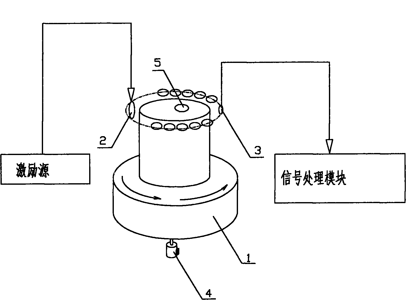
[0047] Such as figure 1 , 7 As shown, the resonant magnetic induction bioelectrical impedance tomography method adopts multi-channel resonance signal detection, through signal processing and reconstruction, to realize tomographic imaging of biological tissue electrical impedance, and its specific steps include:

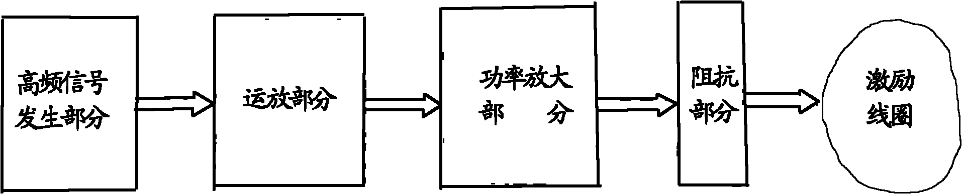
[0048] (1) Place the measured object in the exciting magnetic field;

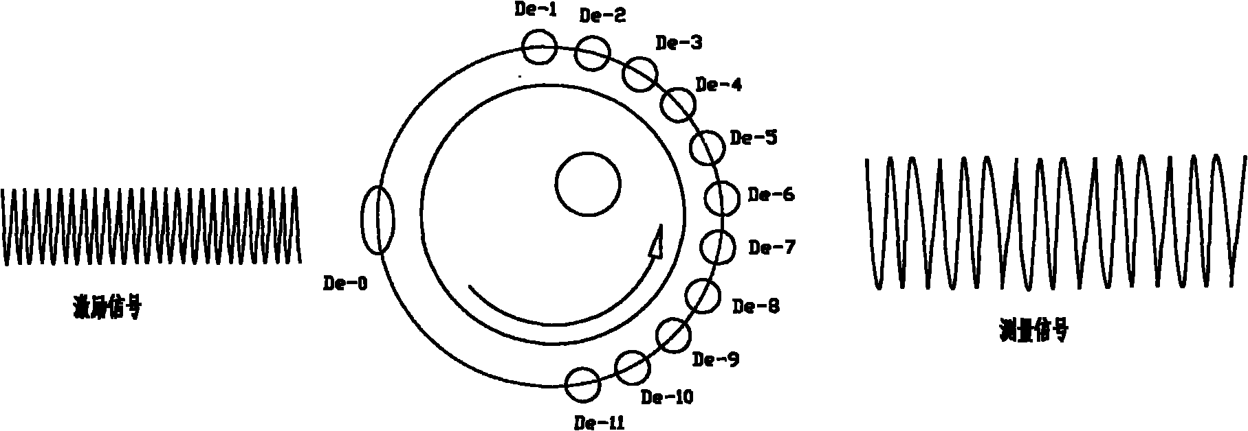
[0049] (2) Detect the disturbance magnetic field generated by the measured object;

[0050] (3) Perform nonlinear data processing and image reconstruction.

[0051]In the described step (1), an excitation coil that feeds a sinusoidal alternating current is adopted to generate an excitation magnetic field; the excitation coil adopts a single-layer coil that adds a yoke in the middle; The coil is the acquisition terminal to realize the detection of the disturbance magnetic field; the step (3) realizes image reconstruction by using the signal amplitude and phase obtained by the detection coil.

[...


PUM


Abstract

The invention belongs to the field of bioelectrical impedance tomography, and particularly relates to a resonant magnetic induction bioelectrical impedance measuring method and equipment matched the same. The method adopts a multi-channel resonance signal for detection, and comprises the following steps: (1) placing a detected object in a resonance magnetic field; (2) detecting a disturbed magnetic field produced by the detected object; and (3) performing non-linear data processing and image reconstruction. The equipment comprises an excitation part, a detected object bearing platform (1) and a detection part; the excitation part comprises an excitation source and an excitation coil (2); the output end of the excitation source is connected with the input end of the excitation coil (2); the detection part comprises a detection coil (3) and a signal processing module; the excitation coil (2) of the excitation part and the detection coil (3) of the detection part are placed on the detected object bearing platform (1); and the output end of the detection coil (3) is connected with the input end of the signal processing module. The equipment has high sensitivity and ideal imaging effect, and can be used for bioelectrical impedance section analysis and imaging analysis.

Description

technical field [0001] The invention belongs to the field of biological electrical impedance imaging, and in particular relates to a resonant magnetic induction electrical impedance measurement, a nonlinear image reconstruction biological tissue electrical impedance tomography method and the equipment used. Background technique [0002] Due to the different types of cells in the tissue, the way the cells are arranged (such as the degree of density), the electrolyte concentration in the intercellular substance, and the permeability of the cell membrane, etc., different biological tissues and the same tissue in different states have different appearances. Different electrical impedance characteristics. In addition to realizing functions similar to X-ray imaging, computed tomography (CT), magnetic resonance imaging (MRI), and ultrasound imaging, bioelectrical impedance imaging can also obtain images that reflect changes in the physiological state of biological tissues. It has ...

Claims


Application Information

Patent Timeline
Patent Type & Authority Applications(China)
IPC IPC(8): A61B5/05
Inventor 柯丽杜强
Owner SHENYANG POLYTECHNIC UNIV
Why Eureka
Try Eureka