Cerebral edema simulator

A simulation device and brain edema technology, which is applied in the field of biomedical medical devices, can solve the problems of inconvenient adjustment of geometric size, change of electrical conductivity and irregular shape, inability to simulate the irregularity of brain edema and the formation of arbitrary positions, etc., to achieve The effect of simple structure, reasonable design and low product cost

Inactive Publication Date: 2011-11-23
ARMY MEDICAL UNIV
View PDF0 Cites 0 Cited by
  • Summary
  • Abstract
  • Description
  • Claims
  • Application Information

AI Technical Summary

Problems solved by technology

[0005] At present, in the research of brain edema monitoring technology, the plexiglass ball model and agar block are used to simulate brain edema. The production of agar block for simulating brain edema is not convenient to adjust the geometric size, the change of electrical conductivity and the formation of irregular shapes in the experiment.
Scholars in Mexico and the United States used the experimental model of double-layer plexiglass spheres to simulate brain edema, which realized the convenient adjustment of the volume of brain edema in the experiment, but it could not simulate the irregularity of the actual region of brain edema and the formation of arbitrary positions.

Method used


Image

  • Cerebral edema simulator
  • Cerebral edema simulator
  • Cerebral edema simulator

Examples


Embodiment 1

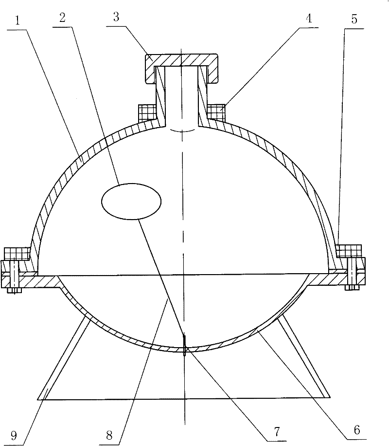
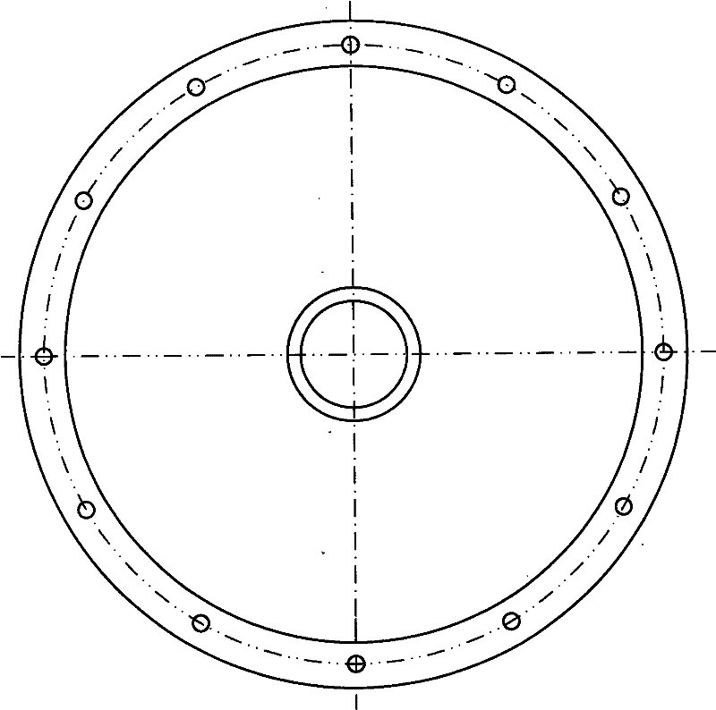
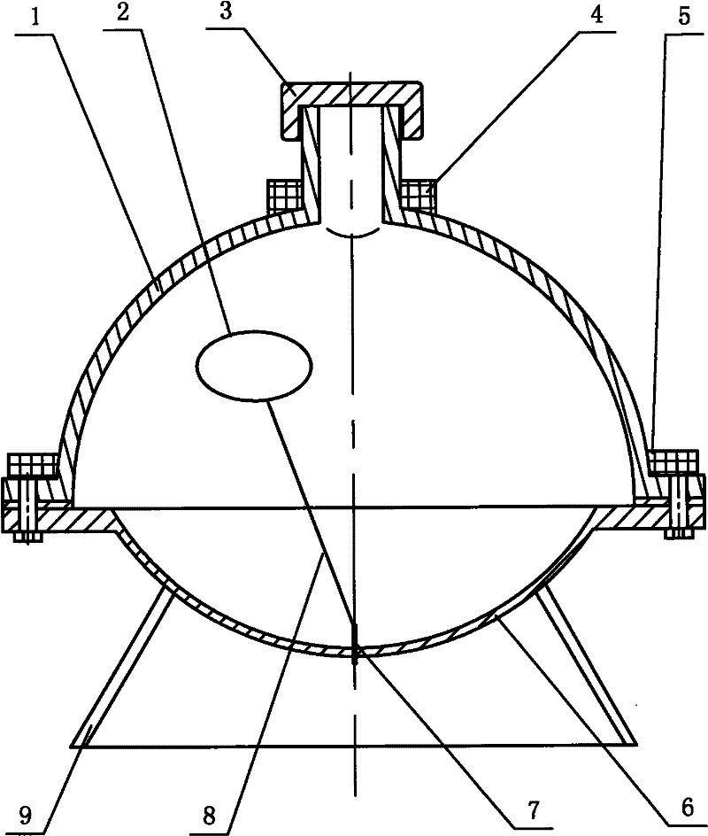
[0017] exist figure 1 Among them, the cerebral edema simulation device of this embodiment is composed of an upper casing 1, a skin expander 2, a top cover 3, an excitation coil 4, a detection coil 5, a lower casing 6, a syringe 7, a communication tube 8, and a support frame 9. .

[0018] The support frame 9 of the present embodiment is a plexiglass product, and its shape is trapezoidal. The upper end of the support frame 9 is bonded with a lower casing 6. The shape of the lower casing 6 is spherical, and the lower casing 6 is transparent. Body is pressed with plexiglass, and the upper end film pressing molding of lower housing 6 is connected as a whole with lower housing 6 and has lower connecting plate. The upper casing 1 is in the shape of a hemispherical crown, and the upper casing 1 is a transparent body, which is made of organic glass. The lower end of the upper casing 1 is molded and connected with the upper casing 1. The radius of the upper housing 1 is the same as th...

Embodiment 2

[0020] The volume of the upper case 1 in this embodiment is equal to the volume of the lower case 6 . Other components and the coupling relationship of the components are the same as in Embodiment 1.

[0021] The working principle of the present invention is as follows:

[0022] The sodium chloride aqueous solution with the same conductivity as that of brain tissue is added into the spherical shell to simulate the electrical parameters of brain tissue, and the sodium chloride aqueous solution with the same conductivity as that of brain edema is injected into the skin expander 2 to simulate the electrical parameters of brain edema tissue. Electrical parameters. Regulating the concentration of the sodium chloride aqueous solution in the skin dilator 2, i.e. the conductivity, through the syringe 7 can simulate the severity of cerebral edema, and adjusting the volume of the sodium chloride aqueous solution in the skin dilator 2 can simulate the geometric size of the cerebral edem...


PUM


Abstract

The invention discloses a cerebral edema simulator. The upper end of a support frame is provided with a lower shell on which an injector is arranged; the upper end of the lower shell is provided with a spherical shell formed by an upper shell; the top of the upper shell is provided with a head cover, the upper part is provided with an exciting coil, and the lower part is provided with a detectioncoil; the injector is communicated with a skin expander arranged in the spherical shell by a communication pipe, sodium chloride solution with the same electrical conductivity as normal brain tissuesis filled in the spherical shell, and sodium chloride solution with the same electrical conductivity as cerebral edema tissues is injected in the skin expander. The skin expander forms irregular cerebral edema zones in different positions in a skull model to truly stimulate the existing situation of cerebral edema in skull, the severity of cerebral edema can be stimulated by regulating the concentration (namely electrical conductivity) of sodium chloride solution in the skin expander, and the geometric size of cerebral edema can be stimulated by regulating the volume of sodium chloride solution in the skin expander. The invention can be applied to the research of a cerebral edema monitoring technology.

Description

technical field [0001] The invention belongs to the technical field of biomedical medical devices, and in particular relates to a brain edema simulation device. Background technique [0002] Intracerebral surgical diseases such as various craniocerebral trauma, brain tumor resection, cerebral ischemia, brain tumor, encephalitis, and high altitude cerebral edema have entered the top three common fatal lesions in terms of morbidity and mortality. These intracerebral surgical diseases are accompanied by symptoms of cerebral edema. Monitoring the severity of cerebral edema is an important basis for doctors to understand the true condition of the disease and guide treatment. [0003] At present, there are mainly two routine detection methods for cerebral edema patients at home and abroad. One is to diagnose with CT, MRI and other equipment. This method is not only expensive, but also not suitable for patients who are critically ill and cannot be moved repeatedly. The other is ma...

Claims


Application Information

Patent Timeline
Patent Type & Authority Patents(China)
IPC IPC(8): G09B23/28
Inventor 秦明新李文哲马珂
Owner ARMY MEDICAL UNIV
Why Eureka
Try Eureka