Processing method and system of CT image

A CT image and processing system technology, applied in the field of medical image analysis, to achieve the effect of reducing the misdiagnosis/missing diagnosis rate

Active Publication Date: 2010-12-15
SHENZHEN INST OF ADVANCED TECH CHINESE ACAD OF SCI
View PDF1 Cites 52 Cited by
  • Summary
  • Abstract
  • Description
  • Claims
  • Application Information

AI Technical Summary

Problems solved by technology

However, for subarachnoid hemorrhage with small bleeding volume and diffuse distr...

Method used


Image

  • Processing method and system of CT image
  • Processing method and system of CT image
  • Processing method and system of CT image

Examples


Embodiment Construction

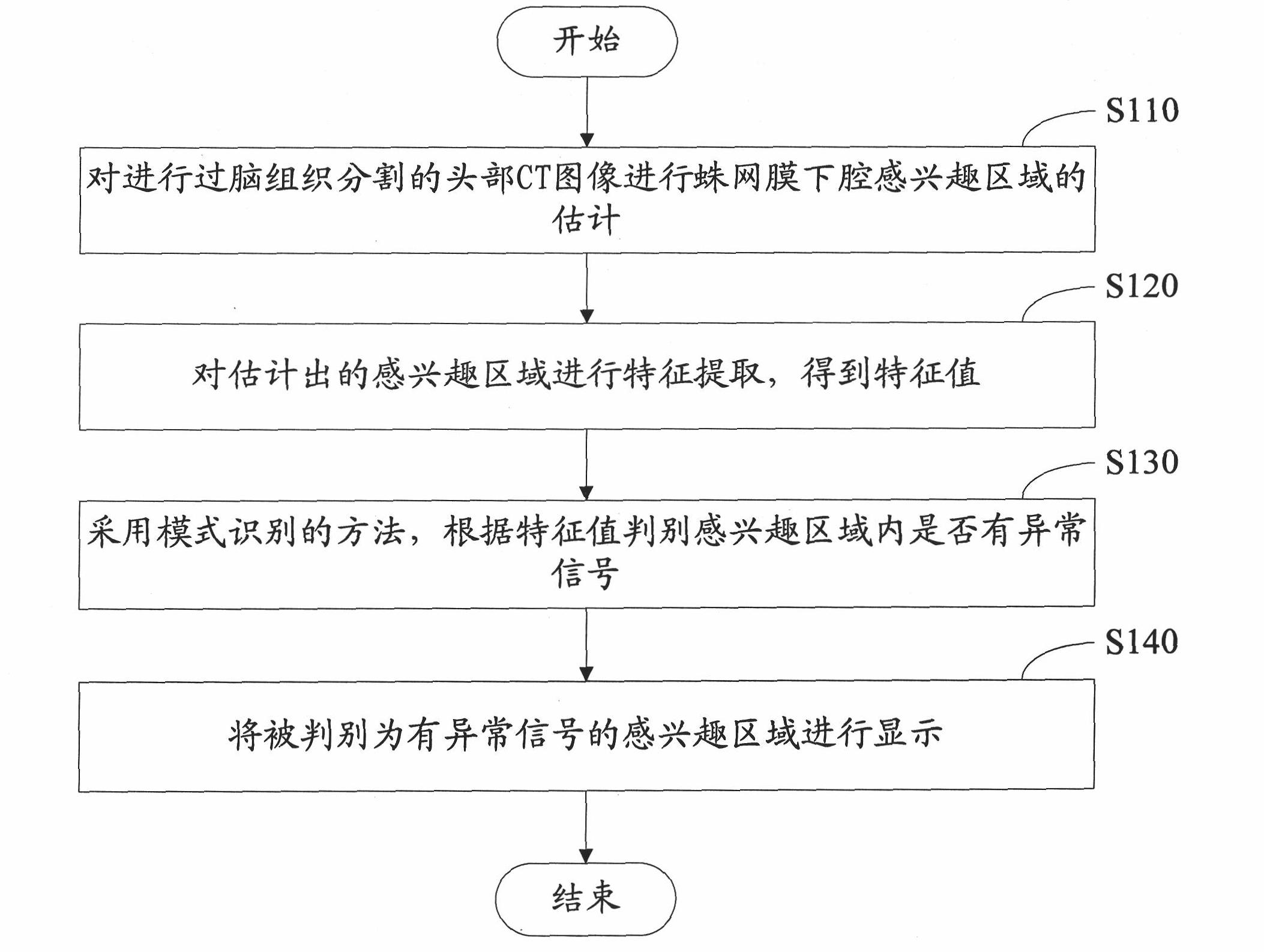
[0045] figure 1 It is a flow chart of the processing method of CT image in an embodiment, comprising the following steps:

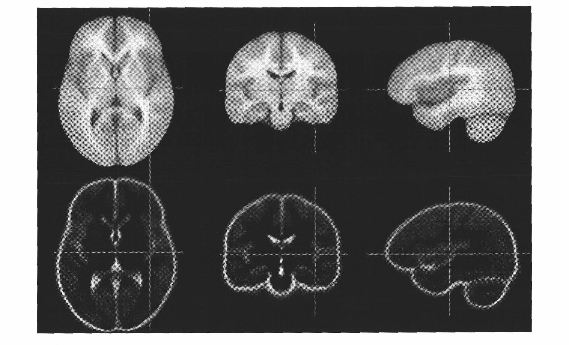
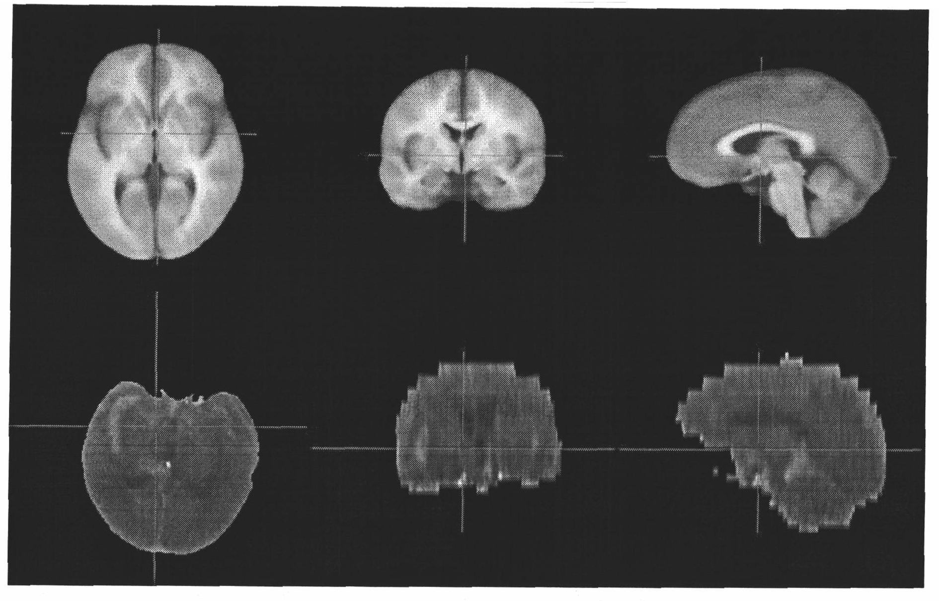
[0046] S110 , estimating a region of interest (ROI) in the subarachnoid space on the head CT image that has undergone brain tissue segmentation.

[0047] In this embodiment, an atlas-based method is used to divide and estimate the cerebrospinal fluid (CSF) region, and then the region of interest in the subarachnoid space. In other embodiments, segmentation and estimation based on grayscale, deformation model and level set may also be used. The map-based method is selected, which has more prior information and better segmentation accuracy. In particular, methods based on probabilistic atlas registration can be used to delineate CSF regions. Because the atlas not only contains the grayscale and texture information of each tissue in the object to be segmented, but also contains shape information and relative positional relationship. The probability map i...


PUM


Abstract

The invention relates to a processing system of a CT image, which comprises a CT image acquiring module, an interesting region estimating module, a characteristic extracting module, an abnormal signal identifying module and a displaying module. The CT image acquiring module is used for acquiring a head CT image subjected to brain tissue segmentation; the interesting region estimating module is used for estimating an interesting region of subarachnoid space to the head CT image; the characteristic extracting module is used for extracting the characteristic to the head CT image subjected to the estimation of the interesting region to acquire a characteristic value; the abnormal signal identifying module is used for identifying whether an abnormal signal is included in the interesting region according to the characteristic value by using a method of mode identification; and the displaying module is used for displaying the identified result and the interesting region in which the abnormal signal exists. The invention also relates to a processing method of the CT image. The invention can display a position in which the abnormal signal exists, which is referred by medical staffs, so as to reduce misdiagnosis/missed diagnosis rate of the subarachnoid space hemorrhage.

Description

【Technical field】 [0001] The invention relates to the field of medical image analysis, in particular to a method for processing CT images, and also to a processing system for CT images. 【Background technique】 [0002] The use of medical image analysis to implement computer-aided diagnosis is one of the research hotspots in modern medical image analysis. Especially for major diseases with high misdiagnosis rates, there is an urgent need to study medical image analysis and processing methods and systems to provide reference information and information for disease diagnosis. Technical Support. [0003] When the subarachnoid space is normal, it is hypointensity of cerebrospinal fluid (Cerebro-Spinal Fluid, CSF) on CT (X-ray computed tomography) image, if it is not low signal, there may be abnormalities such as lesions, especially subarachnoid hemorrhage (Subarachnoid hemorrhage, SAH). For non-low abnormal signals in the subarachnoid space, it is difficult for doctors to make j...

Claims


Application Information

Patent Timeline
IPC IPC(8): G06T7/00A61B6/03
Inventor 胡庆茂
Owner SHENZHEN INST OF ADVANCED TECH CHINESE ACAD OF SCI
Why Eureka
Try Eureka