Human tissue autofluorescence detection system based on excitation of light sources with different wavelength

A technology of human tissue and autofluorescence, applied in fluorescence/phosphorescence, material excitation analysis, etc., can solve the problems of ineffective excitation, information overlapping analysis, low sensitivity and specificity, etc., and achieve high sensitivity and specificity detection , Simplify the process of data processing, the best excitation and detection effect

Inactive Publication Date: 2011-02-16
FUJIAN NORMAL UNIV
View PDF7 Cites 13 Cited by
  • Summary
  • Abstract
  • Description
  • Claims
  • Application Information

AI Technical Summary

Problems solved by technology

[0003] In view of the fact that the existing human tissue autofluorescence detection system uses a single-wavelength or broad-spectrum light source as the excitation light source, it fails to effectively excite the fluorescence information of endogenous fluorescent substances with diagnostic significance, and the sensitivity and specificity are not high in clinical applications. Insufficient, the present invention provides a human tissue autofluorescence detection system based o

Method used


Image

  • Human tissue autofluorescence detection system based on excitation of light sources with different wavelength
  • Human tissue autofluorescence detection system based on excitation of light sources with different wavelength

Examples


Embodiment 1

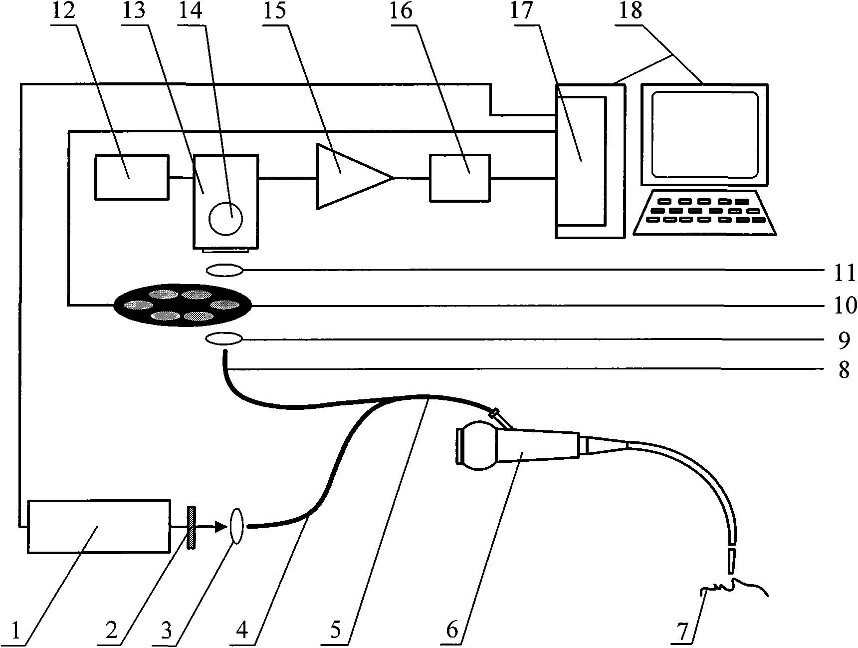
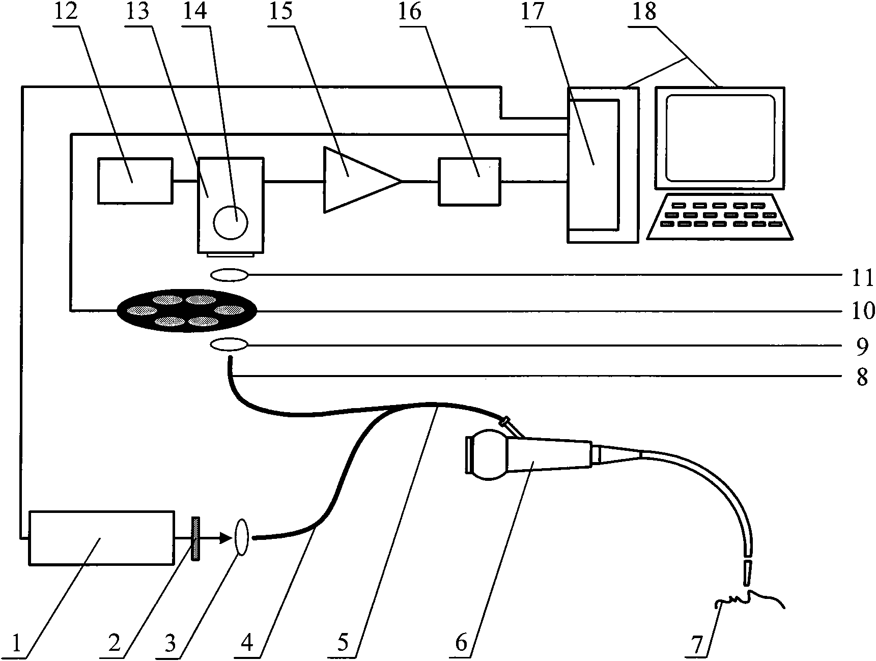
[0014] Such as figure 1 Shown is a diagram of the autofluorescence detection system based on excitation at different wavelengths. A case of human colon tissue was selected for the study of autofluorescence spectral characteristics. Before carrying out the experiment, the cooler (13) (C9144, Hamamatsu Corp., Japan) was opened and cooled for 30 minutes by the cooling voltage controller (12), so that the photomultiplier tube (14) (R928, Hamamatsu Corp., Hamamatsu, Japan) reached a stable working state. According to the experimental requirements, the computer (18) is turned on, and three thresholds I corresponding to the colon tissue have been stored in advance. T1 and I T2 , its numerical value is respectively 30000 and 3, turn on the tunable laser (1) (PIV OPO 355, OPOTEK, USA), adjust the output wavelength of the tunable laser (1) by the control software on the computer (18) to be 400nm, control simultaneously Select a filter with a center wavelength of 635 nm on the filter...

Embodiment 2

[0016] Another case of human colon tissue was selected for the study of autofluorescence spectral characteristics. According to the process of embodiment 1, at first select 400nm excitation light, detect the fluorescent signal I of 635nm 635 , the recorded value is 9000, which is less than the threshold I T1 , the tissue type could not be determined and further testing was required. Next, choose 280nm excitation light to excite, and detect the fluorescence signal I of 335nm 335 , whose value is 75000. Then choose 340nm excitation light, detect the fluorescent signal I of 390nm and 460nm 390 and I 460 , whose values ​​are 70000 and 81000 respectively. Then choose 450nm excitation light to excite, and detect the fluorescence signal I at 535nm 535 , whose value is 10000. Finally, choose 460nm excitation light, detect the fluorescence signal I of 520 520 , whose value is 18000. Calculate I r =(I 460 +I 520 +I 535 )×I 335 / (I 390 ) 2 The result is 1.67, which is les...

Embodiment 3

[0018] Another case of human colon tissue was selected for the study of autofluorescence spectral characteristics. According to the process of embodiment 1, at first select 400nm excitation light, detect the fluorescent signal I of 635nm 635 , the recorded value is 60000, which is higher than the threshold I T1 , indicating that the porphyrin content is relatively high, and it is determined that the tissue to be tested is a colon tumor tissue.


PUM


Abstract

The invention relates to an autofluorescence detection system for exciting specific endogenous fluorescent substances in a human tissue by light sources with different wavelengths and detecting information of fluorescence with specific wavelength corresponding to the fluorescent substances in endoscopy for the human early tumor tissue. The autofluorescence detection system mainly consists of an excitation light source part, an excitation-detection-acquisition part, an optical collection part, a signal preprocessing part and a computer part. The autofluorescence detection system is characterized in that a tunable laser (1) can adjust the needed pulse laser wavelengths according to the specific endogenous fluorescent substances in the human tissue, and a filter wheel (10) is equipped with narrow band pass filters with different center wavelengths. The system can excite the specific endogenous fluorescent substances in the human tissue by a plurality of optimal excitation wavelengths sequentially, thus avoiding the problem of no benefit for analysis owing to insufficient fluorescence information or information overlap and the like caused by excitation of single wavelength or broad spectrum light sources, and improving sensitivity and specificity of early tumor detection.

Description

technical field [0001] The invention belongs to biomedical instruments, and relates to a human tissue autofluorescence detection system, especially an endoscopic examination that can be used for human early tumor tissue, using different wavelength light sources to excite specific endogenous fluorescent substances in human tissue, And an autofluorescence detection system that detects fluorescence information of specific wavelengths corresponding to these fluorescent substances. technical background [0002] The existing human tissue autofluorescence detection system is to use a common endoscope and a single-wavelength or wide-spectrum light source to excite endogenous fluorescent substances in human tissue, and use the difference between the autofluorescence spectra of normal and tumor tissues to detect human tissue. Fluorescence detection system. The autofluorescence of human tissue mainly comes from substances such as amino acids, structural proteins, enzymes, coenzymes, f...

Claims


Application Information

Patent Timeline
IPC IPC(8): G01N21/64
Inventor 李步洪刘丽娜陈德福聂英斌黄志勇李伟华谢树森
Owner FUJIAN NORMAL UNIV
Why Eureka
Try Eureka